Hypothalamus & Tuber Cinereum

Reference Images

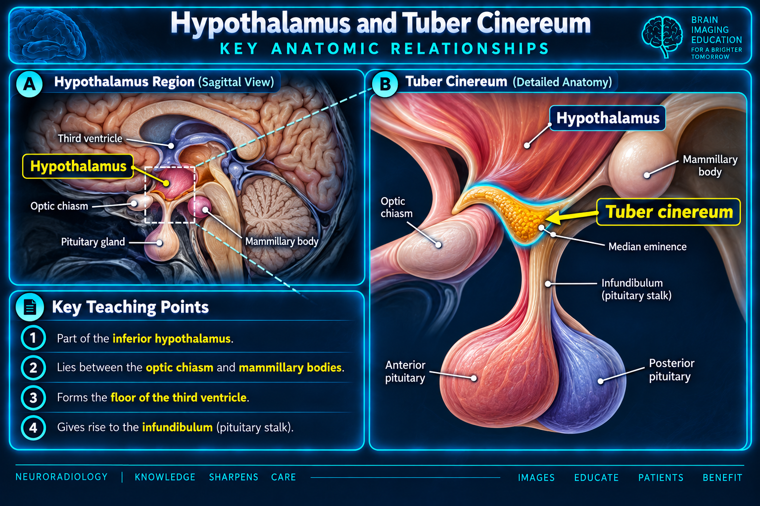
Sagittal hypothalamus and detailed tuber cinereum anatomy
Anatomic RelationshipsClick to enlarge
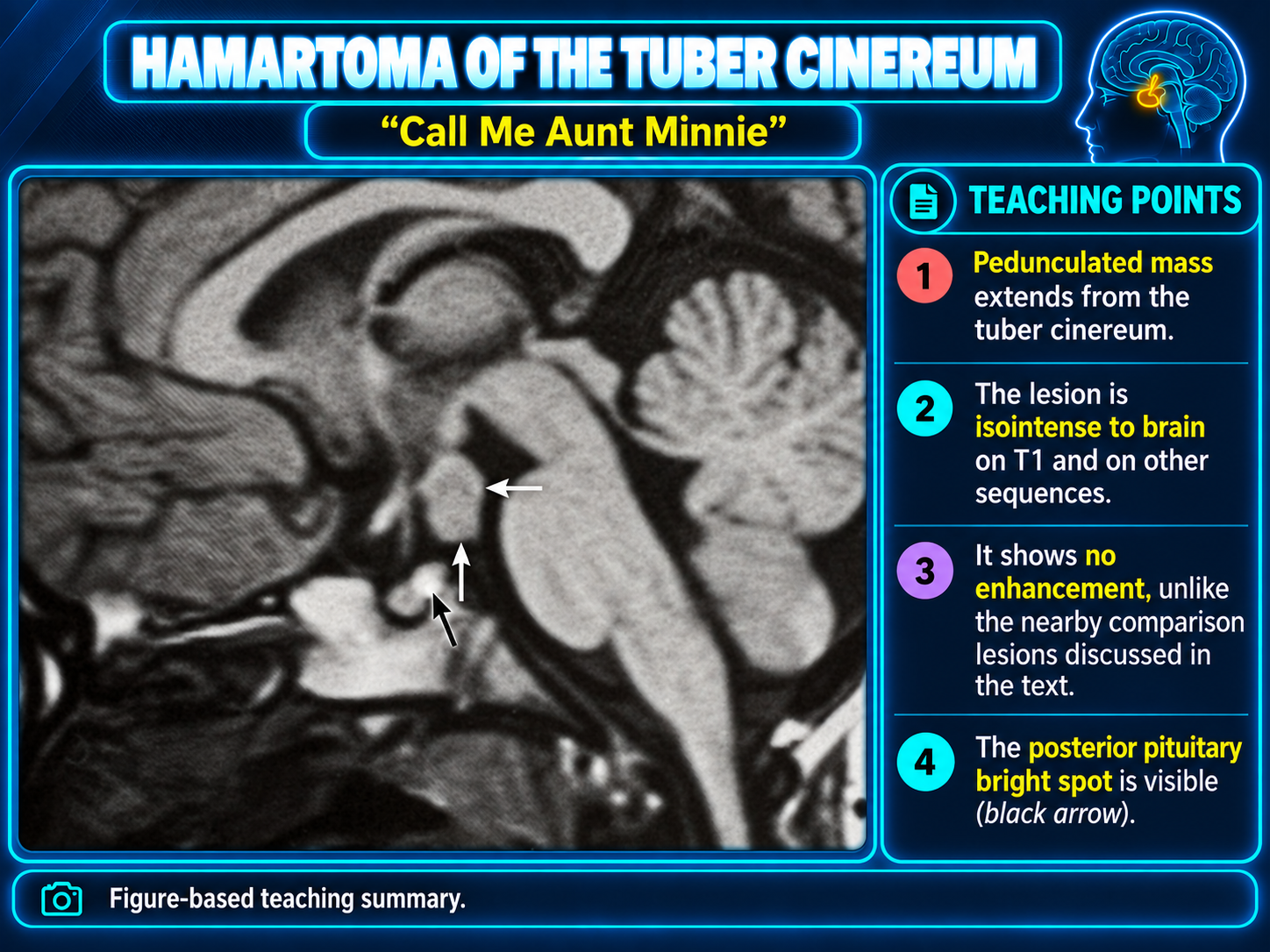
Pedunculated hamartoma arising from the tuber cinereum
Tuber Cinereum HamartomaClick to enlarge
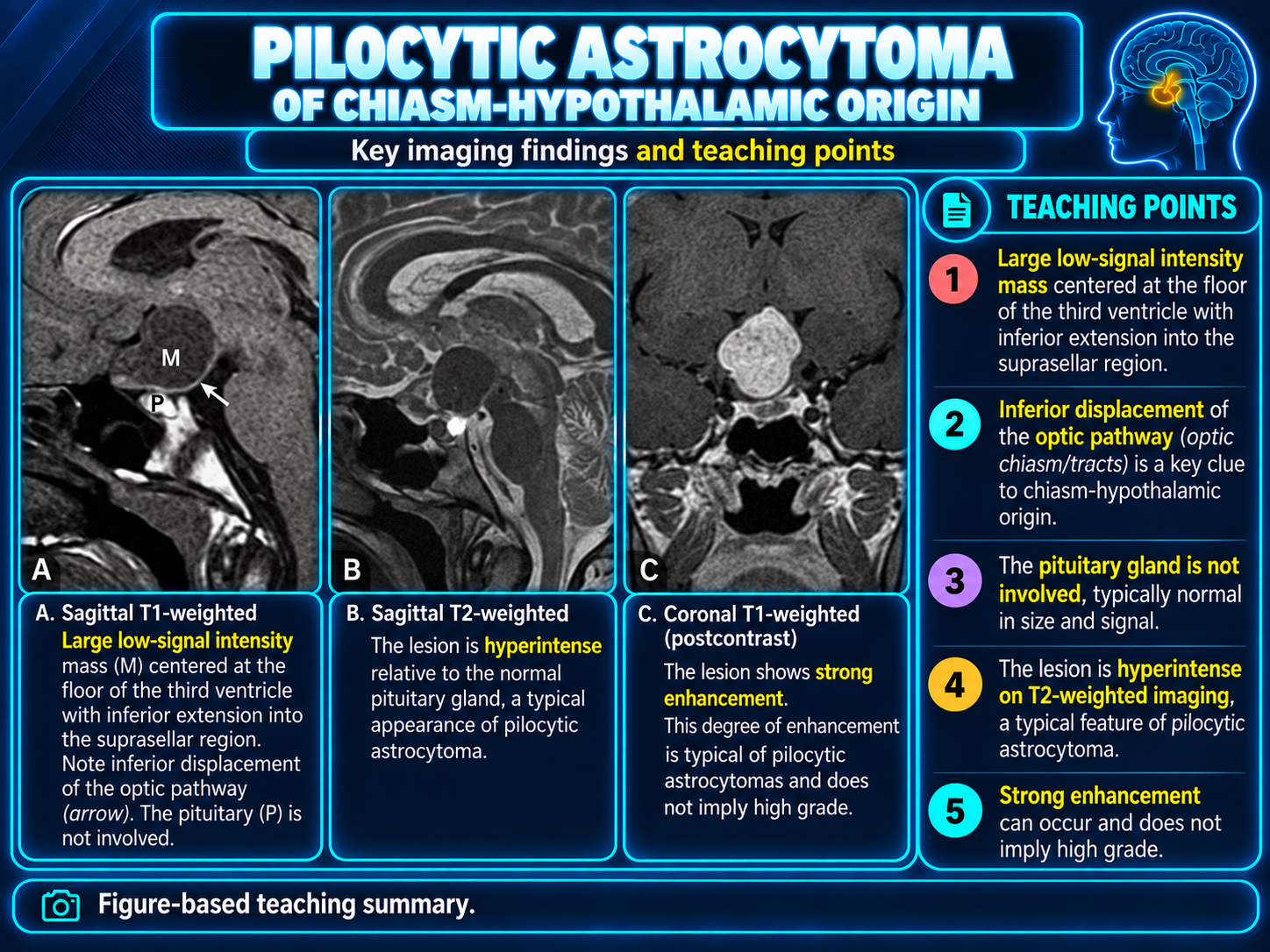
Pilocytic astrocytoma of chiasmatic hypothalamic origin
Chiasmatic Hypothalamic AstrocytomaClick to enlarge

Overview

The tuber cinereum is part of the hypothalamus. It is the thin midline gray-matter prominence forming the inferior hypothalamus and floor of the anterior third ventricle between the optic chiasm and mammillary bodies. The median eminence and infundibulum arise from its central anterior-inferior aspect.

Core imaging conceptA normal tuber cinereum is a thin ventricular-floor structure, not a discrete mass. A sharply marginated, nonenhancing lesion attached here strongly favors hypothalamic hamartoma.

Anatomic Boundaries

BoundaryPractical landmark
AnteriorLamina terminalis; optic chiasm inferiorly and anterior commissure superiorly
SuperiorHypothalamic sulcus or approximate AC-PC plane separating hypothalamus from thalamus
PosteriorPlane from the posterior commissure toward the mammillary bodies
InferiorOptic chiasm, tuber cinereum, median eminence, infundibulum, and mammillary bodies
MedialThird ventricle
LateralOptic tracts, internal capsule and subthalamic region, and basal forebrain interfaces

Regional Organization

Anterior to posterior divisions are preoptic, anterior or supraoptic, tuberal, and mammillary or posterior. Mediolateral zones are periventricular, medial, and lateral.

  • The tuber cinereum is a surface landmark rather than a discrete nucleus.
  • The broader tuberal region contains or abuts arcuate, ventromedial, dorsomedial, lateral hypothalamic, and tuberomammillary territories.

Median Eminence

The median eminence is a specialized neurovascular interface containing portal capillaries and tuberoinfundibular neuronal terminals. It conveys hypothalamic releasing and inhibiting factors to the anterior pituitary.

  • It behaves as a circumventricular structure with an atypical blood-brain barrier.
  • Very thin linear midline enhancement may be physiologic.
  • Mass-like or nodular enhancement is abnormal.

MRI Localization

Routine MRI does not directly resolve most hypothalamic nuclei. Localize disease using:

  • Third ventricle and hypothalamic sulcus.
  • Optic chiasm and tracts.
  • Infundibular recess and pituitary stalk.
  • Mammillary bodies and fornices.
  • Anterior commissure and mammillothalamic tracts.

Functional Map

StructureMajor roleDysfunction pattern
Suprachiasmatic nucleusCircadian pacemaker receiving retinal inputSleep-wake and circadian disturbance
Supraoptic nucleusPredominantly vasopressin productionCentral arginine-vasopressin deficiency
Paraventricular nucleusOxytocin, vasopressin, CRH, and autonomic integrationPituitary-axis and autonomic abnormalities
Arcuate or infundibular nucleusReleasing hormones and energy sensingPituitary-axis and appetite or metabolic disturbance
Ventromedial regionSatiety and metabolic regulationHyperphagia and weight gain after destructive injury
Lateral hypothalamic regionFeeding and arousal networkReduced appetite or altered arousal
Anterior or preoptic regionHeat dissipationHyperthermic tendency when damaged
Posterior hypothalamusHeat conservation and arousalHypothermic tendency when damaged
Tuberomammillary nucleusHistaminergic wakefulnessSomnolence or disturbed arousal
Mammillary bodiesLimbic memory circuitMemory impairment; key adjacency in epileptogenic hamartomas

Targeted MRI Protocol

  • Thin sagittal and coronal T1 before and after contrast.
  • Thin T2 in at least two planes; 3D T2 for attachment and adjacent tracts.
  • Isotropic 3D T1 with approximately 1 mm or smaller voxels for hamartoma or epilepsy.
  • FLAIR, DWI/ADC, SWI, and whole-brain imaging.
  • CT when calcification or osseous change may narrow the differential.

Hypothalamic Hamartoma

A congenital non-neoplastic malformation of mature neurons and glia, usually arising from the tuber cinereum or inferior hypothalamic-mammillary region.

  • Well-circumscribed sessile or pedunculated lesion.
  • No convincing enhancement or restriction.
  • Usually mildly T1 hypointense and T2/FLAIR hyperintense to cortex.
  • No edema, invasion, or progressive enlargement.
  • Thin T2 best defines mammillary body, fornix, and mammillothalamic relationships.

Clinical Clues

  • Gelastic seizures: brief stereotyped laughing episodes, usually from posterior sessile lesions near the mammillary bodies.
  • Central precocious puberty: classic for pedunculated anterior parahypothalamic lesions.
  • Large or mixed lesions may produce both endocrine and epileptic manifestations.
  • Cognitive, behavioral, psychiatric, sleep, and autonomic comorbidities may accompany chronic epilepsy.

Hamartoma Morphology

PatternTypical attachmentMore typical presentation
Intrahypothalamic or sessileBroad posterior attachment near mammillary bodies and third-ventricular floorGelastic seizures, epileptic encephalopathy, and cognitive or behavioral disturbance
Parahypothalamic or pedunculatedProjects into the suprasellar cistern from anterior tuber cinereum or stalk regionCentral precocious puberty, often without epilepsy
MixedComponents of both patternsCombined neurologic and endocrine manifestations

Lesion Differential

EntityFeatures favoring diagnosis over hamartoma
Hypothalamic or optic pathway gliomaT2-hyperintense expansile lesion, optic pathway involvement, variable enhancement, and possible growth
CraniopharyngiomaMixed solid-cystic suprasellar lesion with calcification or proteinaceous cysts
GerminomaEnhancing stalk or suprasellar mass with diabetes insipidus and possible loss of the posterior pituitary bright spot
Inflammatory or infiltrative diseaseStalk thickening and enhancement, hypothalamic enhancement, bright-spot loss, or systemic findings
Metastasis or lymphomaEnhancing infiltrative lesion involving the stalk or hypothalamus, with diffusion or systemic clues
Posterior pituitary-region tumorSolid enhancing sellar, suprasellar, or stalk-centered mass
Vascular lesionFlow signal, pulsation artifact, or arterial continuity; obtain CTA or MRA if aneurysm is possible

Hypothalamic Syndrome

  • Obesity, cachexia, hyperphagia, or anorexia.
  • Hypopituitarism or precocious or delayed puberty.
  • Arginine-vasopressin and thirst disorders.
  • Sleep and circadian disruption.
  • Temperature or autonomic dysregulation.
  • Memory, behavioral, or mood changes.

Reporting Checklist

  • Exact epicenter and three-plane dimensions.
  • Sessile versus pedunculated morphology.
  • Relationship to mammillary bodies, fornices, mammillothalamic tracts, stalk, optic pathways, and third ventricle.
  • T1, T2, and FLAIR signal relative to gray matter.
  • Enhancement, restriction, susceptibility, cystic change, calcification, edema, and growth.
  • Hydrocephalus, posterior bright spot, and associated malformations.

Diagnostic Redirects

A classic hamartoma is nonenhancing and structurally attached to the tuber cinereum or mammillary region.

  • Enhancement or interval growth.
  • Restricted diffusion.
  • Calcification or substantial cystic change.
  • Edema or destructive behavior.
  • A stalk-centered or optic-pathway epicenter.

These features should redirect the differential toward neoplastic, inflammatory, infiltrative, or vascular disease.

Secondary Links