Pituitary Gland

Reference Images

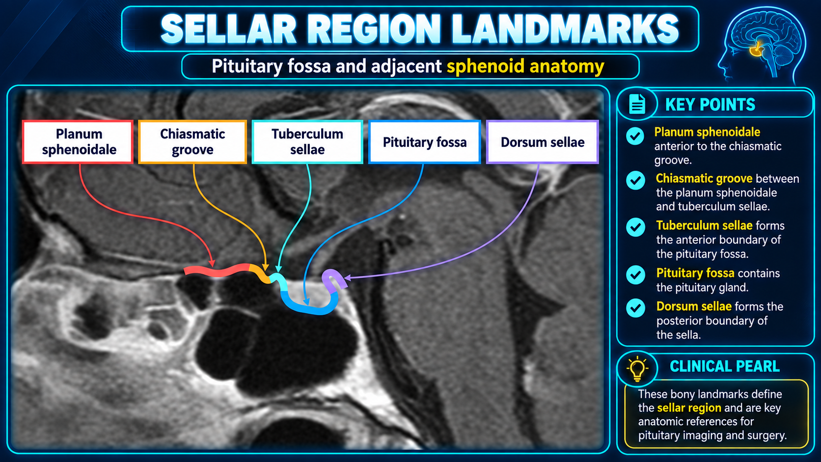
Sagittal MRI landmarks of the sellar region
Sellar Region LandmarksClick to enlarge
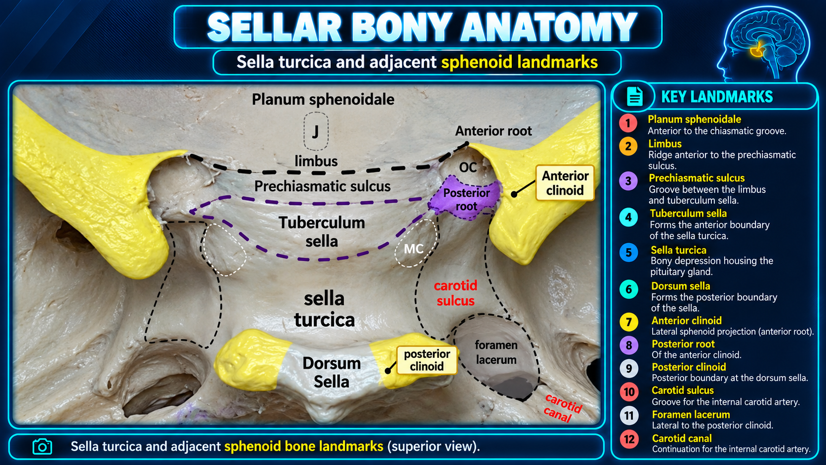
Sella turcica and adjacent sphenoid bony landmarks
Sellar Bony AnatomyClick to enlarge
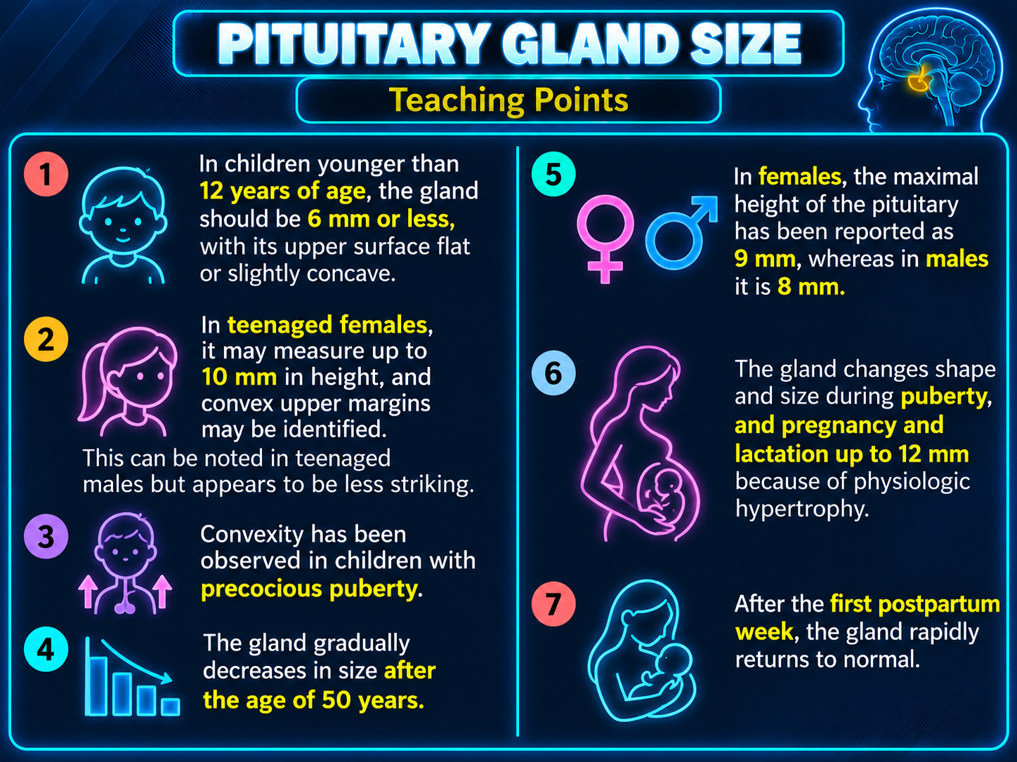
Normal pituitary gland size by age sex and physiologic state
Normal Gland SizeClick to enlarge
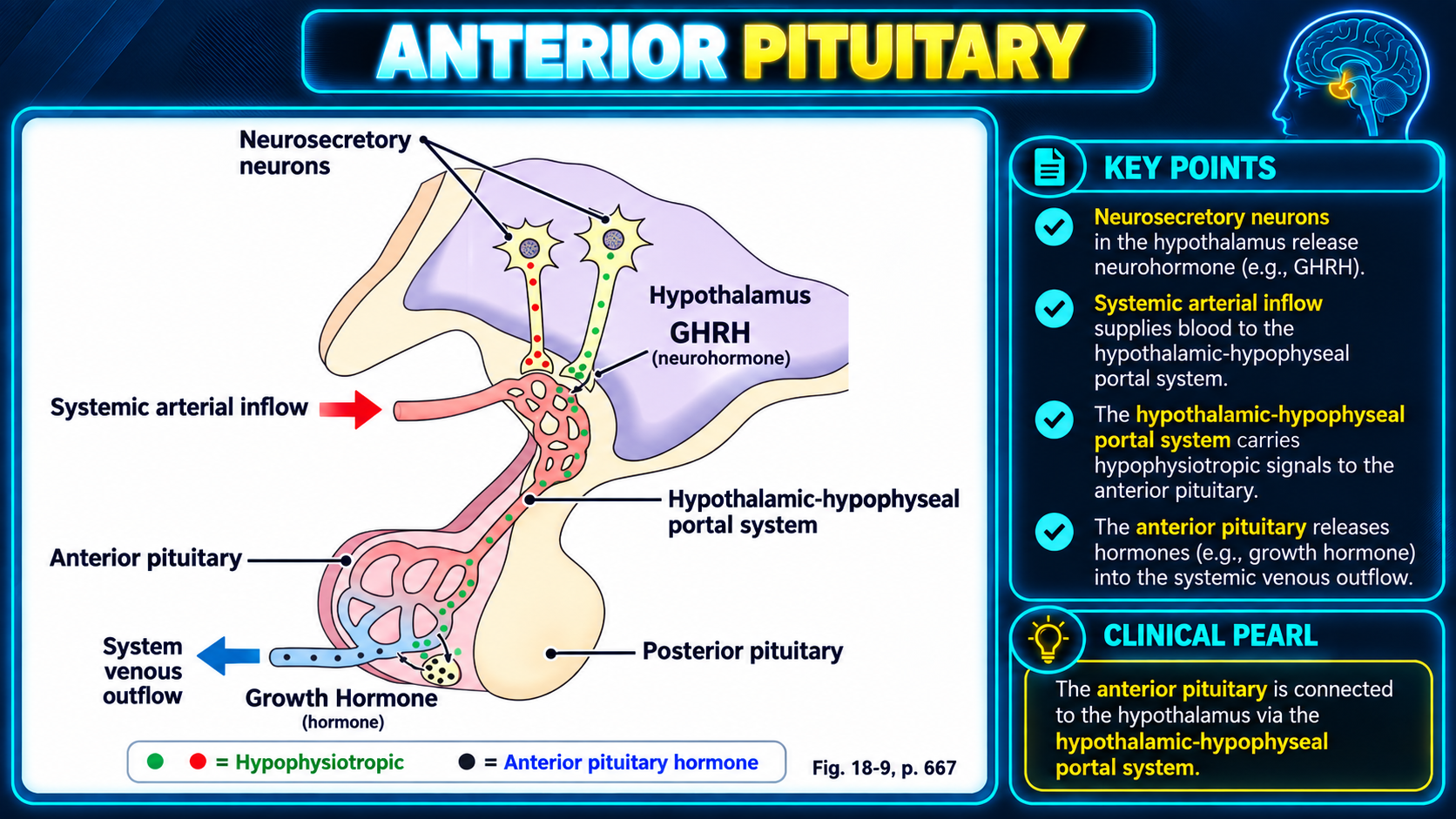
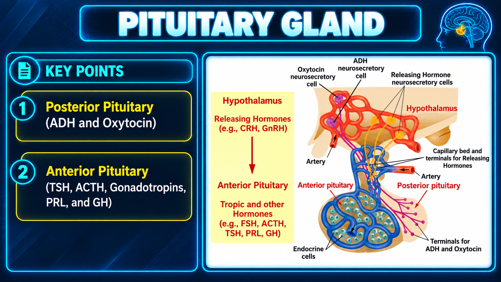
Anterior pituitary and hypothalamic hypophyseal portal system
Anterior PituitaryClick to enlarge
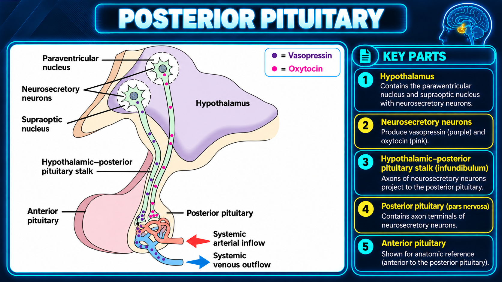
Posterior pituitary anatomy and neurosecretory pathways
Posterior PituitaryClick to enlarge
Anterior and posterior pituitary hormone pathways
Hormone PathwaysClick to enlarge
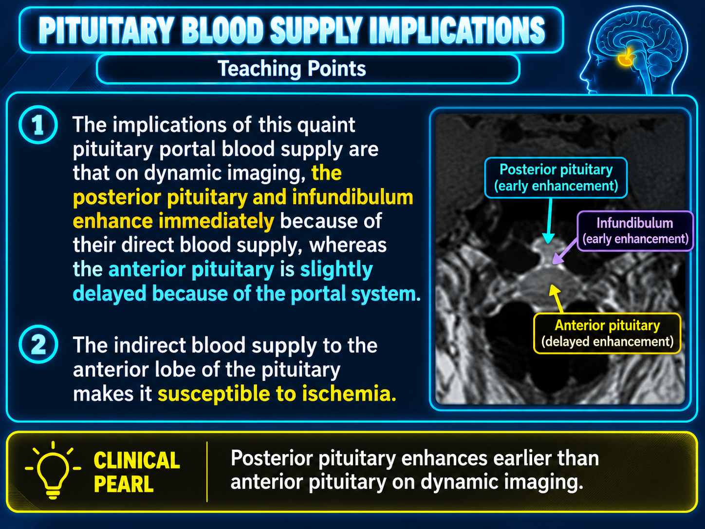
Pituitary blood supply and dynamic enhancement implications
Blood Supply and EnhancementClick to enlarge
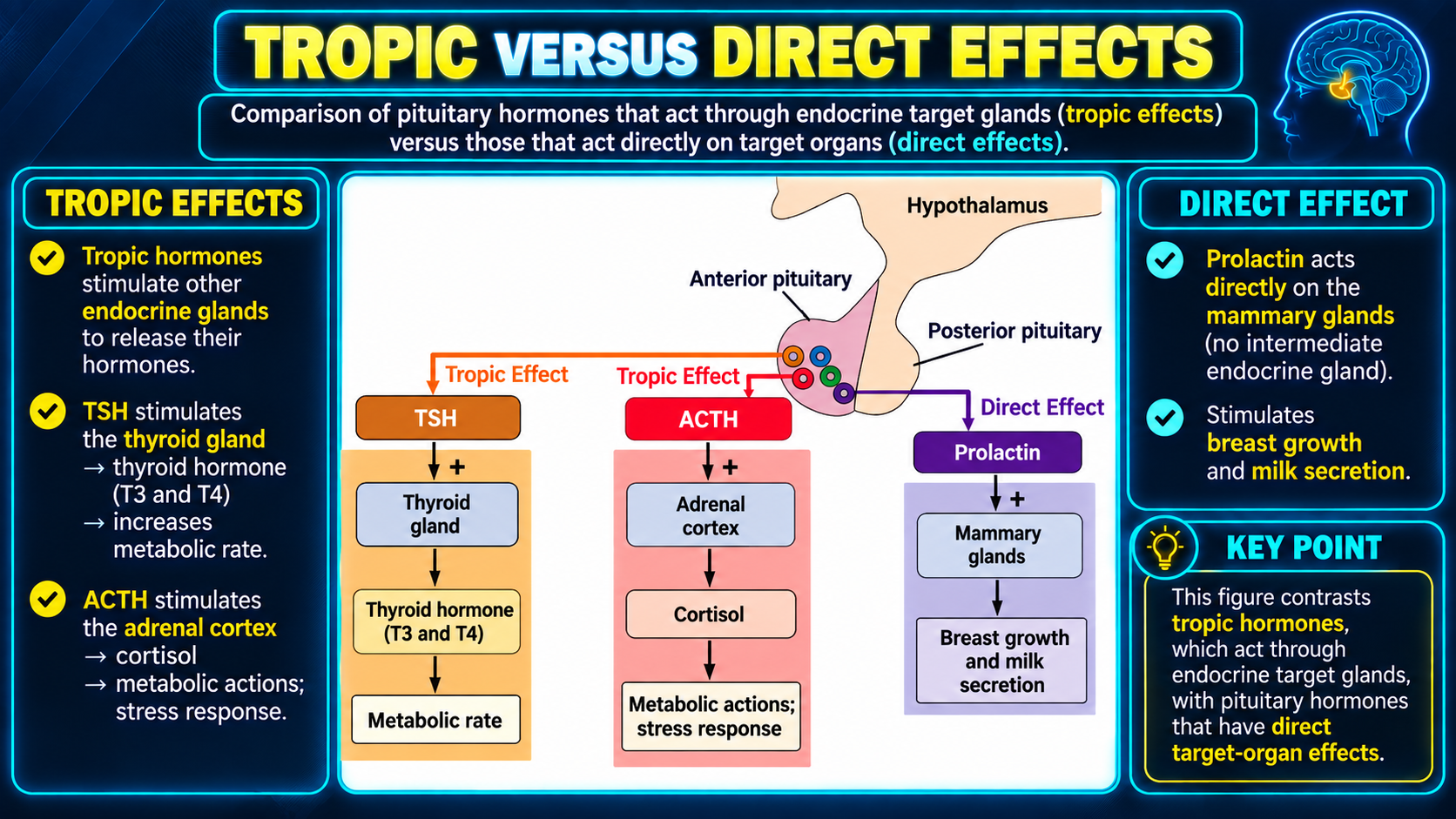
Comparison of tropic and direct pituitary hormone effects
Tropic and Direct EffectsClick to enlarge
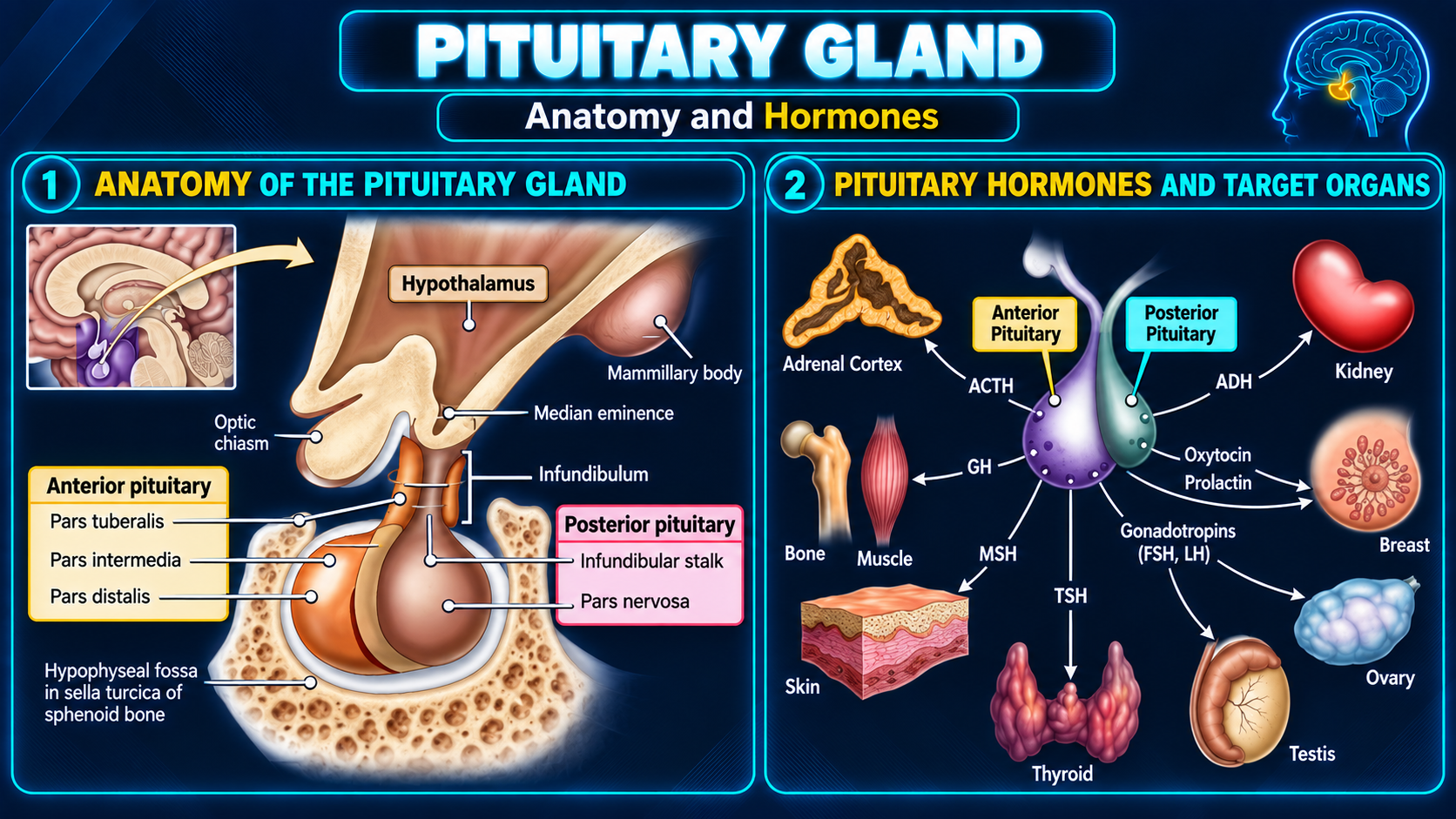
Pituitary anatomy hormones and target organs
Anatomy and HormonesClick to enlarge
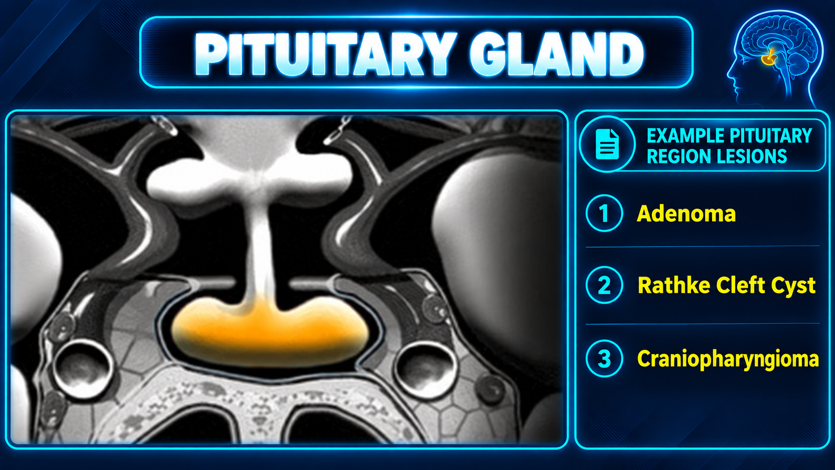
Pituitary region and example sellar lesions
Pituitary Region LesionsClick to enlarge

Overview

The pituitary gland is a small, pea-sized structure at the base of the brain, often called the “master gland” because it regulates many other hormone systems. It lies in the sella turcica below the hypothalamus and connects to it through the infundibulum.

Clinical importancePituitary lesions can produce symptoms by hormone excess, by compressing normal gland and causing hypopituitarism, or by extending superiorly to compress the optic chiasm and cause visual-field loss, classically bitemporal hemianopsia.

Anatomy

Anterior pituitary

The adenohypophysis produces hormones under hypothalamic control and receives its blood supply primarily through the portal system.

Posterior pituitary

The neurohypophysis contains hypothalamic axon terminals and stores and releases hormones made in the hypothalamus.

Relationships

  • Optic chiasm superiorly.
  • Sphenoid sinus inferiorly.
  • Cavernous sinuses and internal carotid arteries laterally.
  • Hypothalamus connected through the infundibulum.

Hormones

Anterior pituitary

  • ACTH: Stimulates adrenal cortisol production.
  • TSH: Stimulates the thyroid gland.
  • FSH and LH: Regulate gonadal function and sex hormones.
  • GH: Drives growth and metabolism through IGF-1.
  • Prolactin: Stimulates milk production.

Posterior pituitary

  • ADH: Regulates water balance and kidney function.
  • Oxytocin: Participates in childbirth and lactation.

MRI Protocol

  • Dedicated pituitary protocol with thin 2–3 mm sections and a small field of view.
  • Sagittal and coronal T1 before and after contrast, plus coronal T2.
  • Dynamic contrast-enhanced imaging improves microadenoma detection because normal gland enhancement precedes adenoma enhancement.
  • Assess the optic apparatus, infundibulum, cavernous sinuses, internal carotid arteries, sphenoid sinus, and sellar floor.

Normal Imaging

  • Anterior pituitary is generally isointense to gray matter on T1 and T2 and enhances avidly and relatively uniformly.
  • The posterior pituitary bright spot is T1 hyperintense from vasopressin-containing neurosecretory granules. Its absence can suggest central diabetes insipidus or stalk pathology.
  • Gland height varies with age and physiology. A practical adult upper limit is approximately 8–10 mm, with enlargement up to 10–12 mm during pregnancy and postpartum lactotroph hyperplasia.
  • The stalk should be midline and approximately 3–4 mm, with the basilar artery serving as a rough upper-size comparison.

Adenomas

Microadenoma (<10 mm)

  • Focal relative hypoenhancement on early dynamic postcontrast images.
  • Secondary signs include a convex gland margin, stalk deviation away from the lesion, and focal sellar-floor depression.

Macroadenoma (≥10 mm)

  • Usually isointense to gray matter with possible cystic, hemorrhagic, or heterogeneous change.
  • Report suprasellar extension, optic-chiasm relationship, and cavernous-sinus involvement.
  • Knosp grades 3–4 suggest cavernous-sinus invasion and affect surgical planning.

Pituitary Apoplexy

Acute hemorrhage or infarction, often within a pre-existing adenoma, is a clinical and neurosurgical emergency.

  • Look for T1-hyperintense hemorrhage and fluid-fluid levels.
  • Identify an underlying enhancing mass.
  • Correlate with sudden headache, visual loss, or ophthalmoplegia.
Hormone Axes

  • HPA: CRH → ACTH → cortisol. Cushing disease often arises from an ACTH-secreting microadenoma that may be MRI occult.
  • HPT: TRH → TSH → thyroid hormones. Primary hypothyroidism can produce thyrotroph hyperplasia that mimics a mass.
  • HPG: GnRH → FSH/LH → gonads. Gonadotroph adenomas are often clinically silent macroadenomas.
  • GH: GHRH → GH → hepatic IGF-1. Excess causes acromegaly after puberty or gigantism before epiphyseal closure.
  • Prolactin: Dopamine is tonically inhibitory. Stalk disruption causes mild-to-moderate elevation; marked elevation supports prolactinoma.

Selected Pathologies

  • Rathke cleft cyst: Well-defined, nonenhancing or thin-rim-enhancing cyst with variable T1 signal and sometimes an intracystic nodule.
  • Craniopharyngioma: Often suprasellar, cystic, and calcified, particularly the adamantinomatous pediatric subtype.
  • Lymphocytic hypophysitis: Symmetric gland enlargement, homogeneous enhancement, and stalk thickening; consider pregnancy, autoimmune disease, or checkpoint inhibitors.
  • Empty sella: CSF-filled sella with flattened gland; may be primary or secondary.
  • Metastasis: Consider rapid growth, known malignancy, and diabetes insipidus from posterior gland or stalk involvement.

Secondary Links