Overview
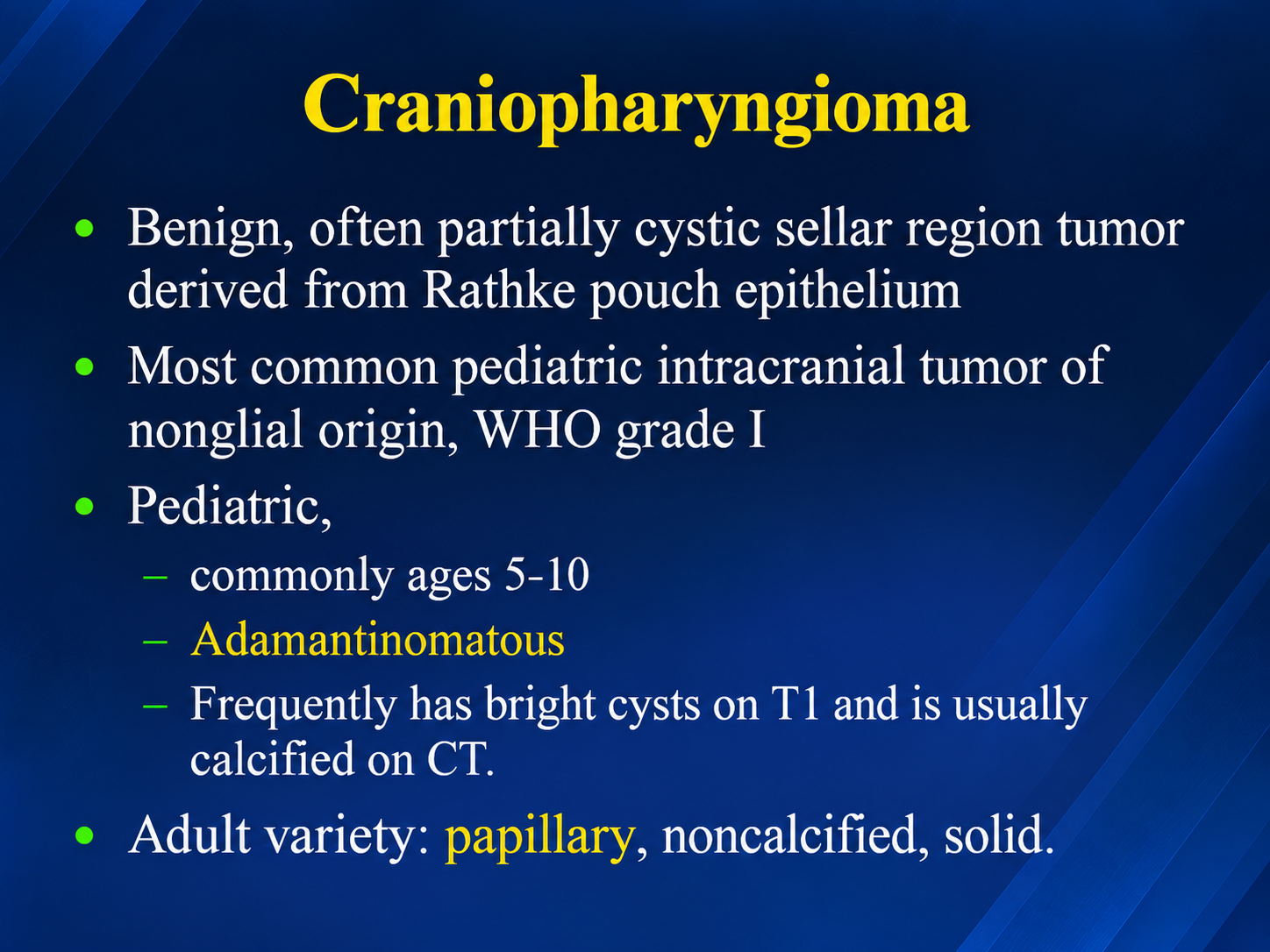
Craniopharyngioma is a histologically benign (WHO grade 1) but locally aggressive epithelial tumor of the sellar and suprasellar region, arising from remnants of Rathke's pouch. Despite benign histology, its intimate relationship with the optic apparatus, hypothalamus, and pituitary gland drives substantial lifelong morbidity. It accounts for roughly 1–3% of intracranial tumors in adults and 5–10% in children, with a bimodal age distribution peaking at 5–14 and 45–74 years. A major recent development is molecularly targeted therapy for the papillary subtype, which is reshaping upfront management. If a mass looks bizarre at the skull base, think Craniopharyngioma
Two Distinct Entities — WHO 2021
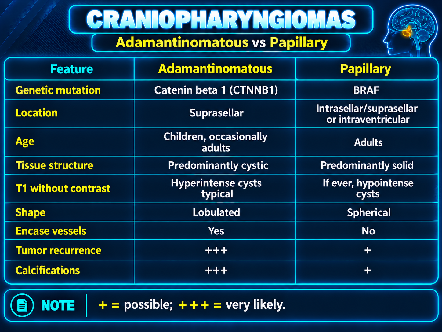
The 5th-edition WHO classification recognizes adamantinomatous (ACP) and papillary (PCP) craniopharyngioma as separate tumors, distinguished by clinical, imaging, histologic, and molecular features.
| Feature | Adamantinomatous (ACP) | Papillary (PCP) |
|---|
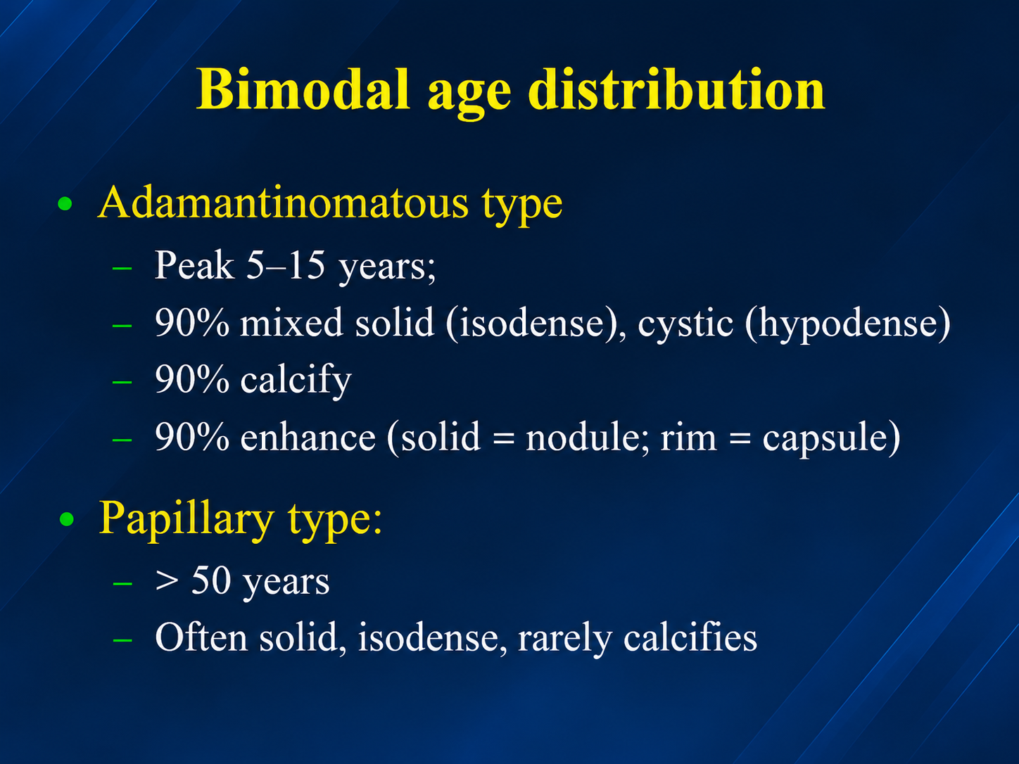
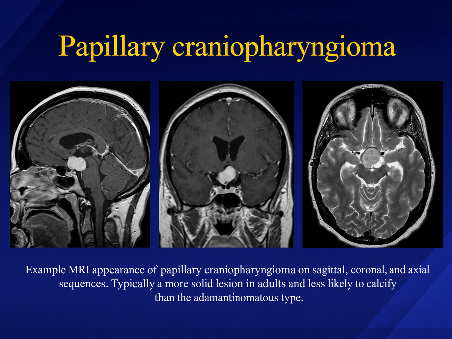
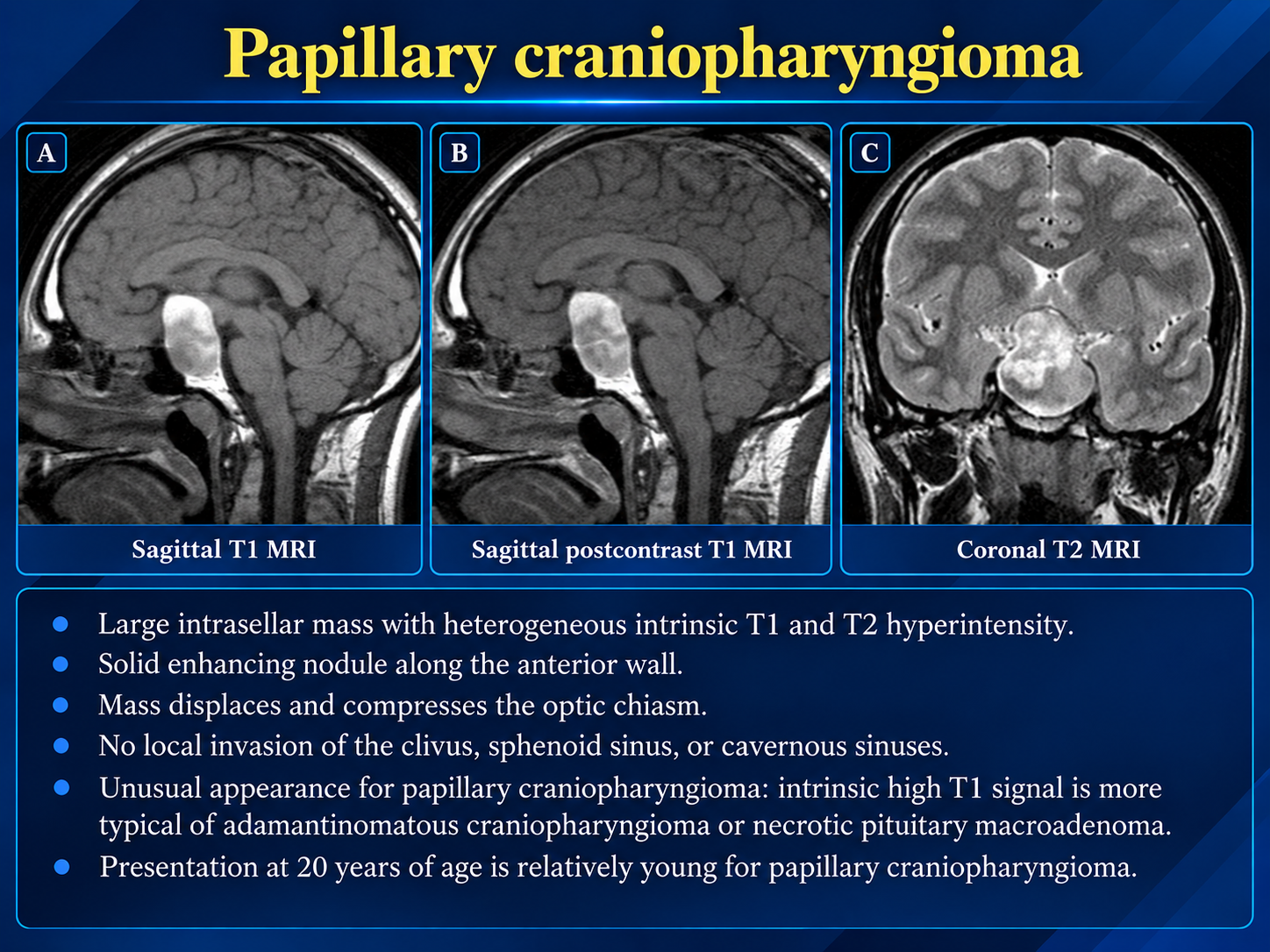
| Age | All ages; nearly all pediatric cases | Almost exclusively adults, usually in the 5th–6th decade; approximately 20–40% of adult craniopharyngiomas |
| Driver mutation | CTNNB1 (β-catenin) | BRAF V600E (>90–95%) |
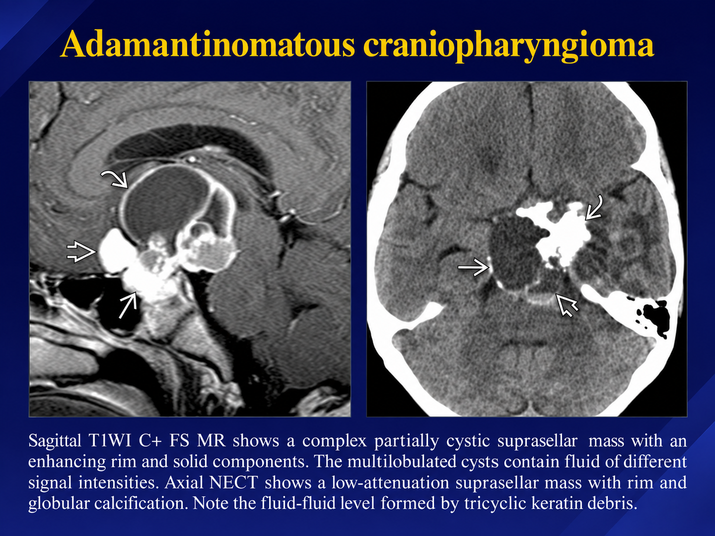
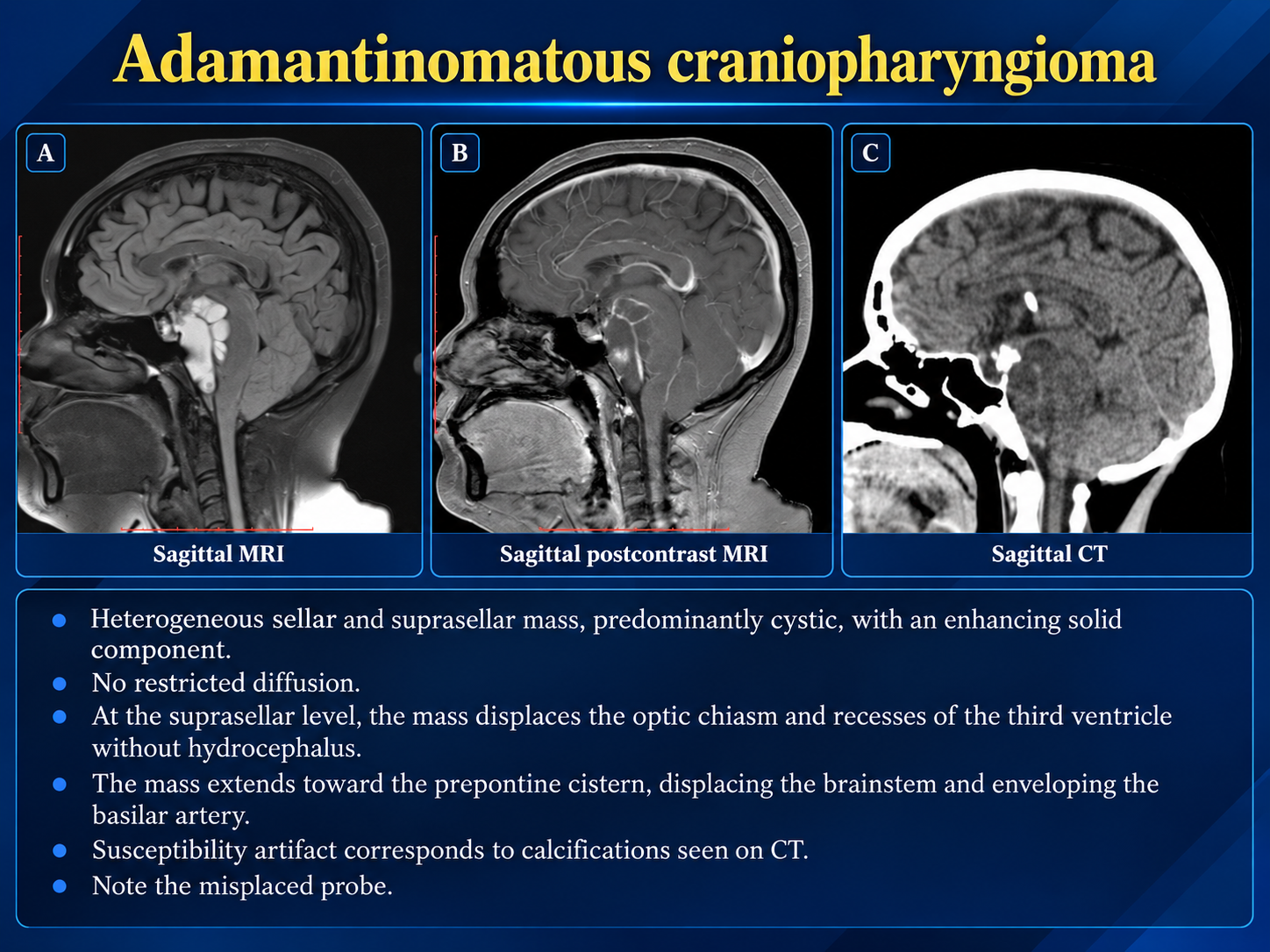
| Morphology | Cystic or multiloculated, with “motor-oil” fluid and calcification | Solid or mixed solid-cystic and often round |
| Calcification | Approximately 90% | Uncommon |
| Behavior | More infiltrative, with higher recurrence | Often more circumscribed and medically targetable |
Radiology Findings
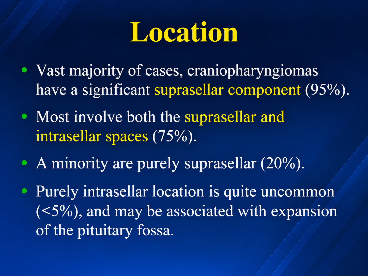
- Location: Usually suprasellar, often with an intrasellar component. Assess extension toward the third ventricle and the relationship to the optic chiasm, pituitary stalk, hypothalamus, and vessels.
- CT: Best for identifying calcification. Adamantinomatous tumors commonly contain cyst-wall or intratumoral calcifications; papillary tumors are usually noncalcified and may appear predominantly solid.
- MRI: Cysts can be T1 hyperintense when their contents are protein-rich and have variable T2 signal. Solid portions and cyst walls typically enhance, producing a heterogeneous appearance. Contrast-enhanced MRI is particularly useful for mapping tumor extent and adjacent anatomy.
- Secondary effects: Look for optic apparatus compression, hypothalamic or third-ventricular involvement, and obstructive hydrocephalus in a large lesion.
Reporting Priorities
Characterize the lesionDescribe the solid and cystic components, enhancement, and calcification. Specify whether the lesion is primarily sellar, suprasellar, or extends into the third ventricle.
Map critical relationshipsDocument the relationship to the optic pathways, stalk and gland, hypothalamus, and major vessels, plus any hydrocephalus. These findings matter for treatment planning.
Use complementary modalitiesContrast-enhanced MRI supplies the anatomic detail, while CT can clarify equivocal calcification.
Subtype Clues
Adamantinomatous
- Common in children but can occur at any age.
- Frequently mixed solid and cystic, often multiloculated.
- Calcification is common, and proteinaceous cysts may be T1 hyperintense.
Papillary
- Occurs almost exclusively in adults.
- Often predominantly solid and more circumscribed.
- Calcification is uncommon.
Clinical Presentation
Symptoms reflect local mass effect and are often present for months to years before diagnosis.
- Raised intracranial pressure: Headache, nausea or vomiting, and sometimes obstructive hydrocephalus.
- Visual impairment: Chiasmal compression causing visual-field defects, decreased acuity, or blindness in 62–84%.
- Endocrine deficits: Present in 52–87%. Growth hormone deficiency is most common, with possible gonadotropin, TSH, and ACTH deficiencies and central diabetes insipidus. Children may have growth failure or abnormal pubertal timing; adults may present with sexual dysfunction.
- Hypothalamic involvement: Weight gain or hypothalamic obesity, disturbed thermoregulation and circadian rhythm, and psychiatric or behavioral changes.
Diagnosis
Workup combines MRI for lesion characterization with CT for calcification detection, full endocrine-axis testing, and formal visual-field assessment.
Sellar mass evaluation
- Morning hormone panel: FSH, LH, ACTH, cortisol, testosterone, GH, IGF-1, prolactin, TSH, and T3/T4.
- Endocrinology evaluation and formal visual-field testing.
- Sellar MRI and CT when calcification requires assessment.
- Proceed to subtype-directed treatment after appropriate tissue and molecular evaluation.
Framework: SELLAR-2, Sellar Tumors: Evaluation and Treatment for Sellar Mass — NCCN Guidelines®, Central Nervous System Cancers, p. 44.
Conventional Treatment
Management combines surgery and radiotherapy, individualized by subtype, location, and hypothalamic involvement.
- Surgery: First-line for tissue diagnosis, symptom relief, and cytoreduction. Endoscopic endonasal approaches offer comparable or slightly higher gross-total resection rates with better visual preservation than transcranial routes; NCCN identifies endoscopic transsphenoidal surgery as preferred.
- Extent of resection: Practice has shifted away from routine aggressive gross-total resection toward maximal safe, hypothalamus-sparing resection. Gross-total resection improves progression-free survival compared with subtotal resection alone, but subtotal resection plus radiotherapy can achieve comparable local control with less hypothalamic morbidity. Tumor size >3.5 cm, retrochiasmatic location, and hypothalamic, optic, or vascular involvement may limit safe resection.
- Radiotherapy: Fractionated radiotherapy is indicated after subtotal resection. Stereotactic radiosurgery may be preferred when lesion geometry and distance from the optic pathways permit it. Close monitoring may be reasonable after gross-total resection.
- Intracystic therapy: Interferon-alpha or peginterferon, bleomycin, or radioisotopes such as P-32 can control predominantly cystic ACP and delay radiotherapy. These treatments do not target solid components and may cause neurotoxicity if cyst contents leak.
Prognosis and Long-Term Sequelae
Overall survival is high at approximately 92%. Reported pediatric survival is approximately 92%, 84%, and 68% at 5, 10, and 20 years, respectively, but quality of life is frequently compromised.
- Endocrinopathy: At least one pituitary axis is affected in 93–100% after treatment, with near-universal need for lifelong hormone replacement, including treatment for diabetes insipidus.
- Hypothalamic syndrome and obesity: Hypothalamic involvement independently reduces long-term survival and drives metabolic syndrome in approximately 50%, cardiovascular risk, and impaired quality of life.
- Neurologic effects: Visual deficits and neurocognitive or behavioral impairment may persist.
- Radiation late effects: Particularly in pediatric patients, these include vasculopathy, moyamoya, stroke, and secondary malignancy.
References