Thickened Infundibulum

Reference Images

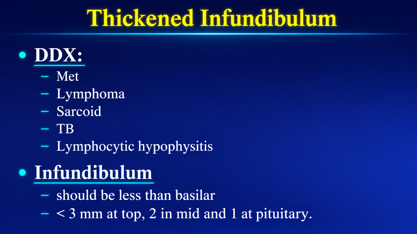
Differential diagnosis and normal measurements for a thickened pituitary infundibulum
Differential and MeasurementsClick to enlarge
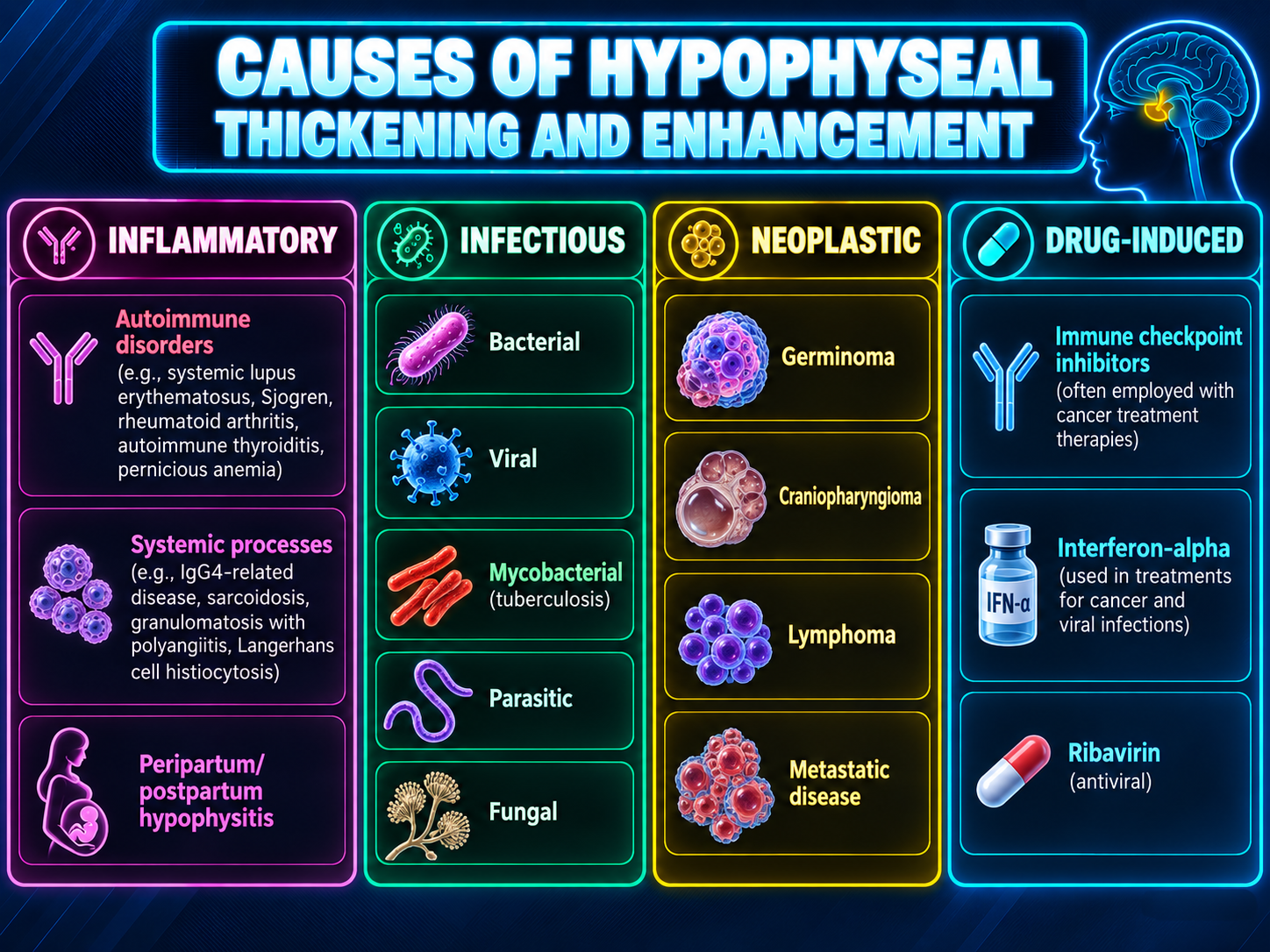
Inflammatory infectious neoplastic and drug-induced causes of hypophyseal thickening
Causes of Hypophyseal ThickeningClick to enlarge

Overview

A thickened pituitary infundibulum is nonspecific. The highest-yield differential includes hypophysitis, neurosarcoidosis, Langerhans cell histiocytosis, germinoma, metastasis, lymphoma, and infection. Age, stalk morphology, central diabetes insipidus, systemic disease, and interval change are key discriminators.

Key limitationNo isolated MRI feature reliably separates inflammatory from neoplastic disease. Indeterminate lesions require endocrine correlation, systemic evaluation, and usually short-interval follow-up MRI; progressive or high-risk lesions may require biopsy.

Radiologic Differential

CategoryImportant causesHelpful clues
InflammatoryLymphocytic or infundibulo-neurohypophysitis, IgG4-related disease, checkpoint-inhibitor hypophysitisSmooth symmetric thickening, homogeneous enhancement, loss of taper, symmetric gland enlargement, intact sellar floor
Granulomatous / infiltrativeSarcoidosis, LCH, Erdheim–Chester disease, granulomatosis with polyangiitisSarcoid may show basilar leptomeningeal or cranial-nerve enhancement; LCH may show skull or mastoid lytic lesions
NeoplasticGerminoma, lymphoma or leukemia, glioma, craniopharyngioma, meningioma, primary stalk tumorsNodular or masslike stalk; germinoma is critical in children or adolescents with CDI and may coexist with a pineal lesion
MetastaticEspecially breast and lung; also renal, melanoma, prostate, and othersOlder patient, known cancer, rapid progression, irregular nodularity, CDI, cavernous-sinus invasion, or bone destruction
InfectiousTuberculosis, fungal infection, syphilis, abscessBasilar meningitis, nodular or ring enhancement, restricted diffusion, systemic infection
Developmental / adjacentRathke cleft cyst, craniopharyngioma, ectopic posterior pituitary, stalk-interruption spectrumAssociated cyst, calcification, ectopic bright spot, small anterior pituitary, or other midline anomaly

Pattern Recognition

  • Smooth diffuse homogeneous thickening: Favors hypophysitis but overlaps with sarcoidosis, LCH, germinoma, and metastasis.
  • Focal nodular or irregular thickening: Raises germinoma, metastasis, lymphoma, LCH, or primary stalk tumor.
  • Absent posterior bright spot: Supports neurohypophyseal involvement but is nonspecific.
  • Pineal mass: Suggests bifocal germinoma in a young patient.

Associated Findings

  • Basilar leptomeningeal or cranial-nerve enhancement favors sarcoidosis or infection.
  • Periventricular enhancing masses raise lymphoma.
  • Calvarial or skull-base lytic lesions raise LCH.
  • Bone erosion without chronic sellar enlargement raises metastasis or another aggressive infiltrative lesion.
  • Restricted diffusion may support abscess or hypercellular neoplasm.

Measurement

A frequently used adult threshold is greater than 3 mm, but the measurement level must be specified because the stalk normally tapers inferiorly.

  • Approximately 2.4 ± 0.6 mm near the optic chiasm.
  • Approximately 1.9 ± 0.3 mm near the gland.
  • Pediatric consensus considers at least 4 mm at the chiasm or 3 mm at the pituitary insertion potentially abnormal.
  • Compare with prior imaging and report interval growth.

Reporting Checklist

  • Maximum diameter and measurement level.
  • Smooth versus nodular morphology.
  • Preservation or loss of normal taper.
  • Enhancement pattern and diffusion restriction.
  • Posterior pituitary bright spot.
  • Gland enlargement or focal pituitary lesion.
  • Hypothalamic, chiasmatic, cavernous-sinus, meningeal, and pineal involvement.
  • Skull-base or calvarial lesions and interval growth.

Clinical Correlation

  • Central diabetes insipidus and anterior pituitary dysfunction.
  • Known malignancy or rapidly progressive symptoms.
  • Pregnancy or postpartum state.
  • Immune-checkpoint inhibitor or interferon therapy.
  • Systemic inflammatory, granulomatous, or infectious disease.
  • Patient age and germ-cell tumor risk.

Secondary Links