Cavernous Sinus Thrombosis

Reference Images

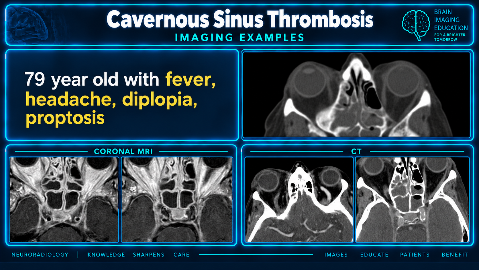
CT and MRI imaging example of septic cavernous sinus thrombosis
Imaging ExampleClick to enlarge
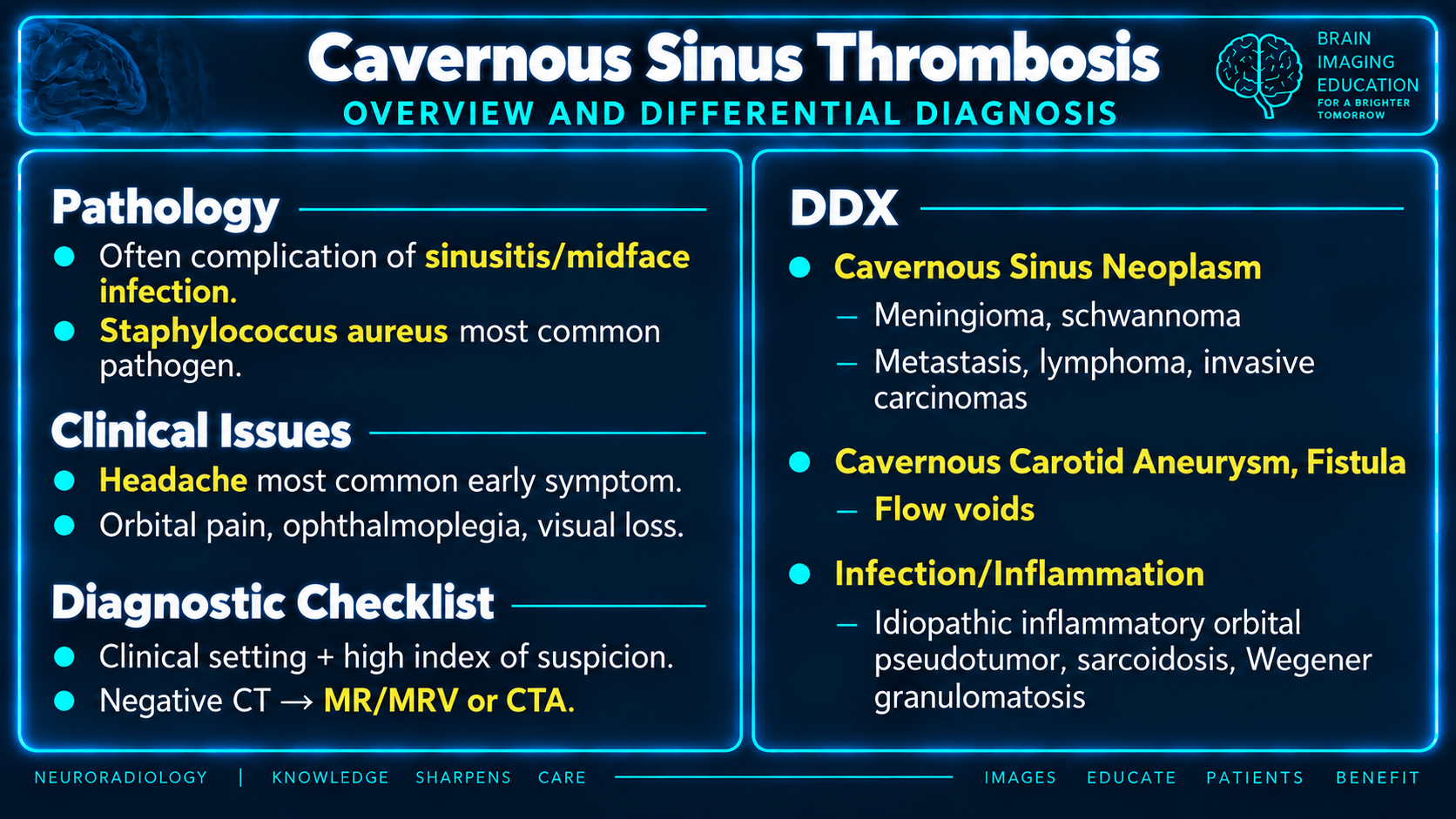
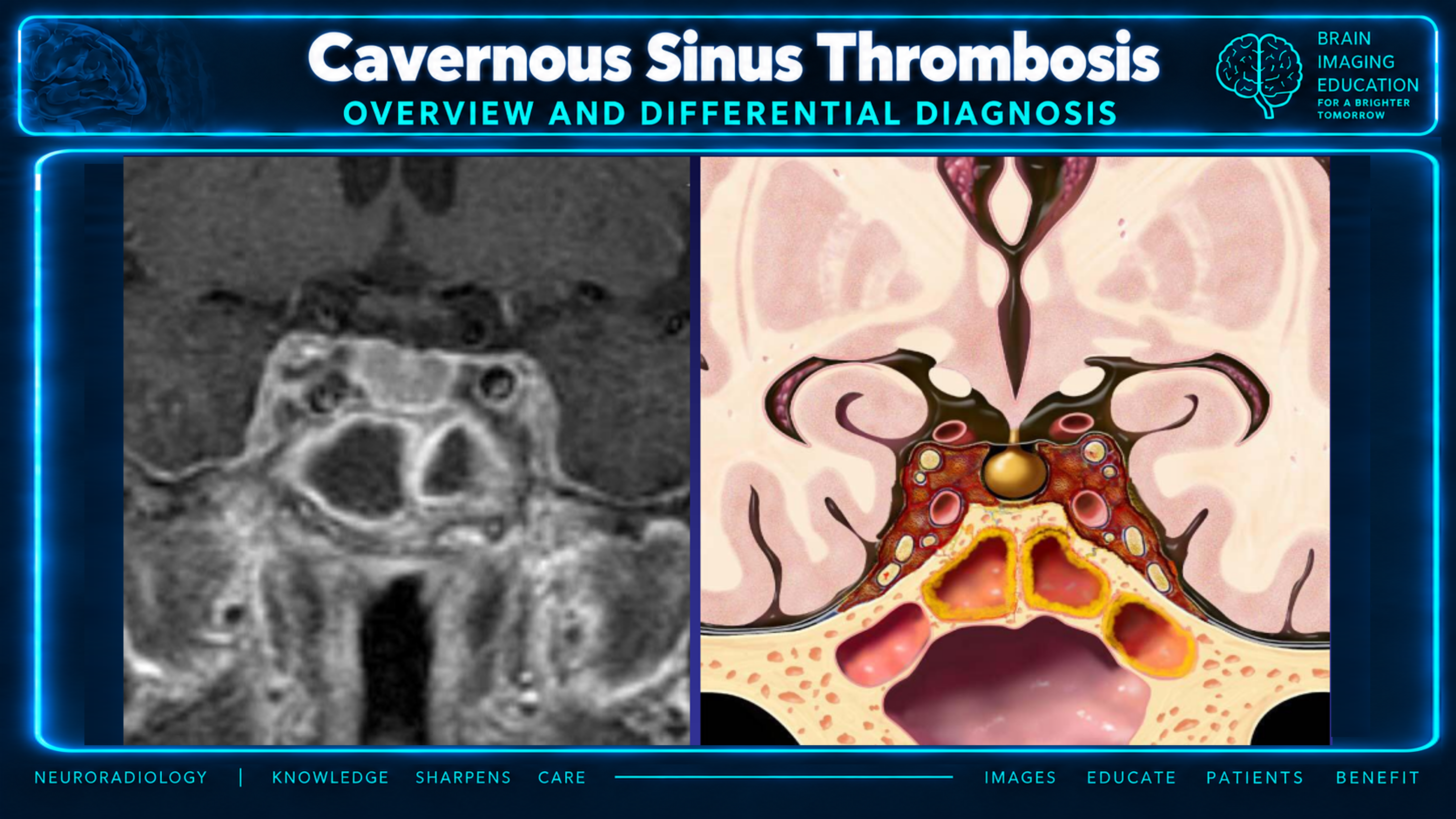
Cavernous sinus thrombosis overview and differential diagnosis
Overview and DifferentialClick to enlarge
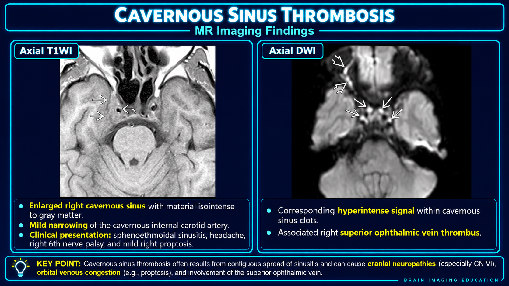
T1 and diffusion MRI findings of cavernous sinus thrombosis
MR Imaging FindingsClick to enlarge
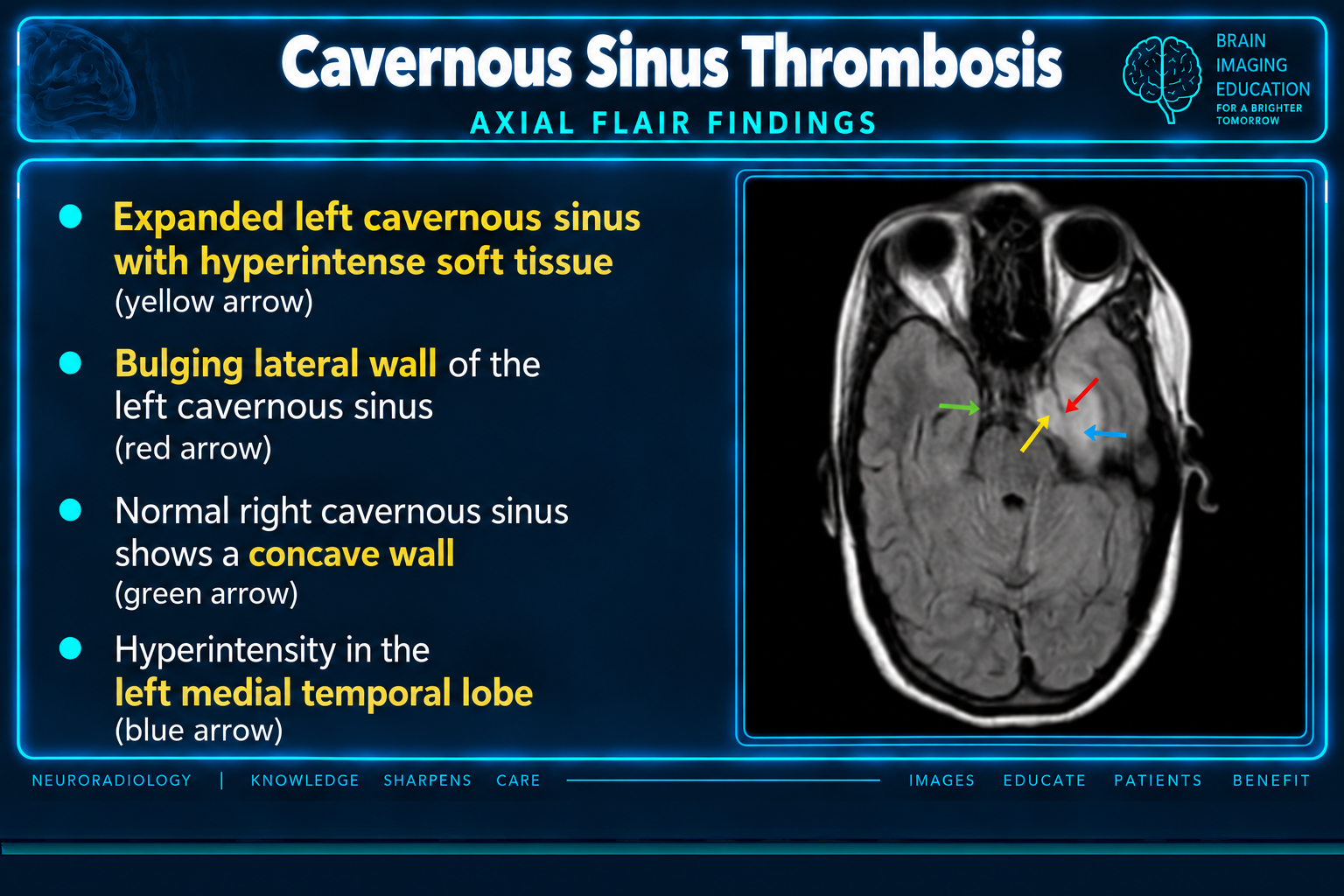
Axial FLAIR findings of cavernous sinus thrombosis
Axial FLAIR FindingsClick to enlarge
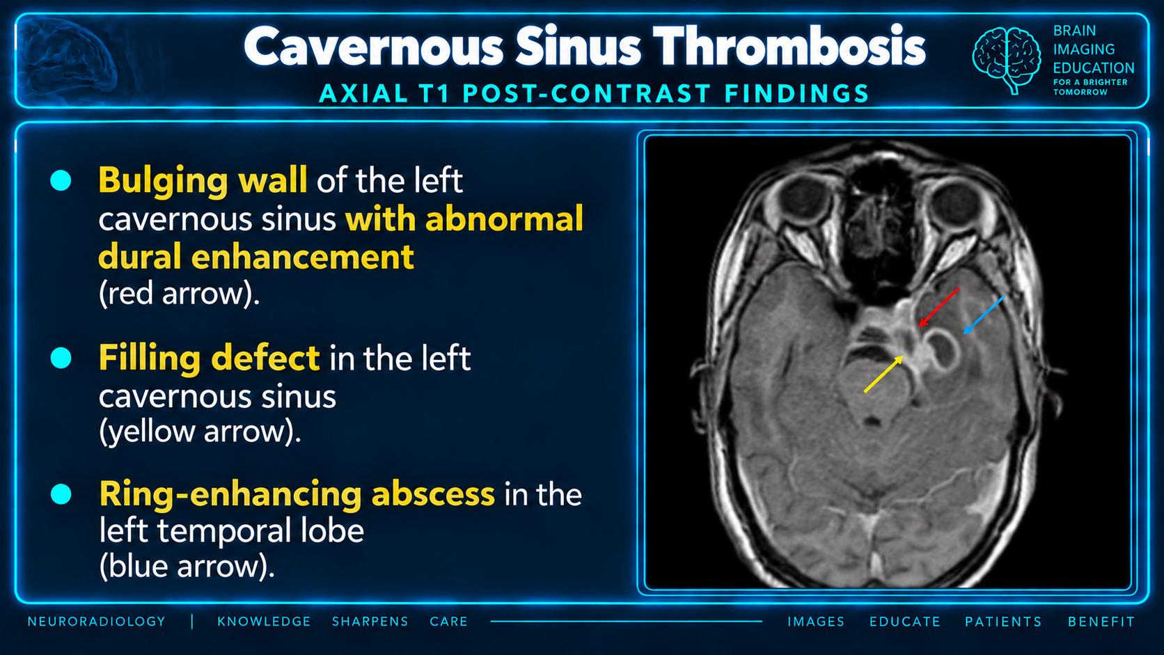
Postcontrast MRI findings and temporal lobe abscess in cavernous sinus thrombosis
Postcontrast FindingsClick to enlarge
Coronal MRI and graphic of cavernous sinus thrombosis
Coronal MRI and GraphicClick to enlarge

Overview

Cavernous sinus thrombosis is thrombus within the cavernous sinus. It is rare, most often septic, and can be subtle on early imaging despite high morbidity and mortality.

Diagnostic priorityIn the appropriate clinical setting, a negative or equivocal noncontrast CT does not exclude CST. Contrast-enhanced MRI with venographic imaging is preferred, and the remaining dural sinuses and cerebral veins must also be assessed.

Epidemiology

  • Approximately 4.5 cases per million people annually.
  • Least common dural venous sinus thrombosis.
  • Accounts for fewer than 1.5% of dural venous sinus thromboses.
  • Septic cases predominate.

Clinical Presentation

  • Headache is the most common symptom.
  • Painful ophthalmoplegia and cavernous sinus syndrome.
  • CN III palsy may be an early manifestation; CN VI is commonly involved.
  • Chemosis, periorbital swelling, ptosis, and proptosis.
  • Papilledema, retinal venous distension, visual loss, or seizures.
  • Fever or systemic toxicity suggests septic thrombosis.

Septic Etiology

Contiguous infection spreads through valveless facial, orbital, and emissary veins. Staphylococcus aureus is the most frequent pathogen.

  • Acute paranasal sinusitis, especially sphenoid or ethmoid disease.
  • Midface infection.
  • Dental abscess.
  • Orbital cellulitis.
  • Bacterial embolization initiates thrombosis and impaired venous drainage.

Aseptic Etiology

  • Trauma or tumor causing cavernous sinus compression.
  • Protein C or protein S deficiency and other thrombophilias.
  • Oral contraceptive use.
  • Malignancy.
  • Pregnancy and postpartum state.
  • Idiopathic cases occur.

Core Imaging Pattern

  • Enlarged cavernous sinus with a convex or bulging lateral wall.
  • Abnormal internal signal replacing expected venous flow voids.
  • Filling defect or heterogeneous enhancement.
  • Narrowing or compression of the cavernous ICA.
  • Dilated or thrombosed superior ophthalmic vein.
  • Proptosis and orbital venous congestion.
  • Concurrent cerebral or dural venous thrombosis.

CT Findings

  • Hyperdense thrombus on noncontrast CT is specific but insensitive.
  • Contrast CT may show cavernous sinus expansion and a non-fat-density filling defect.
  • Assess sphenoid, ethmoid, facial, dental, and orbital infection.
  • Look for proptosis, superior ophthalmic vein enlargement, and orbital edema.
  • Identify abscess, empyema, infarction, hemorrhage, or skull-base disease.

MRI and MRV Findings

  • Loss of normal cavernous sinus flow voids.
  • Thrombus signal varies with age on T1 and T2.
  • Restricted diffusion may be present within acute clot or associated abscess.
  • Bulging lateral wall, abnormal dural enhancement, and intraluminal filling defect.
  • MRV may confirm impaired flow or occlusion.
  • Enhancement does not exclude thrombus because organizing clot can enhance.

Complications

  • Meningitis, subdural empyema, or cerebral abscess.
  • Septic pulmonary emboli, pneumonia, lung abscess, or pleural empyema.
  • Propagation to other dural sinuses and cerebral veins.
  • ICA compression, arteritis, infarction, or stroke.
  • Pituitary compression and hypopituitarism.
  • Blindness and persistent cranial neuropathy.

Imaging Differential

AlternativeHelpful distinction
Carotid-cavernous fistula or cavernous carotid aneurysmArterialized flow, early venous opacification, bruit, prominent flow voids, or arterial continuity
Cavernous sinus neoplasmSolid enhancing mass, chronic remodeling, perineural spread, or known tumor; includes meningioma, schwannoma, metastasis, and lymphoma
Tolosa-Hunt syndromePainful ophthalmoplegia with inflammatory cavernous sinus tissue after exclusion of infection, neoplasm, and vascular disease
Orbital cellulitis or superior orbital fissure syndromeOrbital-centered inflammatory change or lesion without primary cavernous sinus thrombus
Granulomatous inflammationSarcoidosis, granulomatosis with polyangiitis, or other systemic inflammatory findings
Lytic skull-base lesion or cavernous venous malformationBone destruction or characteristic slowly enhancing vascular mass

Treatment and Prognosis

  • Prompt broad-spectrum intravenous antimicrobial therapy for septic CST.
  • Antithrombotic therapy is commonly considered with multidisciplinary guidance.
  • Urgent source control for sinus, dental, orbital, or facial infection when indicated.
  • Mortality remains substantial, and survivors may have blindness or cranial nerve deficits.

Reporting Checklist

  • Side, caliber, and lateral-wall contour of each cavernous sinus.
  • Thrombus signal, filling defect, enhancement, and diffusion.
  • Cavernous ICA caliber and wall abnormality.
  • Superior and inferior ophthalmic veins.
  • Orbital congestion, proptosis, and optic complications.
  • Paranasal sinus, facial, dental, and orbital source.
  • Other dural sinus or cerebral venous thrombosis.
  • Abscess, empyema, meningitis, infarction, or hemorrhage.

Practical Points

  • Compare the cavernous sinus lateral wall with the contralateral side; normal is straight or concave.
  • A bulging wall, asymmetric expansion, and ophthalmic venous enlargement are high-yield clues.
  • Search the complete cerebral venous system because multiple thromboses are frequent.
  • Look for the infectious source and intracranial complications on the same examination.

Secondary Links