Cavernous Sinus Pathology

Reference Images

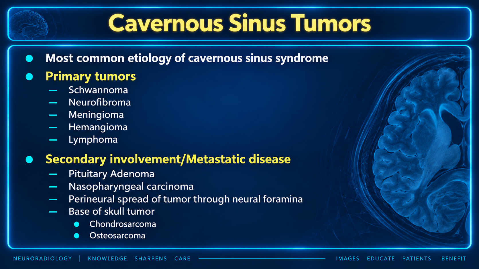
Primary and secondary tumors involving the cavernous sinus
Cavernous Sinus TumorsClick to enlarge
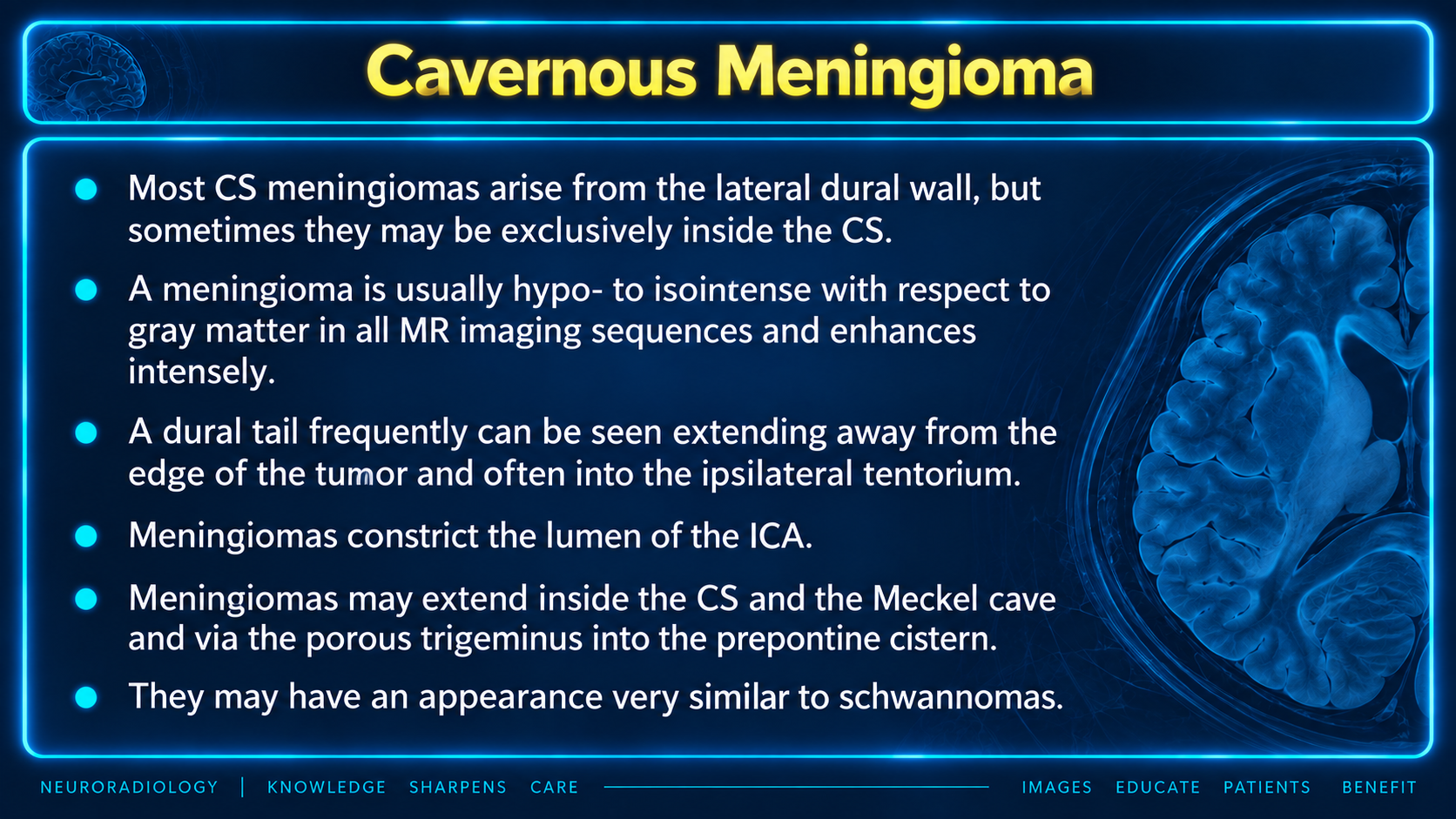
Imaging features of cavernous sinus meningioma
Meningioma FeaturesClick to enlarge
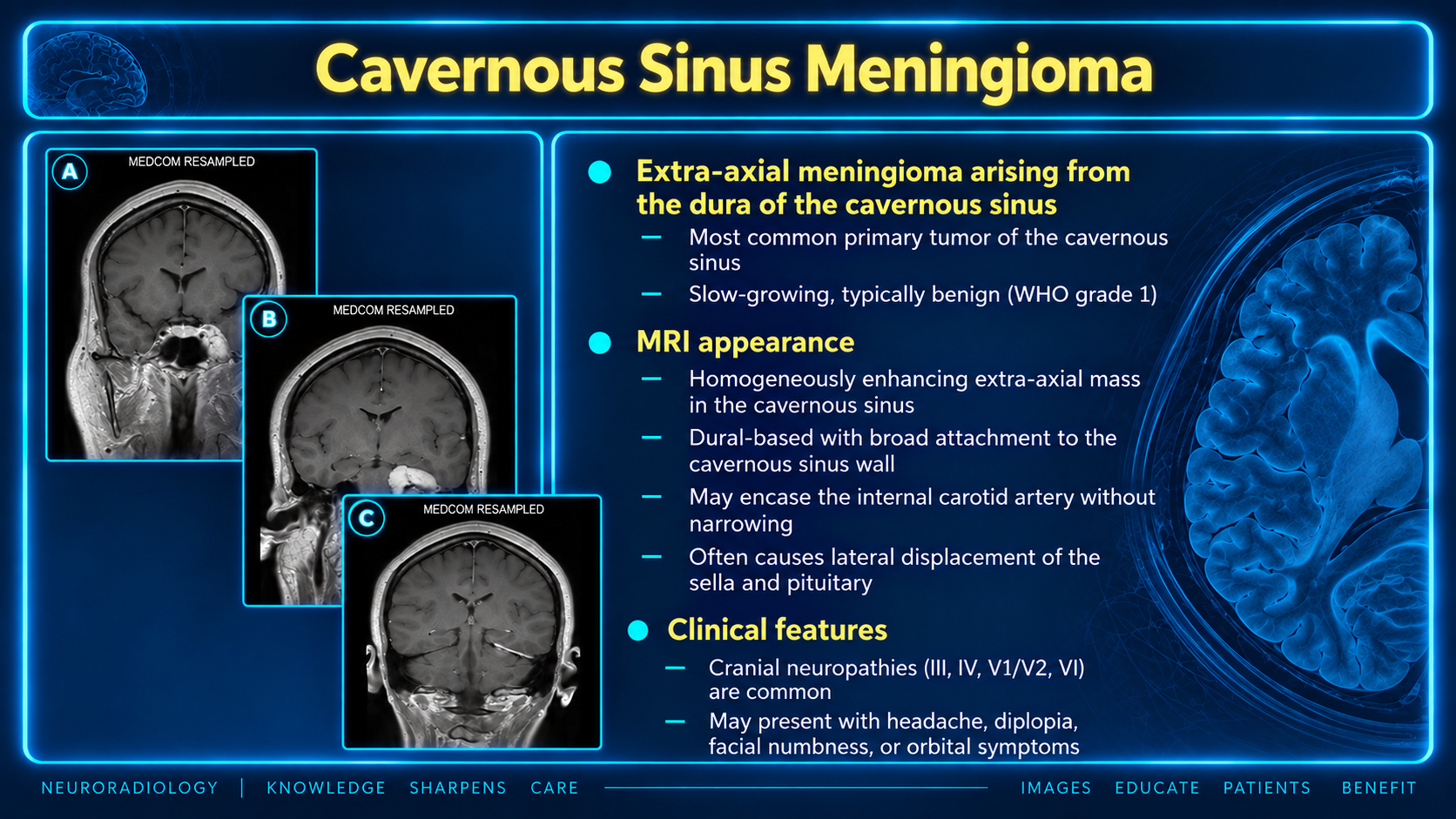
MRI appearance and clinical features of cavernous sinus meningioma
Meningioma OverviewClick to enlarge
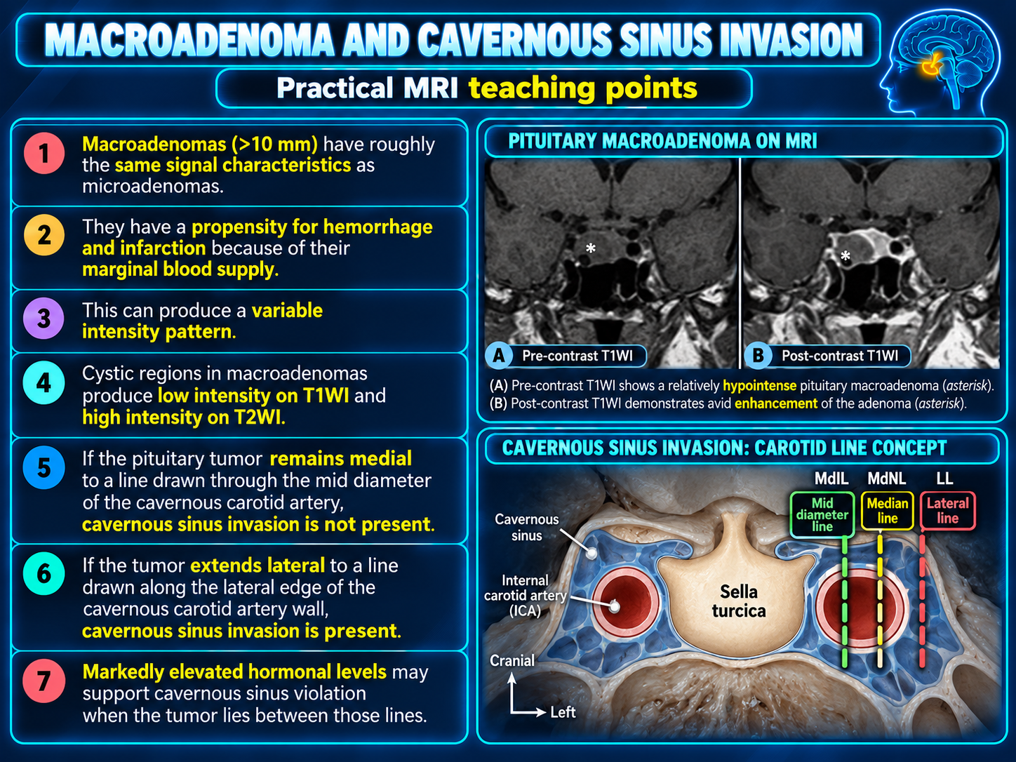
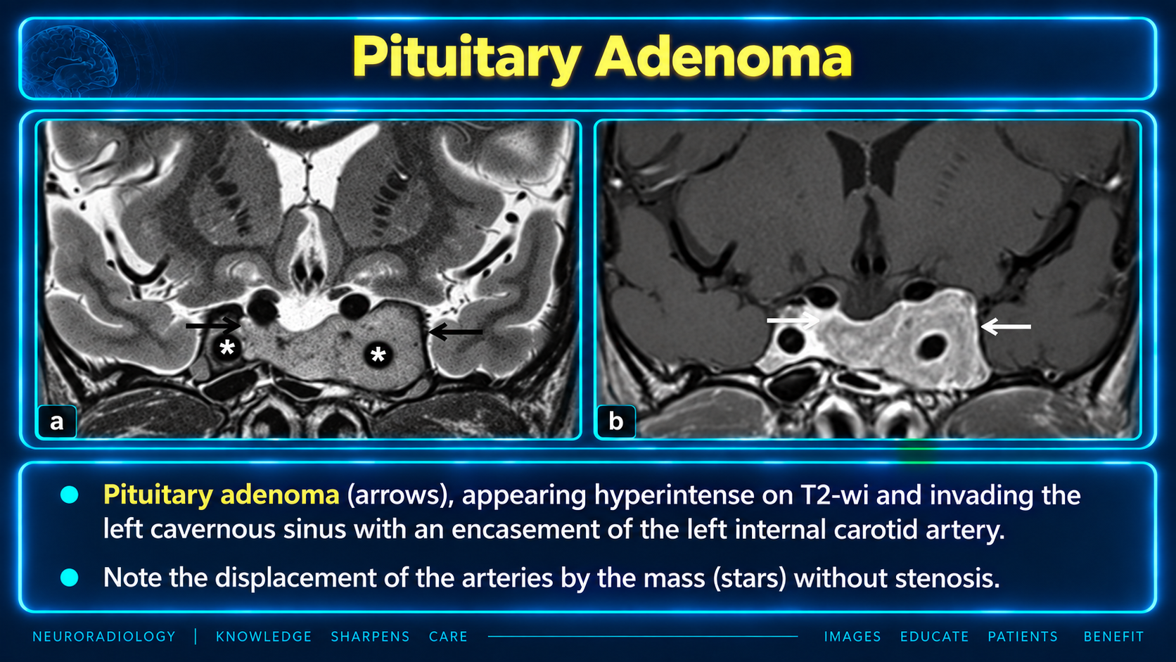
MRI teaching points for pituitary macroadenoma and cavernous sinus invasion
Macroadenoma InvasionClick to enlarge
Pituitary adenoma invading the cavernous sinus and encasing the internal carotid artery
Pituitary AdenomaClick to enlarge
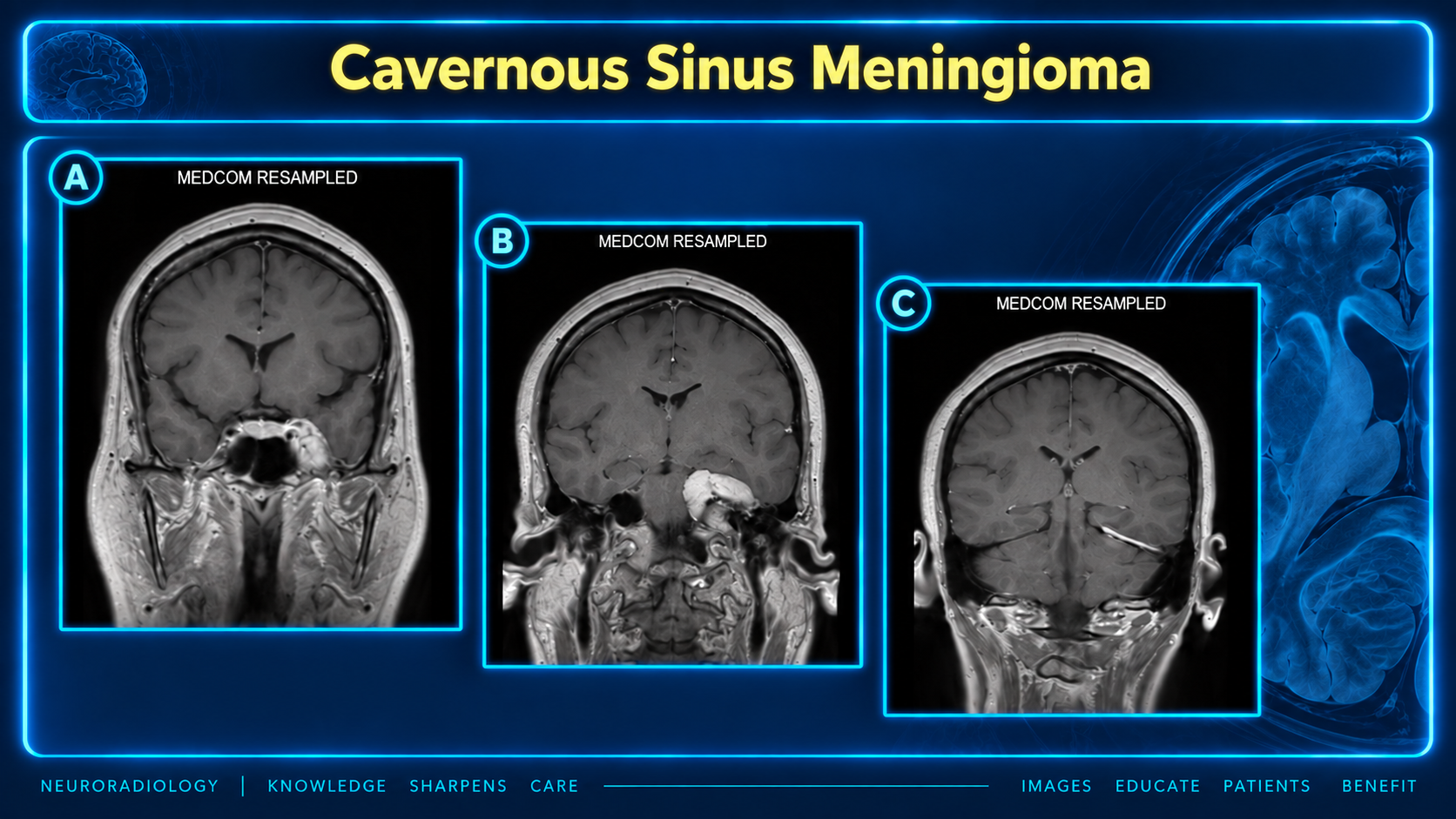
Coronal postcontrast MRI of cavernous sinus meningioma
Meningioma MRIClick to enlarge
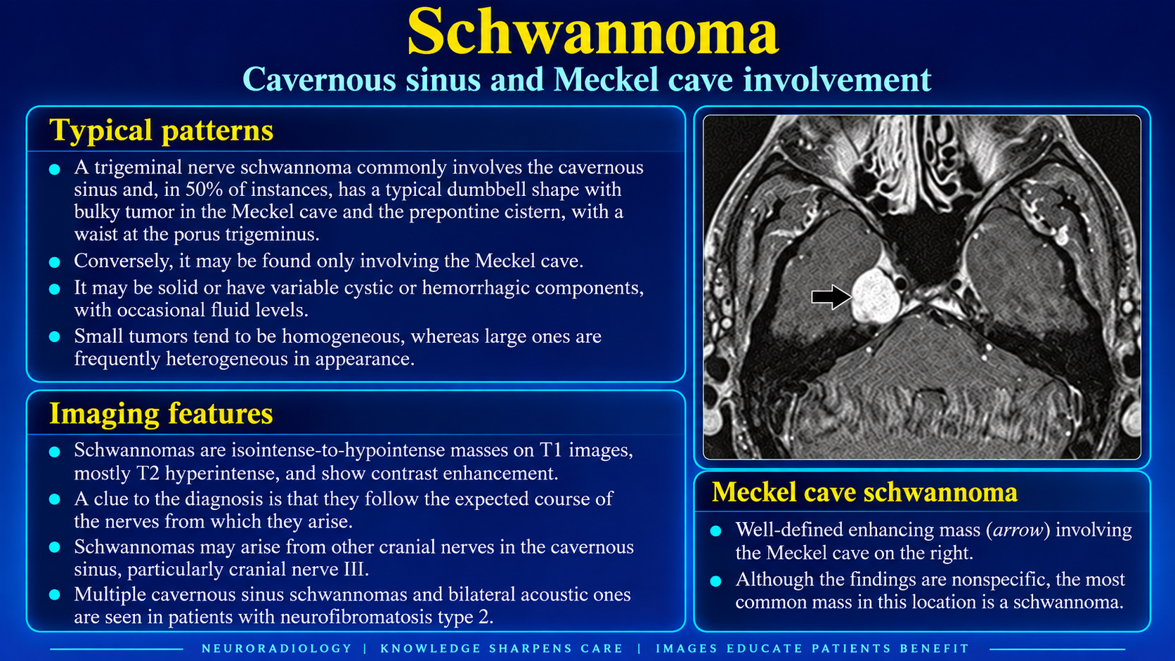
Schwannoma involving the cavernous sinus and Meckel cave
Meckel Cave SchwannomaClick to enlarge
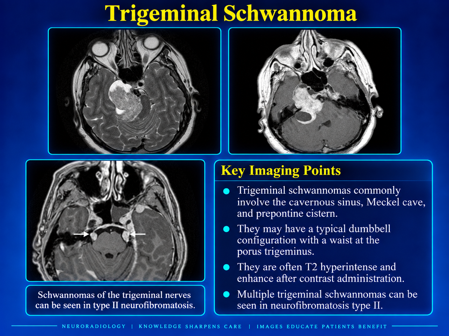
Trigeminal schwannoma involving the cavernous sinus Meckel cave and prepontine cistern
Trigeminal SchwannomaClick to enlarge
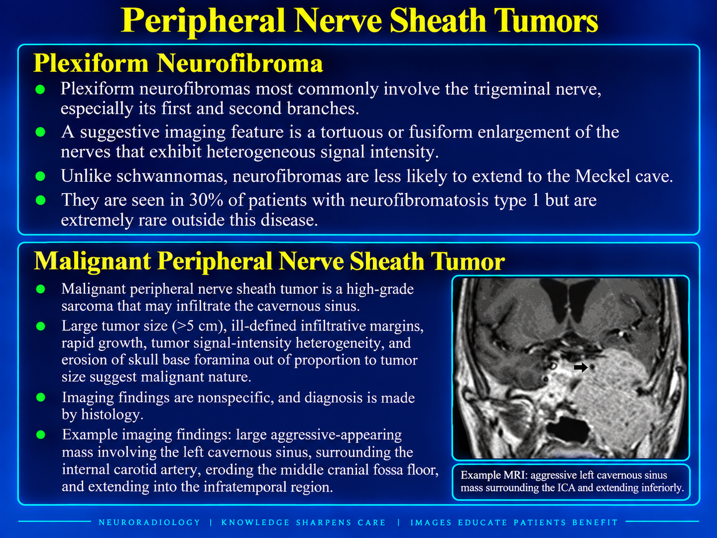
Plexiform neurofibroma and malignant peripheral nerve sheath tumor features
Peripheral Nerve Sheath TumorsClick to enlarge

Overview

Cavernous sinus pathology produces a shared final pathway of compression, infiltration, inflammation, infection, or vascular derangement involving densely packed neurovascular structures. Cavernous sinus syndrome combines variable palsies of CN III, IV, V1/V2, and VI with possible Horner syndrome and orbital venous congestion.

Localization pearlCN VI lies medially beside the internal carotid artery and is often affected earliest. The optic nerve lies outside the sinus; vision loss suggests orbital-apex extension or aggressive infectious disease. Enhancement can be asymmetric. Do not misinterpret it as pathology.

Diagnostic Framework

  • Localize the epicenter: cavernous sinus, sella, Meckel cave, skull base, sphenoid sinus, or orbital apex.
  • Assess the cavernous ICA for encasement, narrowing, displacement, aneurysm, or fistula.
  • Inspect V1 and V2 pathways and skull-base foramina for perineural spread.
  • Evaluate the orbit, superior ophthalmic vein, paranasal sinuses, clivus, and petrous apex.
  • Use contrast-enhanced skull-base MRI with MRA or MRV when vascular disease is possible.

Neoplastic Differential

  • Meningioma.
  • Schwannoma or neurofibroma.
  • Cavernous sinus hemangioma.
  • Pituitary macroadenoma with invasion.
  • Lymphoma, leukemia, or neurolymphomatosis.
  • Hematogenous metastasis or perineural tumor spread.
  • Nasopharyngeal or sphenoid malignancy.
  • Chordoma, chondrosarcoma, osteosarcoma, or juvenile angiofibroma.

Meningioma

  • Avidly and usually homogeneously enhancing dural-based mass, often with a dural tail.
  • Typically hypo- to isointense to gray matter on MRI.
  • Often arises from the lateral dural wall and may extend to the tentorium or Meckel cave.
  • May encase and constrict the cavernous ICA, helping distinguish it from pituitary adenoma.
  • Can resemble a schwannoma when it extends through the porus trigeminus.

Schwannoma and Neurofibroma

  • Trigeminal schwannoma is the most common cavernous-sinus schwannoma.
  • Follow the expected nerve course through Meckel cave and may form a dumbbell lesion with a porus-trigeminus waist.
  • Usually T1 iso- to hypointense, T2 hyperintense, and enhancing; large lesions may be cystic or hemorrhagic.
  • Plexiform neurofibroma produces tortuous or fusiform nerve enlargement and is associated with NF1.
  • Multiple trigeminal and vestibular schwannomas suggest NF2.

Pituitary Adenoma

  • Extends laterally from a sellar epicenter around the cavernous ICA.
  • Soft tissue lateral to the cavernous carotid artery supports invasion.
  • Unlike meningioma, adenoma commonly encases or displaces the ICA without narrowing it.
  • Report lateral extent using carotid reference lines or the Knosp grading system.
  • Assess suprasellar extension, optic chiasm compression, hemorrhage, infarction, and hormonal activity.

Other Tumors

  • Hemangioma: Very T2 bright, strongly enhancing, sometimes with progressive contrast fill-in; important preoperatively because of hemorrhage risk.
  • Lymphoma: Infiltrative enlargement and enhancement, sometimes relatively T2 dark.
  • Metastasis: Enhancing mass or diffuse enlargement; clinical tempo and known primary are important.
  • Perineural spread: Enlarged enhancing V1 or V2 with abnormal foramina.
  • Chordoma/chondrosarcoma: Clival or petroclival epicenter with secondary cavernous extension.

Inflammatory and Infectious

  • Tolosa-Hunt: Painful ophthalmoplegia with enhancing cavernous-sinus or orbital-apex tissue; diagnosis requires exclusion of tumor and infection.
  • Hypertrophic pachymeningitis: Broader dural thickening involving the tentorium, falx, or skull base.
  • Sarcoidosis: Associated meningeal lesions, cranial-nerve thickening, or hypothalamic involvement.
  • Tuberculosis: Often T2-dark enhancing pachymeningitis with basal meningitis.
  • Invasive fungal disease: Sinonasal or orbital source, bone erosion, ICA wall involvement, thrombosis, or orbital-apex spread.

Vascular Lesions

  • Cavernous ICA aneurysm: Flow signal or heterogeneous thrombosis; establish continuity with the ICA using vascular imaging.
  • Carotid-cavernous fistula: Enlarged cavernous sinus, prominent flow voids, dilated superior ophthalmic vein, and proptosis.
  • Cavernous sinus thrombosis: Enlargement, abnormal internal signal or filling defect, peripheral enhancement, and superior ophthalmic vein dilation.

Cystic Mimics

  • Epidermoid: CSF-like on routine sequences but heterogeneous on FLAIR and bright on diffusion-weighted imaging.
  • Dermoid: Rounded T1-bright fat-containing lesion that may displace the ICA without substantial narrowing.

Red Flags and Next Steps

  1. Characterize the ophthalmoplegia and sensory distribution.
  2. Screen for fever, orbital congestion, immunocompromise, and an infectious source.
  3. Obtain dedicated contrast-enhanced skull-base MRI.
  4. Add MRA, MRV, CTA, or catheter angiography for suspected aneurysm, fistula, or thrombosis.
  5. Treat bone erosion, sinus opacification, extrasinus invasion, and ICA involvement as urgent red flags for fungal or malignant disease.

Secondary Links