Carotid-Cavernous Fistula

Reference Images

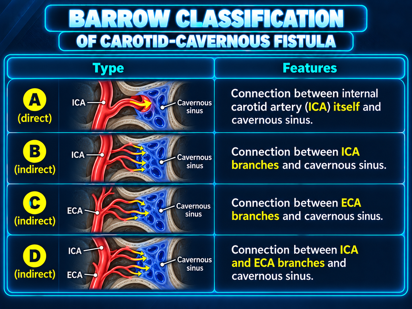
Barrow classification of carotid cavernous fistulas
Barrow ClassificationClick to enlarge
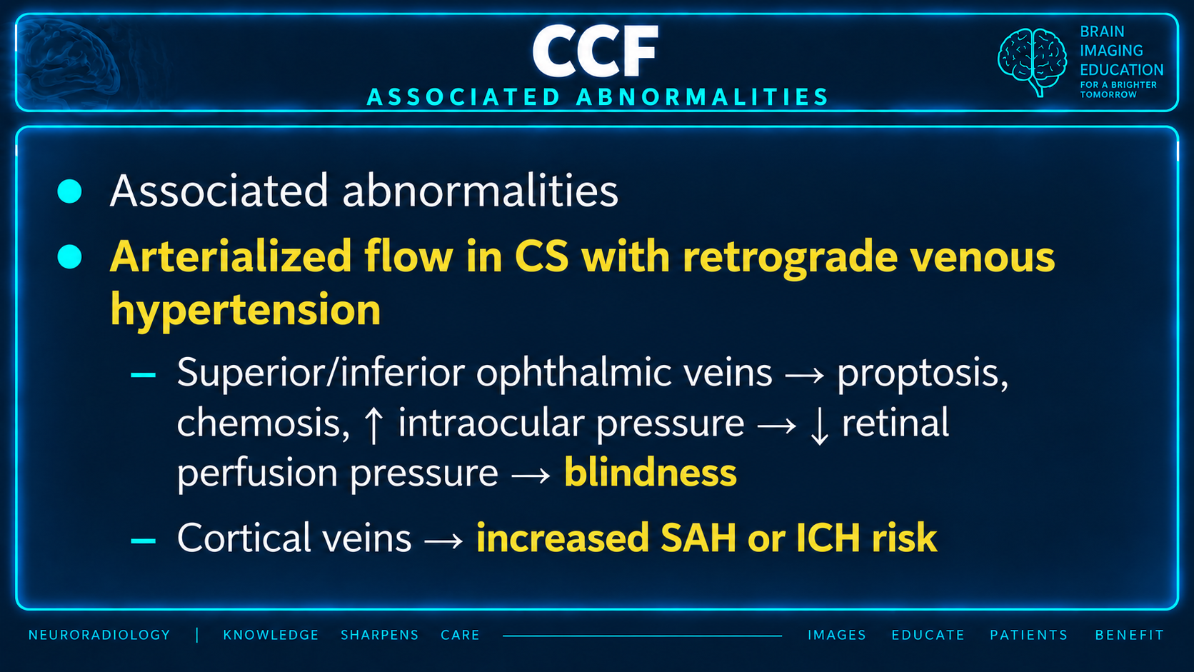
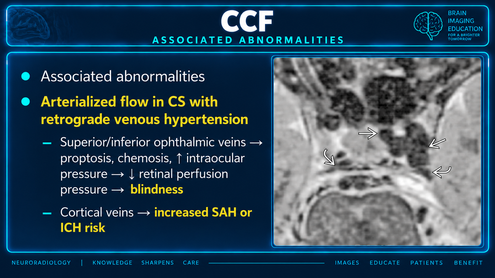
CCF associated abnormalities and retrograde venous hypertension
Associated AbnormalitiesClick to enlarge
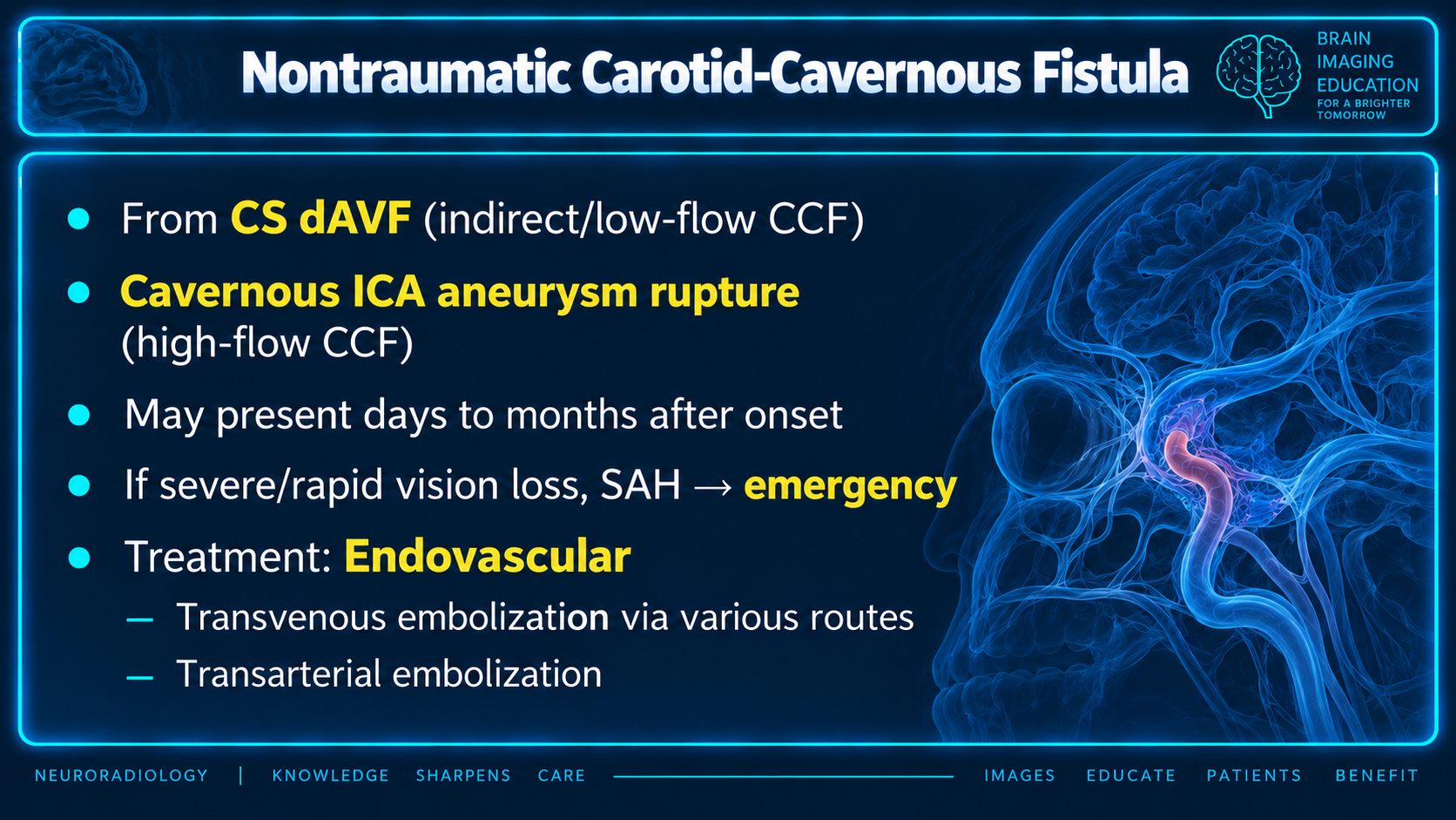
Nontraumatic carotid cavernous fistula teaching points
Nontraumatic CCFClick to enlarge
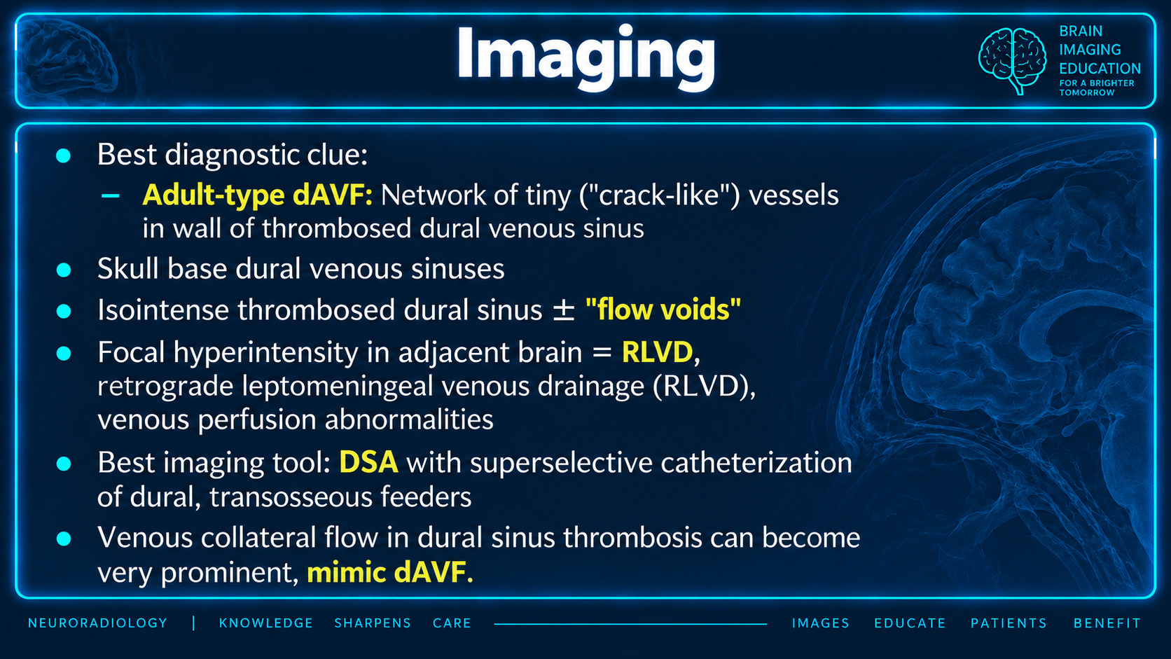
Imaging features of dural arteriovenous fistulas and CCF
Imaging PrinciplesClick to enlarge
MRI findings of retrograde venous hypertension from CCF
Retrograde Venous HypertensionClick to enlarge
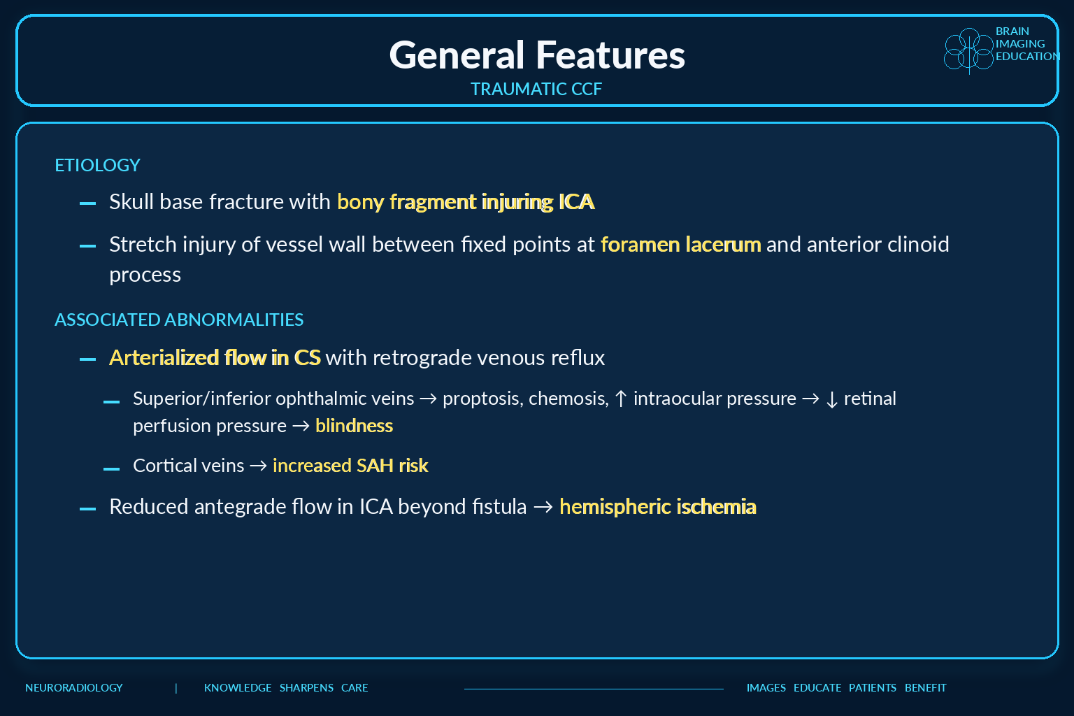
General features of traumatic carotid cavernous fistula
Traumatic CCF FeaturesClick to enlarge
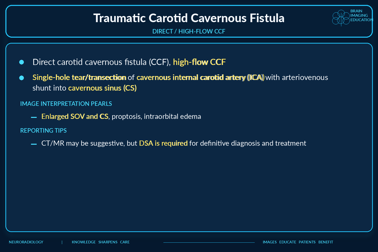
Direct high flow traumatic carotid cavernous fistula
Direct High-Flow CCFClick to enlarge
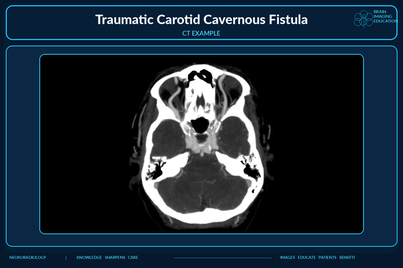
CT example of traumatic carotid cavernous fistula
Traumatic CCF CT ExampleClick to enlarge
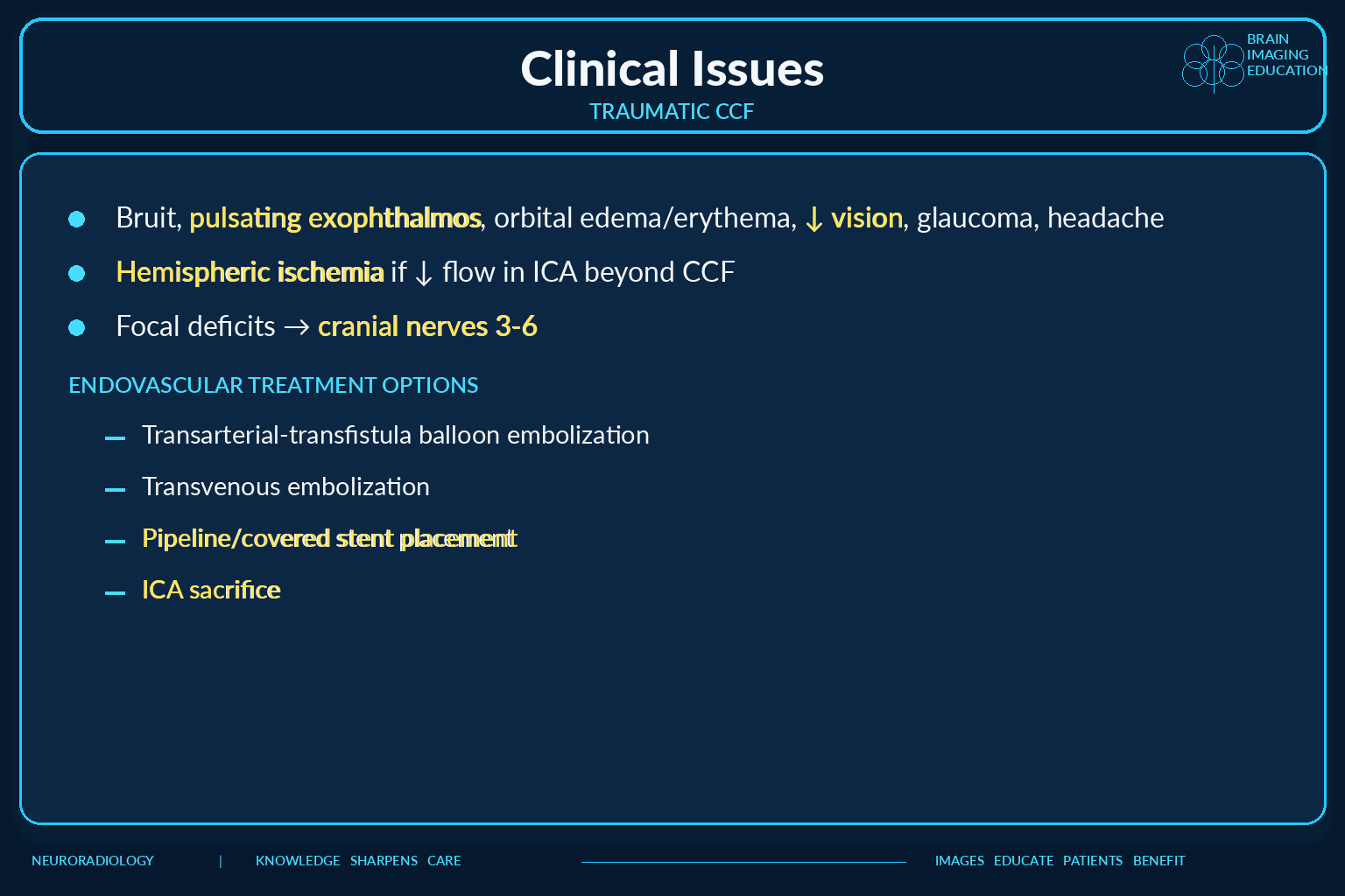
Clinical issues and treatment options for traumatic CCF
Clinical Issues and TreatmentClick to enlarge
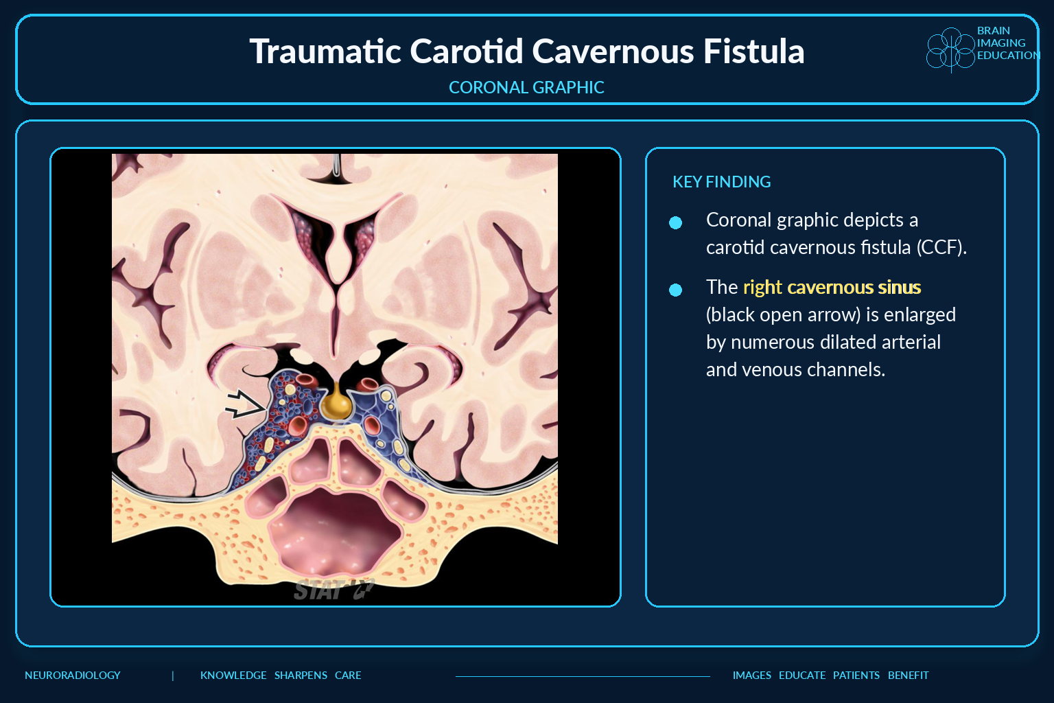
Coronal graphic of enlarged cavernous sinus in CCF
Coronal GraphicClick to enlarge
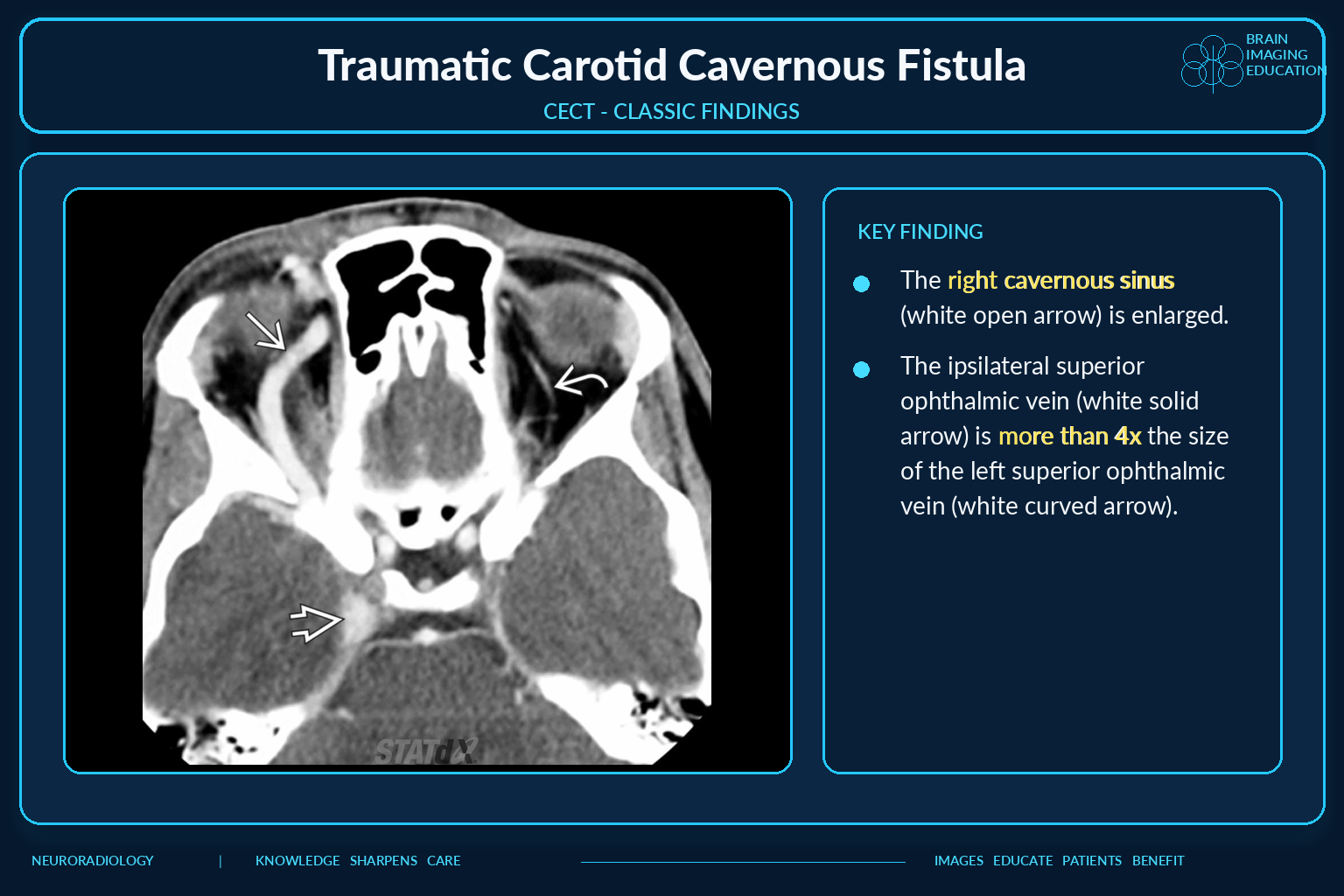
Contrast enhanced CT classic findings of CCF
Classic CECT FindingsClick to enlarge

Overview

A carotid-cavernous fistula is an abnormal communication between the carotid arterial circulation and cavernous sinus. Direct and indirect fistulas differ in anatomy, flow, etiology, clinical tempo, and treatment.

Critical principleClinical risk is determined not only by fistula flow but also by venous drainage. Cortical venous reflux, rapidly declining vision, severe orbital hypertension, or hemorrhage requires urgent neurovascular evaluation.

Barrow Classification

TypeCommunicationTypical category
ADirect connection between intracavernous ICA and cavernous sinusDirect, usually high flow
BDural shunt from intracavernous ICA branchesIndirect
CDural shunt from meningeal ECA branchesIndirect; commonly encountered dural type
DCombined ICA and ECA dural branchesIndirect

Direct CCF

  • Single communication between cavernous ICA and cavernous sinus.
  • Usually high flow with rapid symptom onset.
  • Most often follows trauma, skull-base fracture, surgery, or angiography.
  • May result from rupture of a cavernous ICA aneurysm.
  • Other associations include dissection, fibromuscular dysplasia, and collagen disorders.

Indirect CCF

  • Dural arteriovenous shunt through ICA and/or ECA branches.
  • Usually low flow with insidious presentation.
  • Predilection for postmenopausal women.
  • May follow cavernous sinus thrombosis and revascularization.
  • Associations include pregnancy, regional surgery, sinusitis, Ehlers-Danlos syndrome, and fibromuscular dysplasia.

Clinical Presentation

  • Pulsatile proptosis or exophthalmos and orbital bruit.
  • Chemosis, conjunctival injection, and subconjunctival hemorrhage.
  • Ophthalmoplegia from cranial nerves III, IV, and VI; VI is most commonly affected.
  • V1 or V2 sensory symptoms.
  • Progressive visual loss, glaucoma, headache, or pulsatile tinnitus.
  • Indirect fistulas may be subtle or incidentally detected.

Venous Hypertension

  • Ophthalmic venous reflux causes proptosis, chemosis, elevated intraocular pressure, and reduced retinal perfusion.
  • Cortical venous reflux increases subarachnoid and intracerebral hemorrhage risk.
  • Reduced antegrade ICA flow beyond a high-flow fistula may cause hemispheric ischemia.
  • Drainage toward the petrosal sinuses may produce less conspicuous orbital findings.

CTA Findings

  • Arterial-phase opacification and asymmetric expansion of the cavernous sinus.
  • Enlarged superior or inferior ophthalmic veins.
  • Proptosis, retrobulbar edema, and enlarged extraocular muscles.
  • Cavernous sinus attenuation approaching the ICA.
  • Direct fistula tract or venous pouch when visible.
  • Associated skull-base fracture, aneurysm, or intracranial hemorrhage.

MRI and MRA Findings

  • Abnormal cavernous sinus contour and internal flow voids.
  • Prominent ophthalmic and other draining veins.
  • Orbital or periorbital swelling.
  • Early venous enhancement on time-resolved contrast MRA.
  • Venous congestion, edema, hemorrhage, or abnormal perfusion from cortical reflux.
  • Thin-section 3D postcontrast T1 improves anatomic assessment.

Digital Subtraction Angiography

DSA is the definitive diagnostic and treatment-planning examination because it provides superior spatial and temporal resolution.

  • Defines direct versus dural shunting.
  • Identifies ICA and ECA arterial feeders.
  • Maps venous egress and dangerous cortical venous reflux.
  • Assesses collateral circulation and ICA preservation options.

Direct Versus Indirect Pattern

FeatureDirect CCFIndirect CCF
ShuntICA wall defect directly into cavernous sinusDural branches of ICA and/or ECA
Typical flowHigh flowLow flow, although variable
Typical causeTrauma or cavernous ICA aneurysm ruptureSpontaneous dural fistula, often related to venous thrombosis
Clinical tempoAcute and dramaticInsidious or intermittent
Treatment emphasisClose the ICA tear while preserving the parent artery when possibleOcclude dural feeders and/or cavernous sinus venous pouch

Urgent Features

  • Rapid or severe visual loss.
  • Progressive glaucoma or marked orbital hypertension.
  • Cortical venous reflux.
  • Subarachnoid or intracerebral hemorrhage.
  • Epistaxis or otorrhagia.
  • Hemispheric ischemia or neurologic deficit.
  • Associated feeding-vessel aneurysm.

Treatment

  • Observation may be appropriate for selected low-flow indirect fistulas without high-risk features.
  • Endovascular therapy is the treatment mainstay.
  • Transvenous cavernous sinus embolization is common for indirect fistulas.
  • Transarterial embolization may treat direct tears or dural feeders.
  • Covered or flow-diverting stents may reconstruct the ICA.
  • ICA sacrifice requires adequate collateral assessment.
  • Surgery is reserved for failed or inaccessible endovascular cases.

Reporting Checklist

  • Side and degree of cavernous sinus enlargement.
  • Early cavernous sinus opacification and suspected direct fistula tract.
  • Superior and inferior ophthalmic vein caliber.
  • Orbital congestion, proptosis, and extraocular muscle enlargement.
  • Petrosal, intercavernous, pterygoid, facial, and cortical venous drainage.
  • Cortical venous reflux, edema, hemorrhage, or venous infarction.
  • Associated fracture, ICA injury, dissection, aneurysm, or thrombosis.

Imaging Pitfalls

  • Prominent venous collateral flow from cavernous sinus thrombosis may mimic a fistula.
  • A partially thrombosed cavernous sinus can obscure high-velocity flow voids.
  • Absence of marked proptosis does not exclude posterior or cortical venous drainage.
  • CTA and MRA may suggest the diagnosis but cannot replace angiographic mapping before treatment.

Secondary Links