Epidemiology and Risk
Prevalence rises strongly with age: over 1% at 60–64 years versus 20–40% beyond 85–90 years. Onset before 65 is early-onset disease and represents approximately 5% of patients.
Risk factors
- Advanced age and female sex
- ApoE ε4: one allele ≈3× risk; two ≈8×
- Hypertension, inactivity, smoking, and head trauma
- Family history; APP mutation; Down syndrome
- Chronic inflammation
- Rare iatrogenic acquisition risk
Education, income, occupation, and social/family support influence presentation through cognitive reserve. About 5–10% of early-onset cases reflect autosomal dominant PSEN1/PSEN2 mutations.
Biomarker Diagnosis
Reliable biomarkers may suggest disease before clinical manifestations:
- Amyloid PET
- CSF Aβ42/40
- CSF p-tau181/Aβ42
- CSF t-tau/Aβ42
- Equivalent validated plasma assays
This biomarker-based shift creates a potential presymptomatic therapeutic window.
Historical NINCDS-ADRDA categories
- Definite: clinical diagnosis plus histology
- Probable: typical syndrome without histology (81% sensitive, 73% specific)
- Possible: atypical features without a better diagnosis
Longitudinal clinical criteria are sensitive for dementia generally but less accurate for Alzheimer disease specifically.
Clinical Presentation
Typical disease begins with anterograde episodic-memory impairment, progressing over years to attentional/executive, semantic-memory, praxis, and visuoperceptual deficits.
Neuropsychiatric symptoms eventually affect most patients: apathy, depression, anxiety, agitation/aggression, delusions, and hallucinations.
Early-onset disease
More often nonamnestic, with executive, language, or visuospatial dysfunction and a hippocampal-sparing phenotype. It usually progresses more aggressively with faster global decline and atrophy.
Atypical focal variants
Pathology and Disease Progression
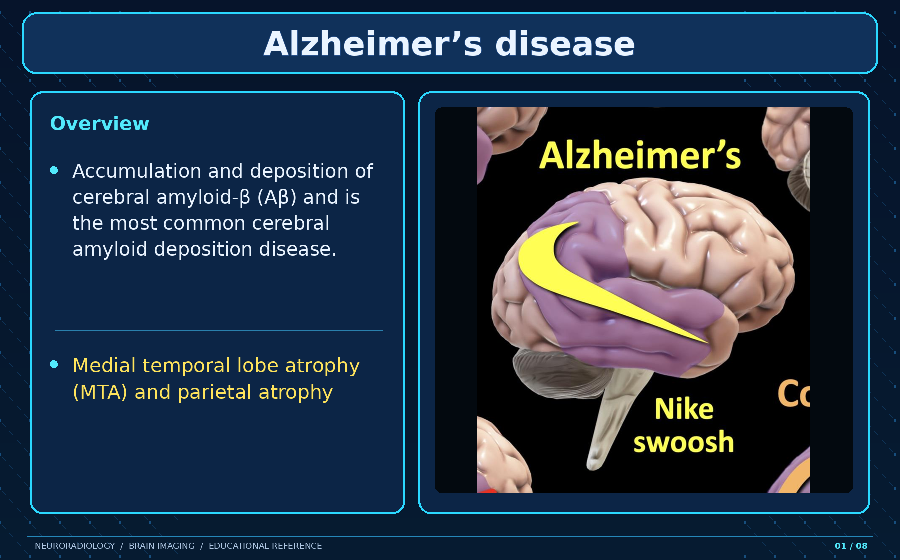
Cerebral Aβ forms neuritic plaques; tau forms neurofibrillary tangles, followed by progressive neuronal loss. Aβ favors neocortical association areas, posterior cingulate, precuneus, and limbic cortex. Chronic microglial activation and inflammatory mediators may contribute to neuronal injury.
Rare iatrogenic Alzheimer disease has been demonstrated after cadaveric pituitary-derived growth hormone exposure. ApoE may influence oligodendrocyte cholesterol processing and myelination.
Braak sequence on imaging
- Stages I–II: earliest entorhinal cortex involvement
- Stages III–IV: limbic system and hippocampus
- Stages V–VI: cortex, especially precuneus and temporal lobes
Structural Imaging
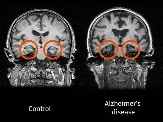
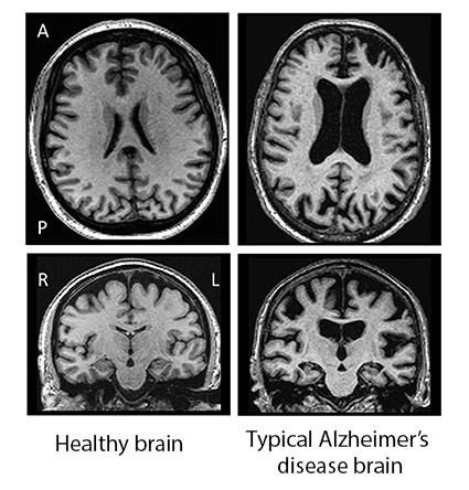
MRI is preferred to CT for detecting characteristic atrophy and excluding alternative causes. Structural diagnosis rests on:
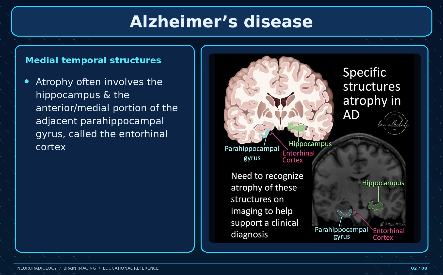
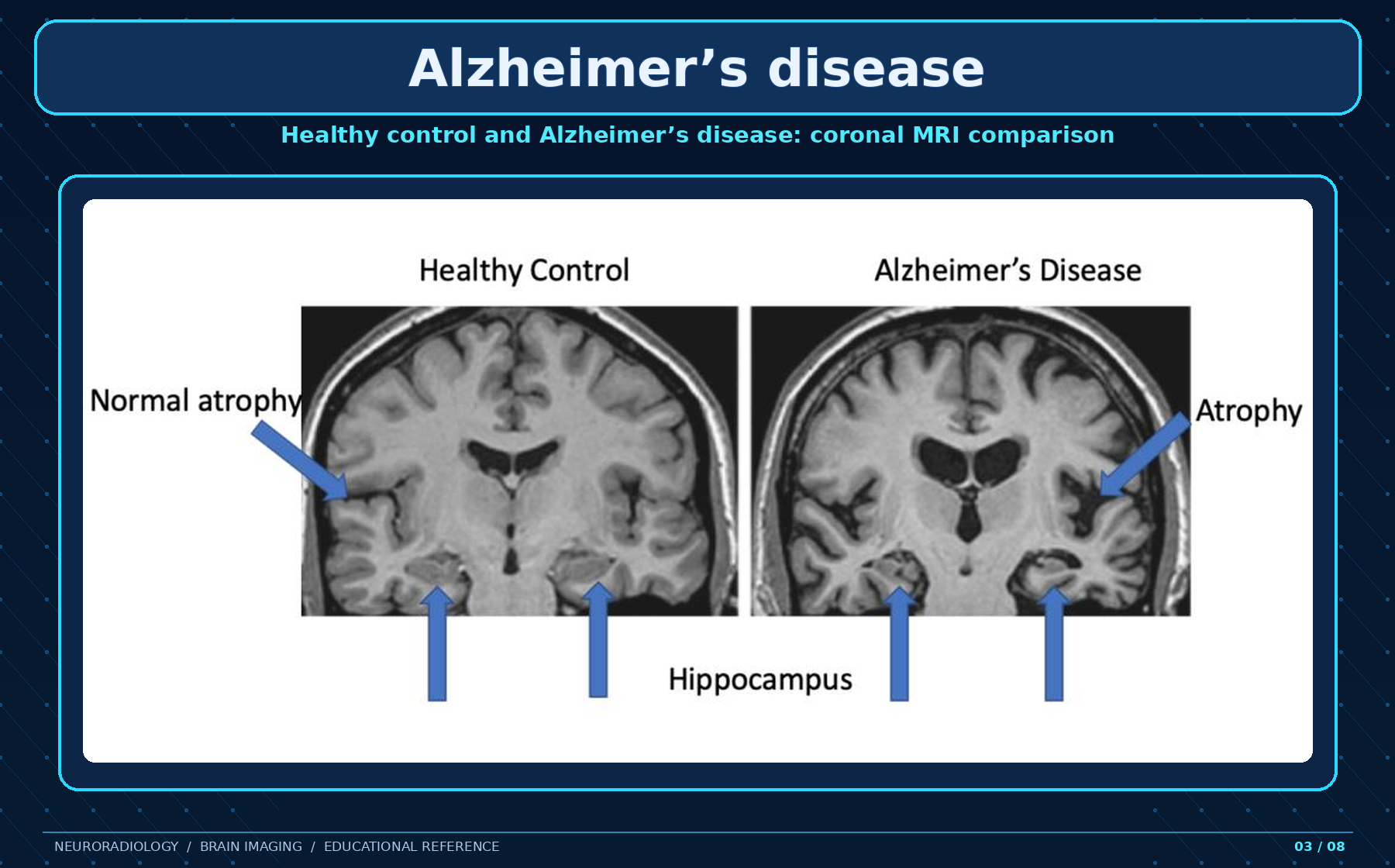
- Mesial temporal atrophy—especially hippocampus, entorhinal cortex, and perirhinal cortex
- Temporoparietal cortical atrophy
Direct hippocampal/parahippocampal volume loss is more accurate than indirect fissural enlargement, but ideally requires volumetry.
Visual scores
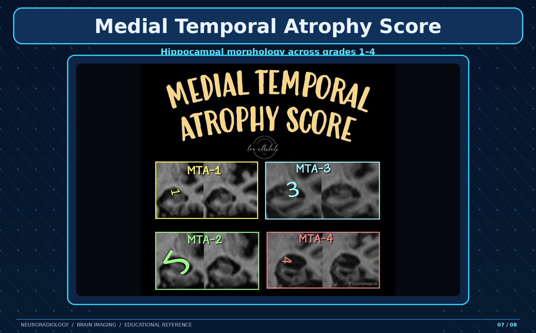
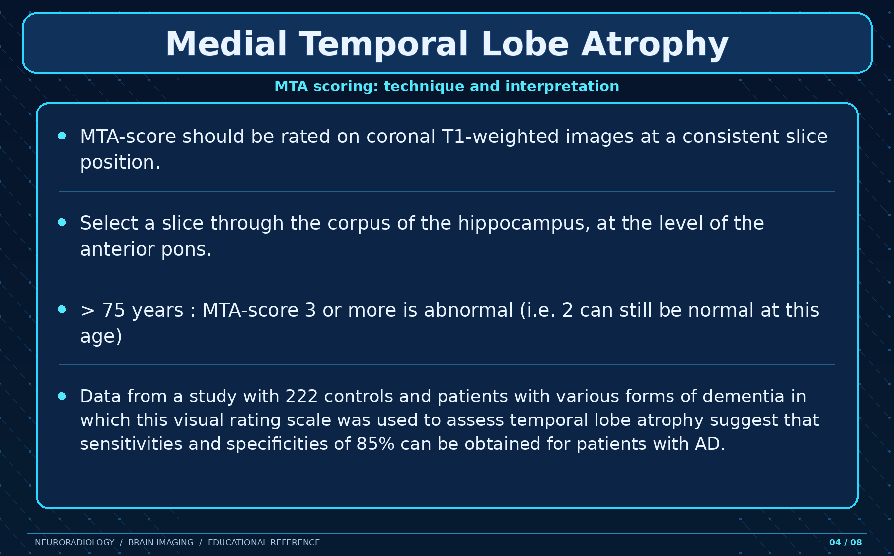
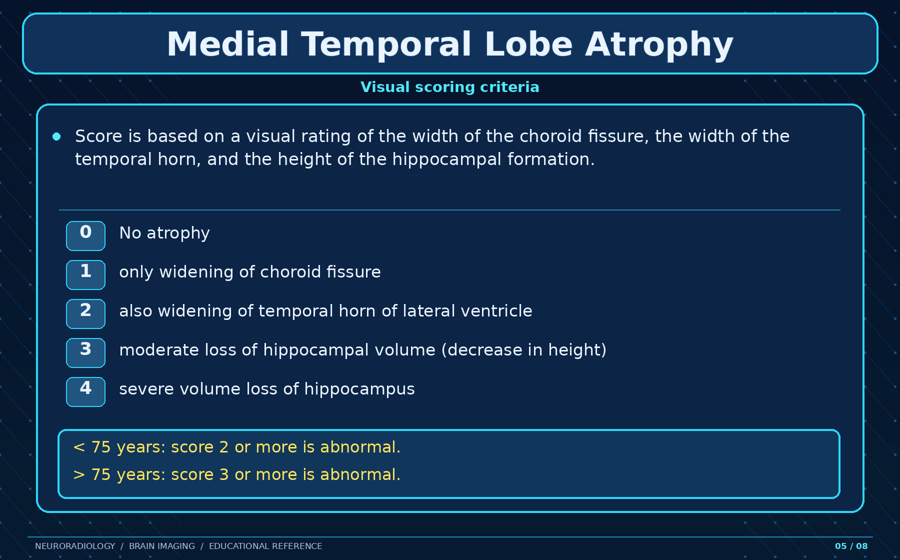
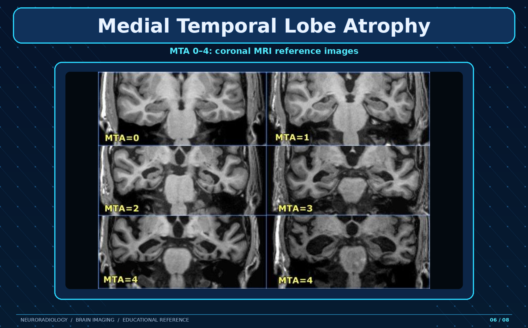
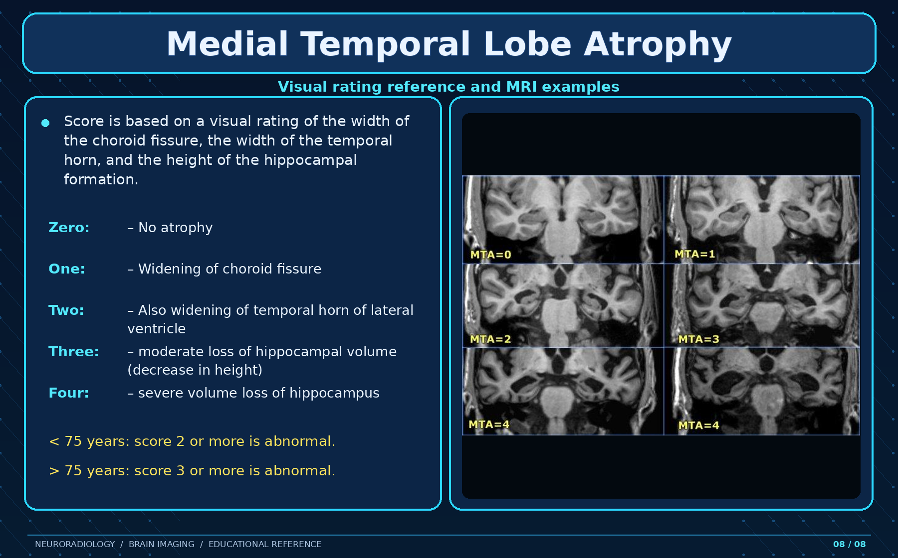
- Medial temporal atrophy (MTA) score predicts MCI-to-dementia progression.
- ERICA evaluates entorhinal cortical atrophy.
- Koedam score assesses parietal/precuneus atrophy, particularly in early-onset disease or posterior cortical atrophy.
Volume Change and WMH
Alzheimer disease produces accelerated whole-brain volume loss—approximately 1% versus 0.5% per year normally. Hippocampal loss is approximately 4.5% versus 1.5% per year.
White-matter T2 hyperintensity, often described as chronic small-vessel ischemic change, correlates with and may precede cognitive impairment.
Limitation: Characteristic volume loss may not be apparent early in the disease course.
Nuclear Medicine
FDG-PET / perfusion
SPECT and FDG-PET demonstrate biparietal and bitemporal hypoperfusion/hypometabolism. FDG-PET typically shows bilateral, usually symmetric temporoparietal, precuneus, and posterior cingulate hypometabolism. Early asymmetry may occur. Anterior cingulate, primary visual and sensorimotor cortices, basal ganglia, thalami, occipital lobes, and cerebellum are relatively spared; frontal involvement may occur late. Early-onset patients often show left-predominant parietal hypometabolism.
Amyloid PET
C-11 Pittsburgh compound B and F-18 florbetapir, flutemetamol, and florbetaben bind beta-amyloid fibrils. Increased cortical activity causes loss of normal gray–white differentiation. A negative scan makes Alzheimer disease unlikely, but 20–25% of cognitively healthy individuals show amyloid deposition, and burden does not correlate well with cognitive severity.
Tau PET
F-18 flortaucipir accumulates in hippocampal, entorhinal, temporal, and parietal regions and correlates with impairment, but is not Alzheimer-specific. Early-onset disease shows greater neocortical tau in the precuneus, inferior parietal lobule, and dorsolateral prefrontal cortex.
Amyloid deposition occurs first, followed by tau accumulation, then reduced glucose metabolism.
Treatment and ARIA
There is no cure; therapy aims to improve symptoms or slow progression.
- Cholinesterase inhibitors: donepezil, rivastigmine, galantamine
- Partial NMDA antagonist: memantine
- Amyloid-lowering antibodies: lecanemab and donanemab; aducanumab was discontinued in 2024
- Symptomatic psychiatric, antiseizure, or antiparkinsonian therapy as appropriate
Monoclonal antibodies can cause amyloid-related imaging abnormalities: hemorrhage (ARIA-H) or edema (ARIA-E). Treatment eligibility and surveillance require susceptibility-sensitive imaging and attention to microbleeds and cortical superficial siderosis.
Differential Diagnosis
LATE
In an elderly amnestic patient with bilateral mesial temporal loss, consider limbic-predominant age-related TDP-43 encephalopathy. Suggestive features include rostrocaudal amygdala-to-hippocampal involvement, profound asymmetric amygdala/hippocampal loss with relative sparing elsewhere, and negative amyloid and tau PET. Definitive diagnosis remains postmortem; LATE may coexist with Alzheimer disease.
Other mimics or co-pathologies
- Primary age-related tauopathy (PART)
- Argyrophilic grain disease (AGD)
- Dementia with Lewy bodies, which may coexist and may have positive amyloid PET
History
In 1910, German psychiatrist Emil Kraepelin included the condition in his psychiatry treatise as “Alzheimer’s dementia.” Italian physician Gaetano Perusini, like Alois Alzheimer, also contributed to understanding its pathophysiology.
Source text last revised by Ashesh Ishwarlal Ranchod on 18 September 2026.