Dementia (Major Neurocognitive Disorder)

Reference Images

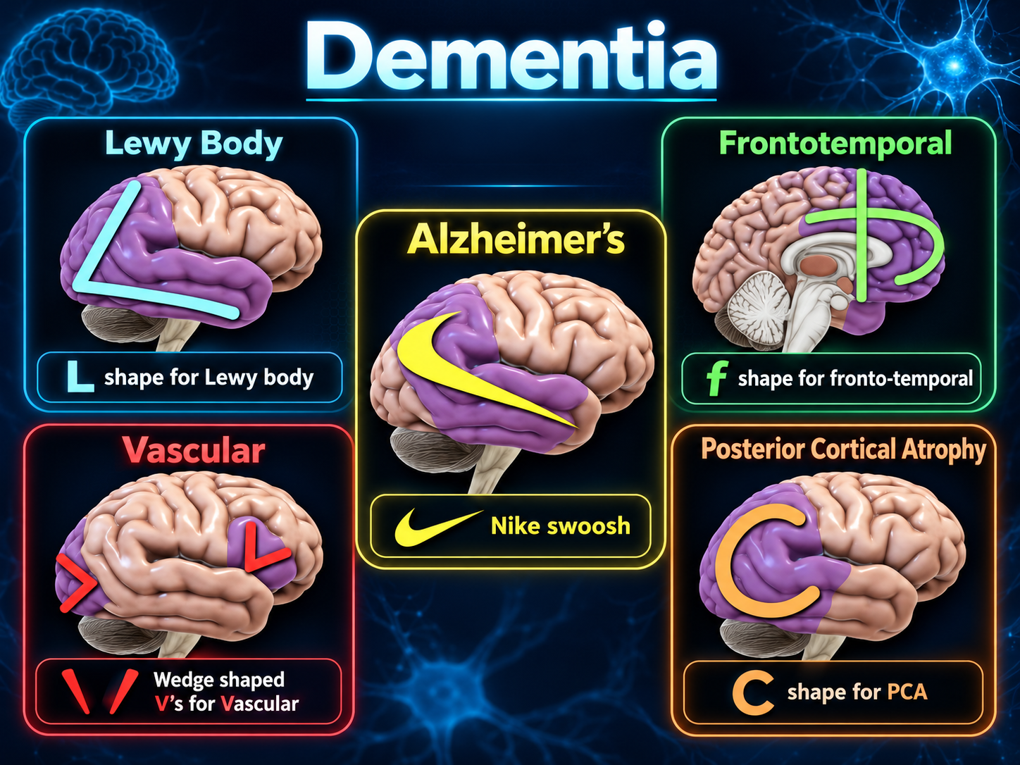
Dementia FDG PET imaging patterns
Dementia FDG-PET PatternsClick to enlarge
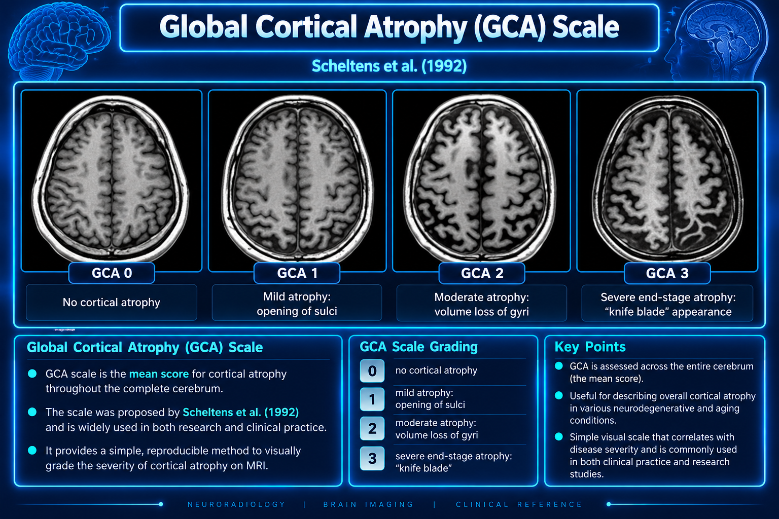
Global Cortical Atrophy scale
Global Cortical Atrophy ScaleClick to enlarge
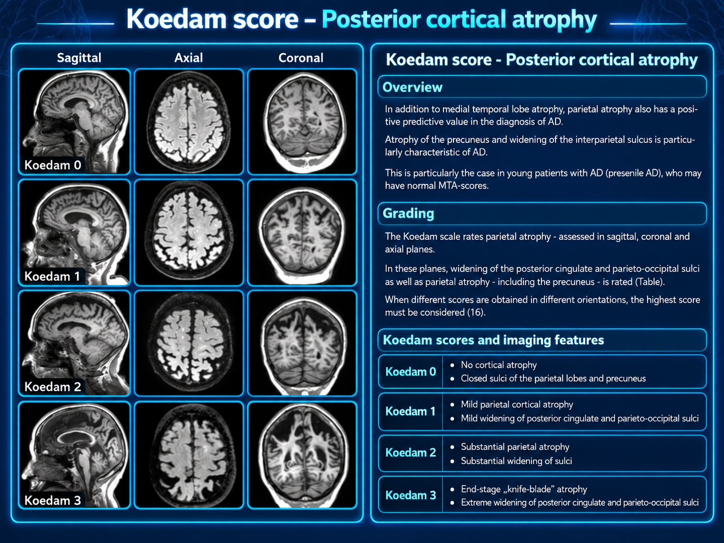
Koedam posterior cortical atrophy score
Koedam Posterior Atrophy ScoreClick to enlarge
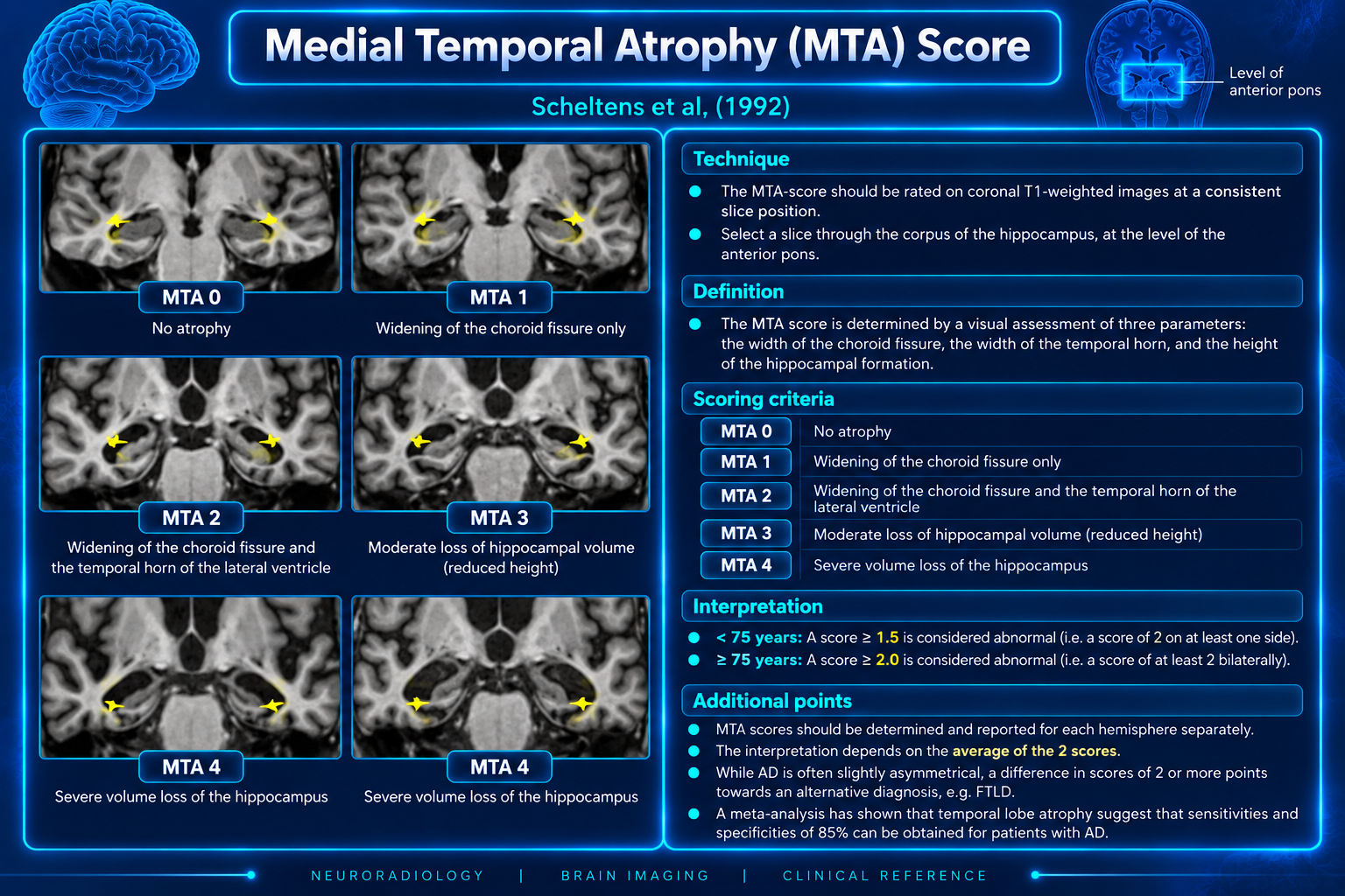
Medial Temporal Atrophy score
Medial Temporal Atrophy ScoreClick to enlarge

Overview

Dementia—relabelled major neurocognitive disorder in the Diagnostic and Statistical Manual of Mental Disorders (DSM-5)—is a clinical syndrome defined by progressive decline in one or more cognitive domains relative to a person’s premorbid functional level. To fulfill criteria for dementia, the resulting cognitive impairment must significantly interfere with independence in performing activities of daily living.1,2

Terminology

The use of dementia in the literature is heterogeneous, with varying definitions.8 Some authors include reversible and irreversible causes of cognitive impairment, while others state that dementia is irreversible6 and exclude reversible conditions. Thus, chronic subdural hematoma and normal pressure hydrocephalus may or may not be included as causes, depending on the definition.

Given this heterogeneity, it is prudent to use the word increasingly sparingly10 and reserve it for the conditions broadly accepted as irreversible neurodegenerative disease, such as Alzheimer disease and frontotemporal lobar degeneration.

Young-onset dementia, also called early-onset dementia and historically presenile dementia, generally refers to onset before age 65 (reported definitions range from 60–70 years).7 Rapidly progressive dementia describes dementia progressing over weeks to months.

FDG-PET Imaging Patterns

These simplified visual pattern-recognition aids derive from a 2006 teaching document.

1. Alzheimer’s disease (AD)Decreased metabolism in the parietal and temporal regions. The distribution resembles the Nike swoosh.
Memory aid: the Nike swoosh pattern is associated with Alzheimer’s disease.
2. Dementia with Lewy bodies (DLB)Temporoparietal hypometabolism similar to Alzheimer disease, with additional occipital involvement; it may extend farther into the anterior temporal cortex. The distribution resembles the letter L.
Memory aid: Lewy begins with L.
3. Frontotemporal dementia (FTD)Hypometabolism involving the frontal and temporal lobes with additional anterior cingulate involvement. The distribution resembles a lowercase letter f.
Memory aid: frontotemporal dementia begins with f.
4. Posterior cerebral atrophyMay be associated with Alzheimer disease or DLB. Hypometabolism involves the occipital and posterior temporal lobes with relative anterior temporal preservation. The distribution resembles a C rather than the L-shaped DLB pattern.
Memory aid: posterior cerebral atrophy (pCa) — C-shaped pattern.
5. Vascular dementiaVariable distribution based on infarct location. Cortical infarctions may create wedge-shaped hypometabolic regions resembling an inverted V.
Memory aid: V represents both vascular and variable.

Clinical Interpretation

Do not use in isolation

FDG-PET findings should be interpreted alongside clinical history, cognitive phenotype, structural imaging, laboratory evaluation, and other diagnostic information rather than used as standalone diagnostic criteria.

Pattern overlap

  • AD and DLB may both show temporoparietal hypometabolism.
  • Occipital involvement favors DLB in the appropriate context.
  • Posterior cerebral atrophy may reflect AD or DLB pathology.
  • Vascular patterns vary with infarct location and burden.

Etiology — Neurodegenerative

  • Alzheimer disease
    • Posterior cortical atrophy
  • Vascular dementia
    • Cerebral small vessel diseases
  • Limbic-predominant age-related TDP-43 encephalopathy (LATE)
  • Primary age-related tauopathy (PART)
  • Argyrophilic grain disease (AGD)
  • Lewy body dementia
    • Dementia with Lewy bodies
    • Parkinson disease dementia
  • Frontotemporal lobar degeneration
    • Behavioral variant FTD
    • Primary progressive aphasia
    • Right temporal variant FTD
    • Corticobasal degeneration
    • Progressive supranuclear palsy
  • Multiple system atrophy
  • Cerebral amyloid angiopathy
  • Chronic traumatic encephalopathy
  • Huntington disease
  • Fahr disease
  • Prion disease, including Creutzfeldt-Jakob disease
  • Leukodystrophies

Other CNS Pathology

These causes are not strictly neurodegenerative and may be reversible:

  • Chronic subdural hematoma
  • Normal pressure hydrocephalus
  • Brain tumor
  • Spontaneous intracranial hypotension (frontotemporal brain sagging syndrome)
  • Wilson disease

Psychoactive substances

  • Alcohol
  • Sedatives, hypnotics, or anxiolytics
  • Other substances, including volatile inhalants

Autoimmune and Infection

Autoimmune disorders

  • Multiple sclerosis
  • Autoimmune encephalitis
  • Cerebral vasculitis
  • Inflammatory cerebral amyloid angiopathy
  • Systemic lupus erythematosus
  • Neurosarcoidosis

Infection

  • HIV-associated dementia
  • Neurosyphilis
  • Progressive multifocal leukoencephalopathy
  • Subacute sclerosing panencephalitis
  • Whipple disease

Systemic, Nutritional, and Toxic

Endocrine

  • Hypothyroidism
  • Hyperparathyroidism

Deficiency states

Thiamine, vitamin B3, vitamin B6, vitamin B12, and folate deficiency.

Other

  • Medication effects and interactions
  • Toxin exposure, including heavy metals

Secondary Links