Thyroid Nodule CT/MRI/PET Triage
Reference Images
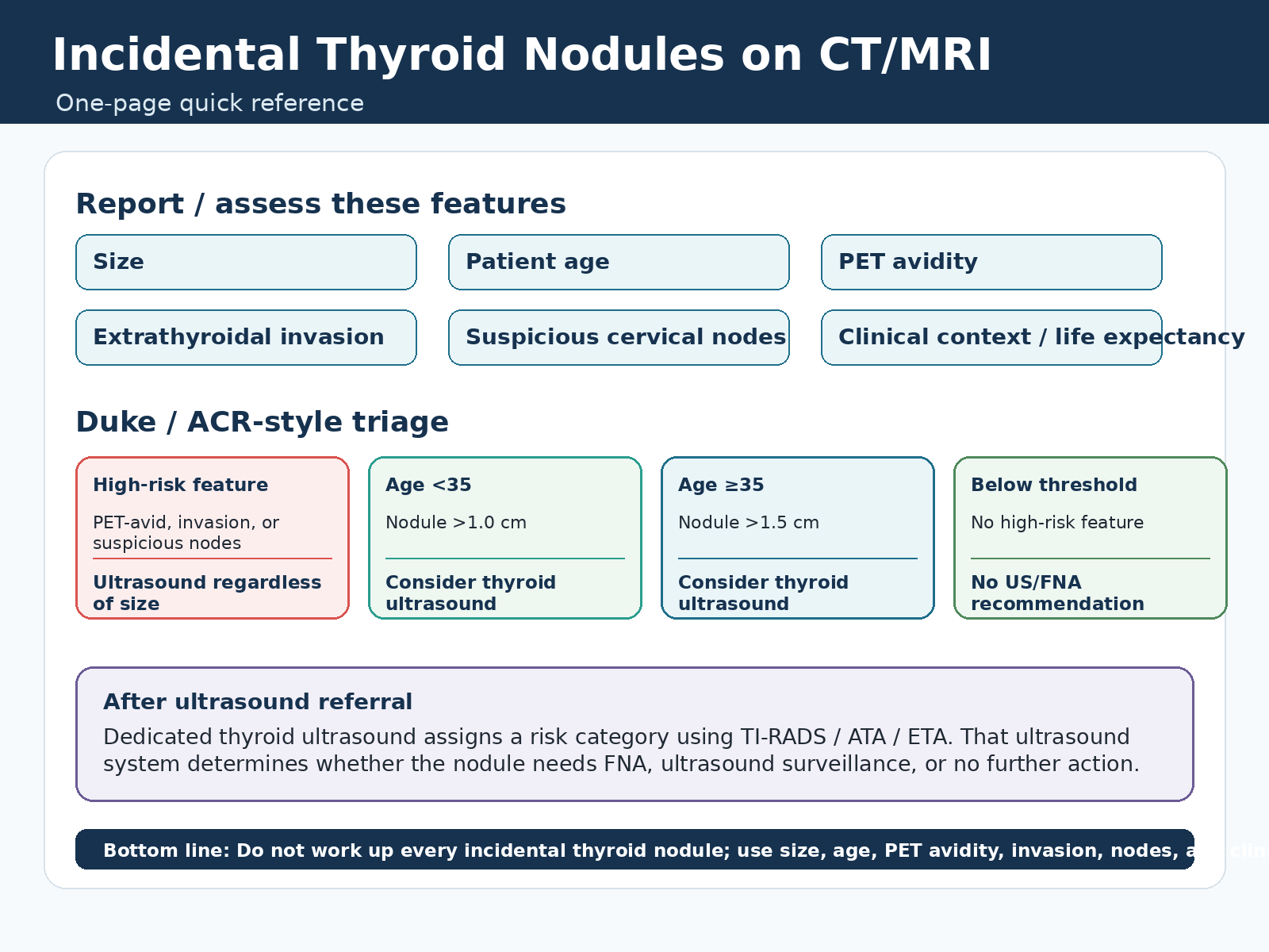
When To Recommend Ultrasound
High-risk feature
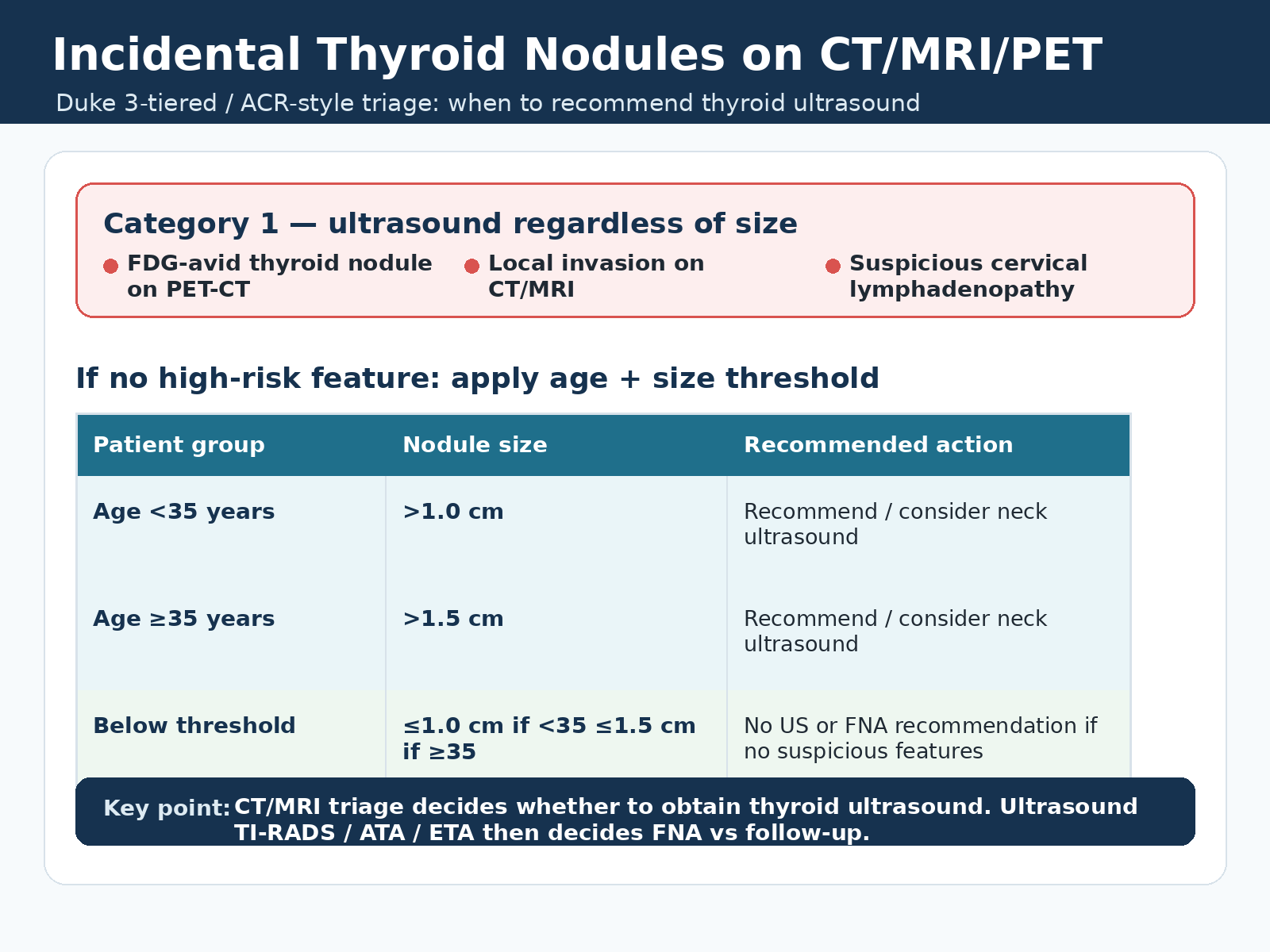
Recommend thyroid ultrasound regardless of nodule size when there is FDG avidity, local invasion, or suspicious cervical lymphadenopathy.
Red flags override size
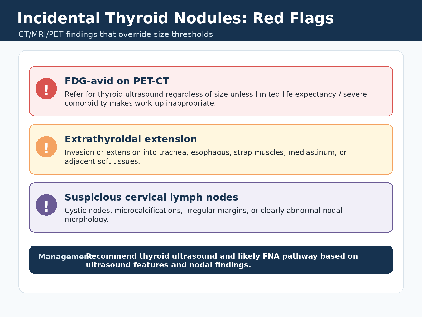
- Focal FDG-avid thyroid nodule on PET/CT.
- Local invasion or extrathyroidal extension on CT/MRI.
- Suspicious cervical lymph nodes.
- Consider clinical context: limited life expectancy or severe comorbidity may make work-up inappropriate.
If no high-risk feature
- Age under 35 years: consider thyroid ultrasound when the nodule is greater than 1.0 cm.
- Age 35 years or older: consider thyroid ultrasound when the nodule is greater than 1.5 cm.
- Below threshold: no thyroid ultrasound or FNA recommendation if there are no suspicious features.
Report / assess
- Maximum nodule size, patient age, PET avidity, extrathyroidal invasion, suspicious cervical nodes, and clinical context.
- Smaller incidental nodules without PET avidity, invasion, or suspicious nodes can be described in the body of the report but usually should not be emphasized in the impression.
Workflow / Red Flags
CT/MRI/PET triage
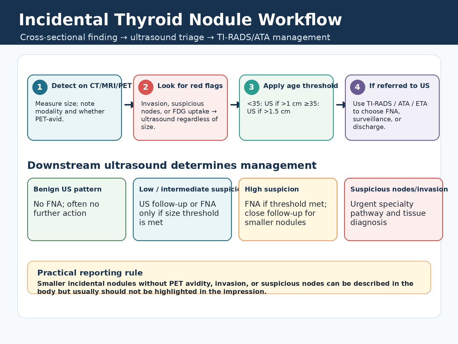
Cross-sectional imaging decides whether dedicated thyroid ultrasound is needed; ultrasound then determines FNA, surveillance, or discharge.
Stepwise approach
- Detect on CT, MRI, or PET/CT; measure the nodule and note modality.
- Look for red flags: FDG uptake, invasion, or suspicious cervical nodes.
- If no red flag is present, apply the age and size threshold.
- If referred to ultrasound, use TI-RADS / ATA / ETA to choose FNA, surveillance, or no further action.
Suspicious features
- Extrathyroidal extension into trachea, esophagus, strap muscles, mediastinum, or adjacent soft tissues.
- Cystic cervical nodes, microcalcifications, irregular margins, or clearly abnormal nodal morphology.
- Suspicious nodes or invasion should move the patient into an urgent specialty pathway with tissue diagnosis as clinically appropriate.
After ultrasound
- Benign ultrasound pattern: no FNA; often no further action.
- Low or intermediate suspicion: follow-up or FNA only if size threshold is met.
- High suspicion: FNA if threshold is met; close follow-up for smaller nodules.
Report Language
No work-up threshold met
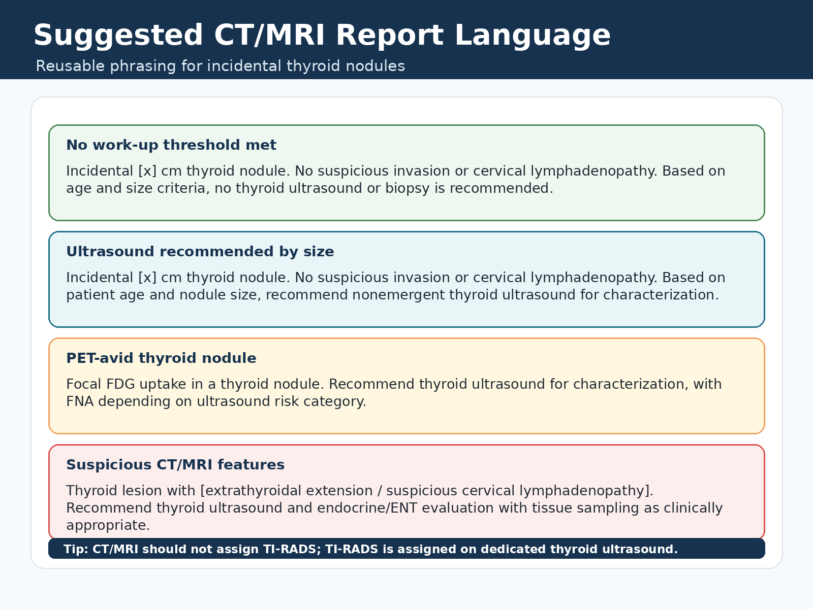
- Incidental [x] cm thyroid nodule. No suspicious invasion or cervical lymphadenopathy. Based on age and size criteria, no thyroid ultrasound or biopsy is recommended.
Ultrasound recommended by size
- Incidental [x] cm thyroid nodule. No suspicious invasion or cervical lymphadenopathy. Based on patient age and nodule size, recommend nonemergent thyroid ultrasound for characterization.
PET-avid or suspicious
- Focal FDG uptake in a thyroid nodule. Recommend thyroid ultrasound for characterization, with FNA depending on ultrasound risk category.
- Thyroid lesion with [extrathyroidal extension / suspicious cervical lymphadenopathy]. Recommend thyroid ultrasound and endocrine/ENT evaluation with tissue sampling as clinically appropriate.
Practical note
- CT/MRI should not assign TI-RADS; TI-RADS is assigned on dedicated thyroid ultrasound.
- Open the thyroid ultrasound TI-RADS page.