External and Middle Ear
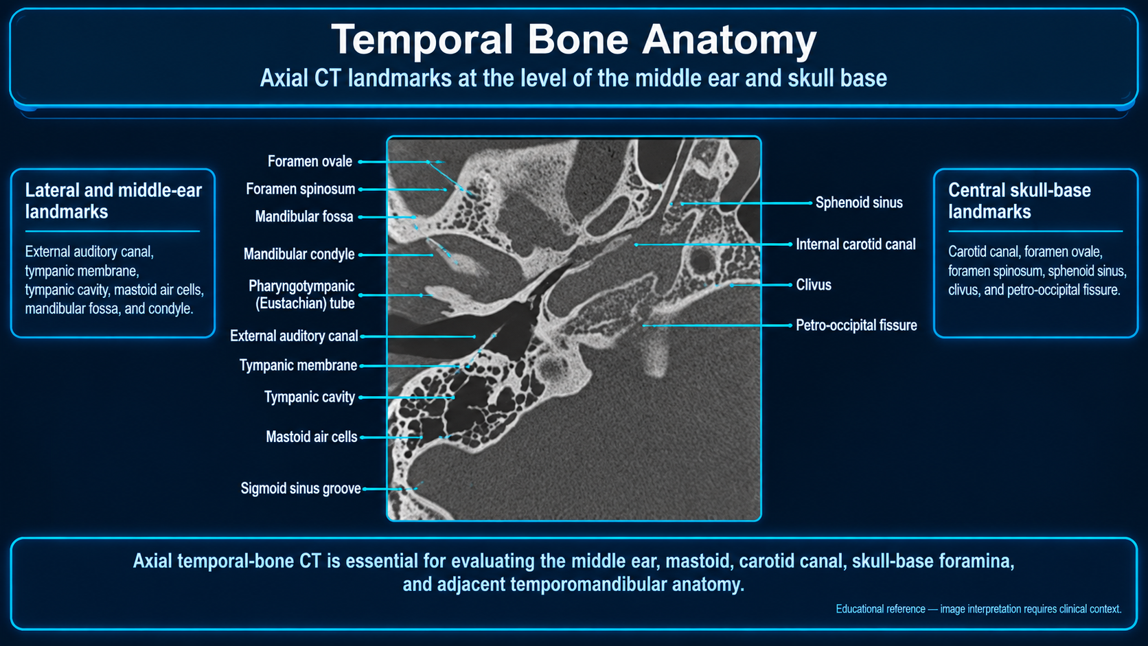
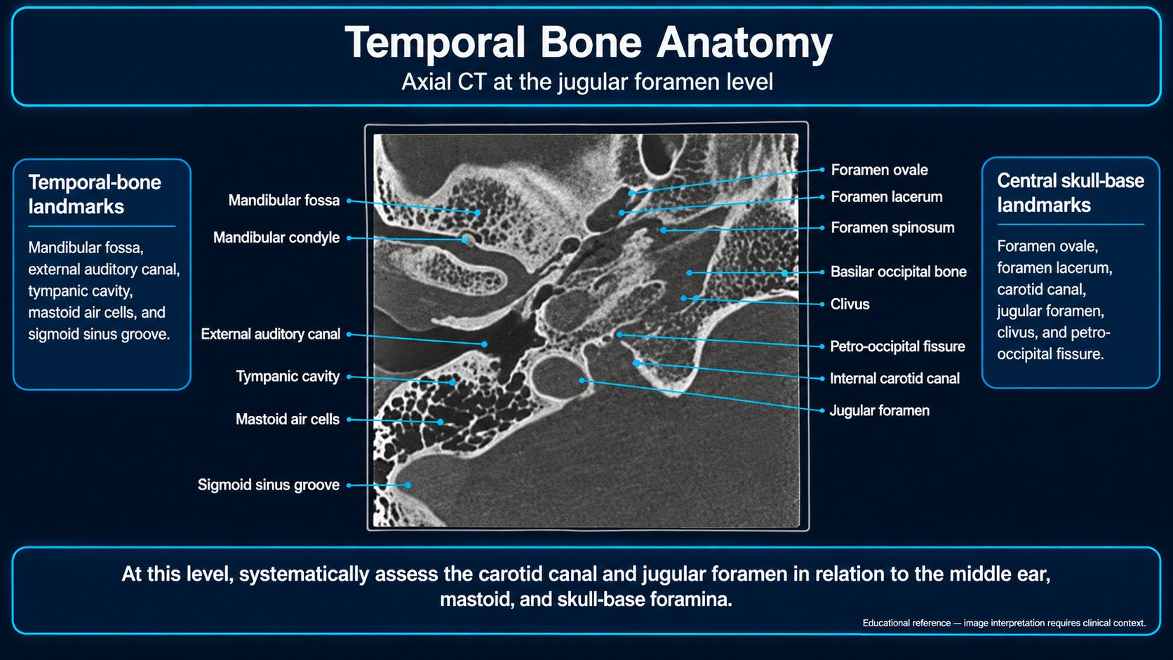
Bony frameworkSquamous, tympanic, petrous, mastoid, and styloid portions form the temporal bone. The petrous portion encloses the otic capsule and inner ear.
External ear
- The lateral one-third of the external auditory canal is cartilaginous and the medial two-thirds are bony.
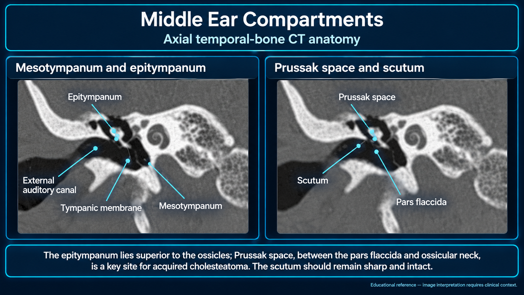
- The tympanic membrane separates the canal from the middle ear. The pars tensa is larger and inferior; the pars flaccida is smaller and superior.
- The scutum is the sharp lateral attic margin adjacent to the pars flaccida.
Middle-ear compartments
- Epitympanum: above the tympanic membrane; contains the malleus head and incus body.
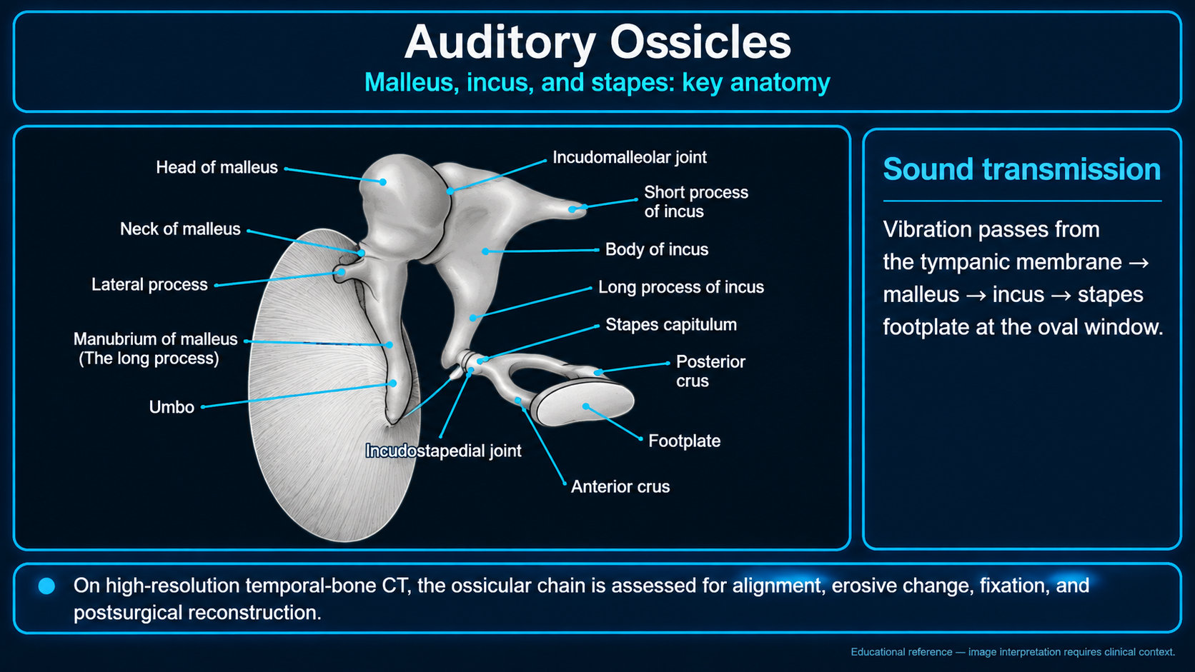
- Mesotympanum: contains the malleus handle, incus long process, stapes, promontory, and oval and round windows.
- Hypotympanum: inferior cavity adjacent to the jugular bulb and inferior petrous carotid region.
- Protympanum: anterior cavity leading to the auditory tube.
- Retrotympanum: posterior recesses include the facial recess and sinus tympani.
Ossicles and spaces
Sound passes from tympanic membrane to malleus, incus, stapes footplate, and oval window. On axial CT, the malleus head and incus body form the “ice-cream cone.” Prussak space lies between the pars flaccida/scutum and malleus neck.
Inner Ear and Neural Pathways
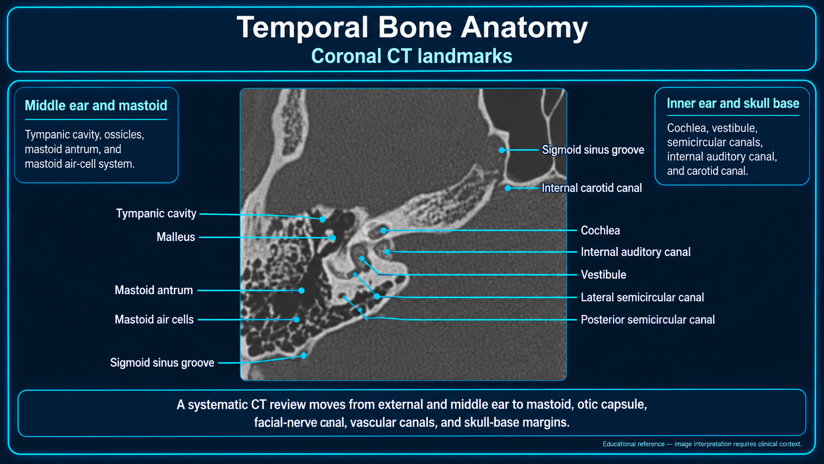
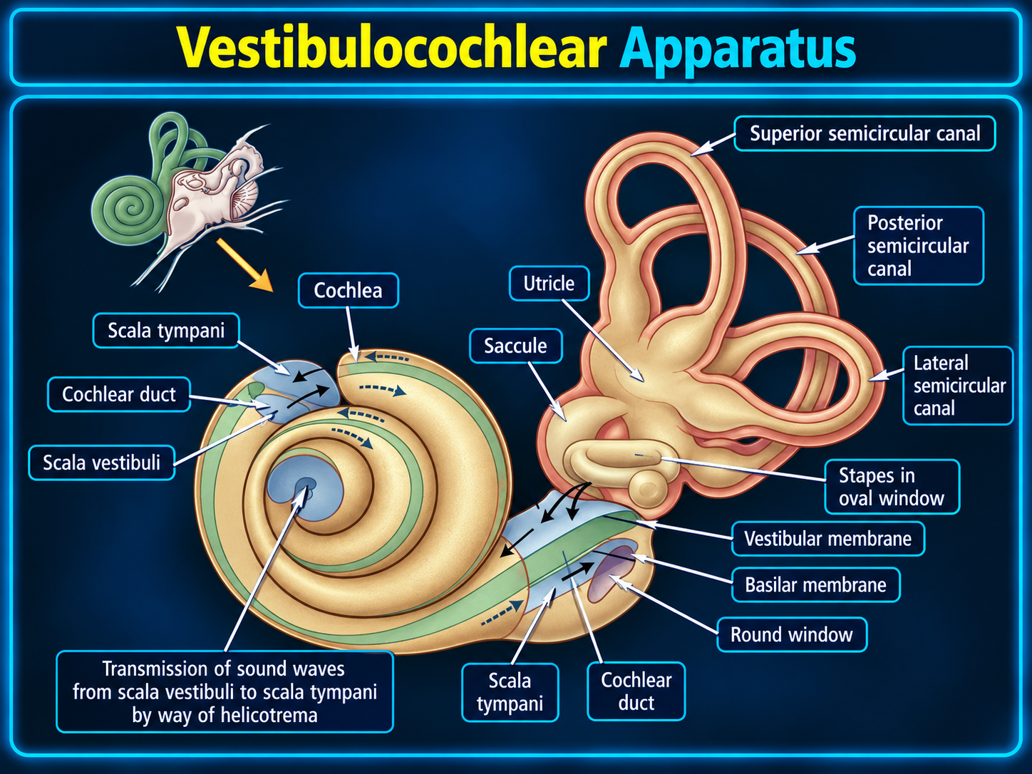
Otic capsuleDense bone surrounds the cochlea, vestibule, and semicircular canals. The membranous labyrinth contains endolymph and is surrounded by perilymph.
Labyrinth
- Cochlea: approximately 2½–2¾ turns around the modiolus; its basal turn forms the cochlear promontory.
- Vestibule: contains the utricle and saccule; the stapes footplate interfaces with it at the oval window.
- Semicircular canals: superior, posterior, and lateral canals sense rotation; the superior and posterior canals share a common crus.
- Aqueducts: the vestibular aqueduct carries the endolymphatic duct; the cochlear aqueduct relates to the perilymphatic duct.
IAC fundus
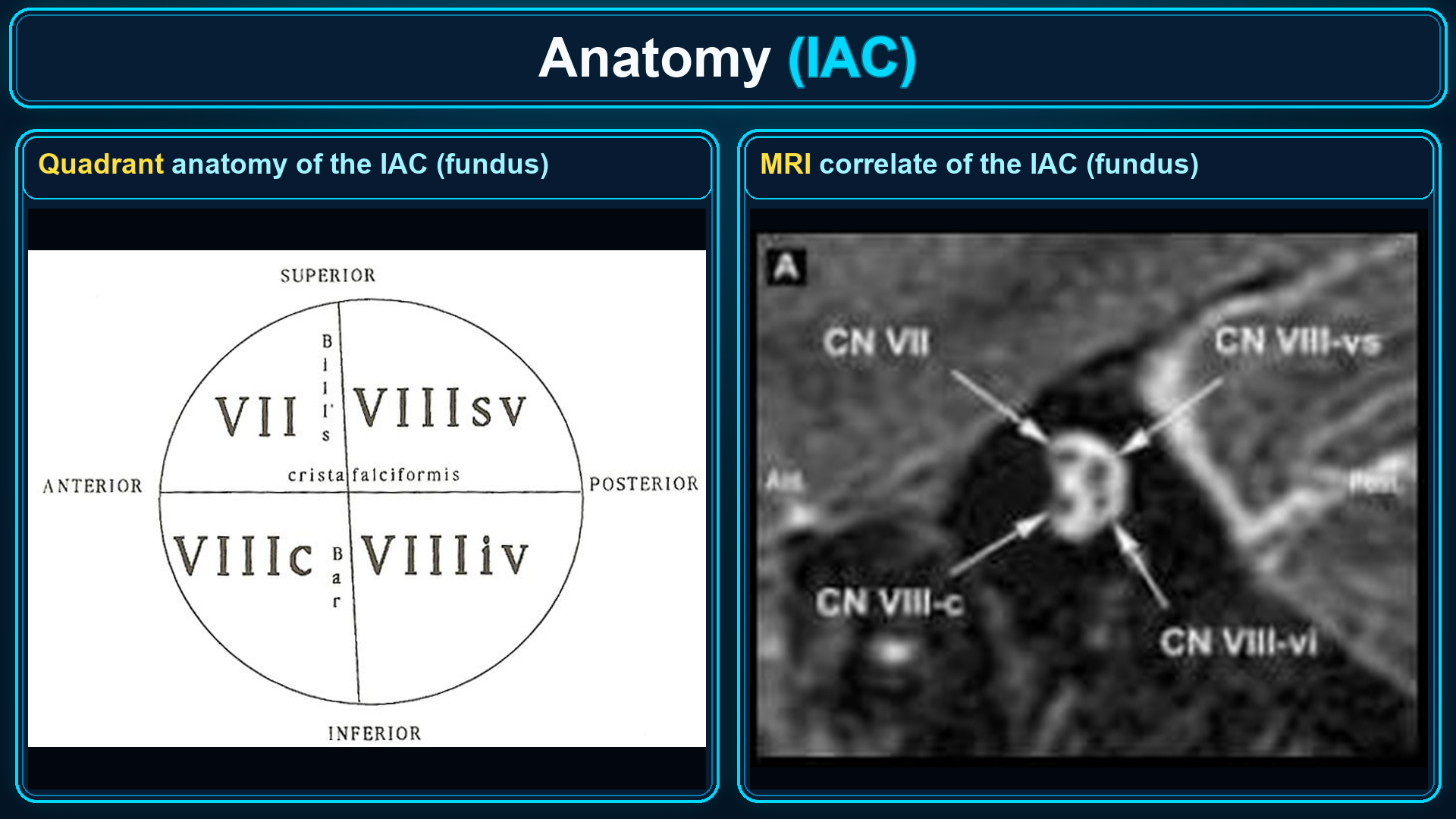
- Anterosuperior: facial nerve (CN VII).
- Anteroinferior: cochlear nerve.
- Posterior: superior and inferior vestibular nerves.
Facial nerve
IAC → labyrinthine segment → geniculate ganglion → tympanic segment → second genu → mastoid segment → stylomastoid foramen. The tympanic segment passes above the oval window and below the lateral semicircular canal.
Mastoid and Imaging
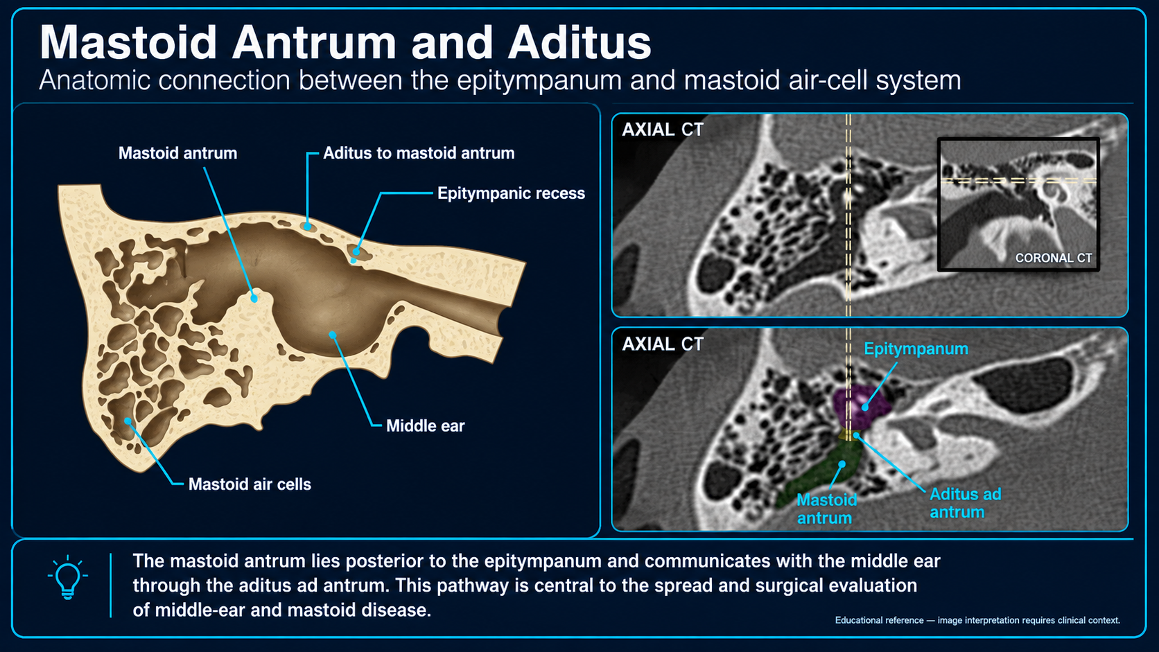
Middle ear to mastoidEpitympanum → aditus ad antrum → mastoid antrum → mastoid air cells.
Mastoid boundaries
- The tegmen mastoideum forms the roof.
- The sigmoid plate borders the sigmoid sinus.
- The posterior external auditory canal wall is a lateral boundary.
- Koerner septum may separate lateral and medial mastoid cell groups.
Modality strengths
High-resolution CT best depicts bone, ossicles, windows, and air-cell pathways. MRI complements CT for cranial nerves, labyrinthine fluid, soft tissue, and selected complications.
Suggested search pattern
EAC and membrane → scutum and middle ear → ossicles and windows → facial nerve canal → otic capsule and labyrinth → IAC → aditus, antrum, and mastoid cells.
Sources