Oval window atresia
Key Images
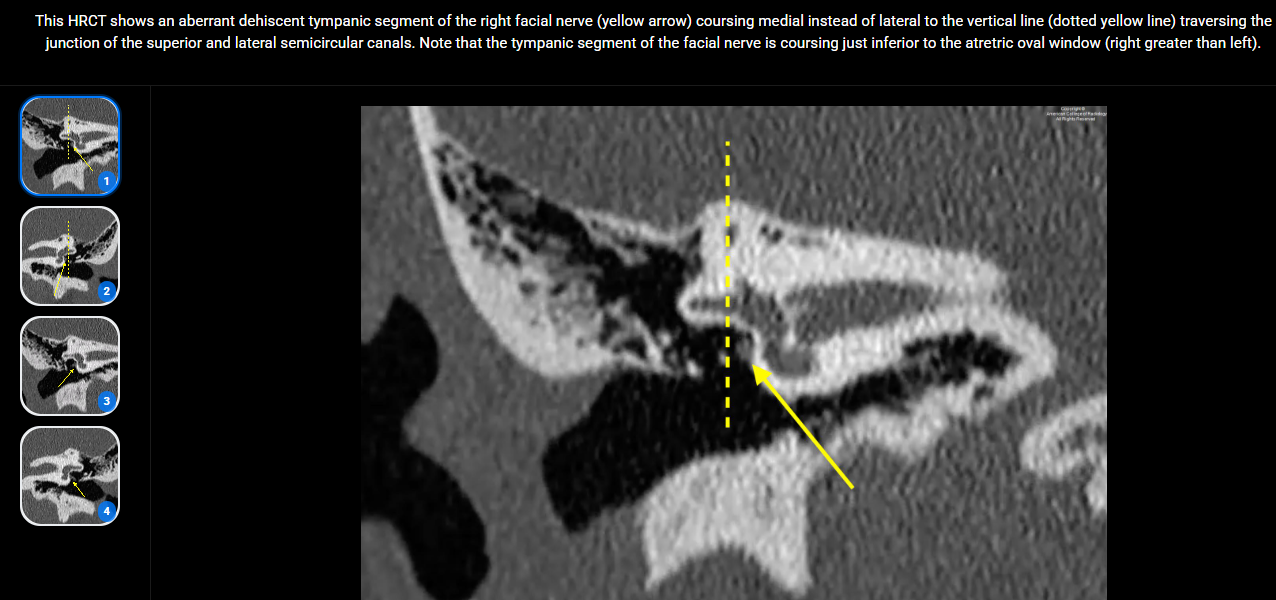
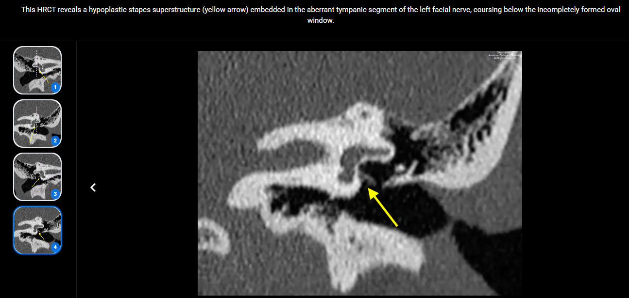
Look For First
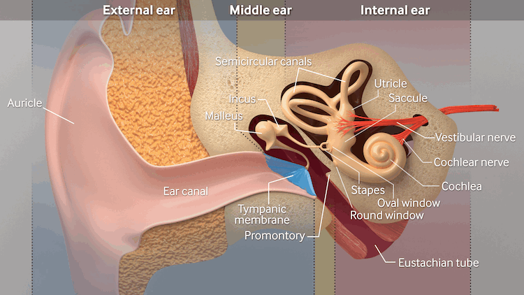
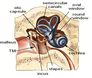
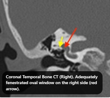
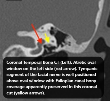
- Absent or atretic oval window on coronal CT images
- Associated stapes superstructure dysplasia on axial CT
- Aberrant facial nerve course with possible interposition between stapes and oval window
Key Image Findings
- HRCT is the preferred imaging modality for characterizing oval window atresia and associated ossicular and otic capsule anatomy.
- Axial CT views optimally visualize the stapes superstructure to assess for dysplasia, which is almost always present with oval window atresia.
- Coronal CT views are ideal for direct visualization of the atretic oval window and the course of the facial nerve in relation to the stapes footplate.
- The stapes footplate may be absent, hypoplastic, or displaced due to aberrant facial nerve interposition between the stapes and oval window.
- Dysplasia of the stapes superstructure is an invariable finding due to embryologic interdependence of the stapes, facial nerve, and oval window during development.
- The facial nerve course is frequently abnormal, with the nerve interposing in the space between the stapes footplate and the atretic oval window.
- Associated ossicular chain abnormalities and anomalies of the otic capsule may be present and require detailed assessment on both axial and coronal planes.
- High-resolution thin-section CT with multiplanar reconstructions is necessary to comprehensively evaluate the severity and morphology of this complex malformation.
Differential Diagnosis
- Otosclerosis: progressive fixation of the stapes footplate with normal oval window anatomy; oval window atresia presents with absent/atretic oval window and stapes superstructure dysplasia rather than footplate stiffening.
- Tympanosclerosis: acquired middle ear ossification and ossicular fixation from chronic infection or trauma; oval window atresia is congenital with absent oval window rather than acquired ossification.
- Stapes fixation from other causes: bilateral involvement and oval window atresia with associated stapes dysplasia distinguish this entity from isolated stapes fixation.
- Congenital stapes fixation without oval window atresia: presence of a patent oval window and intact stapes superstructure help distinguish from oval window atresia.
- Congenital aural atresia: affects the external auditory canal; oval window atresia may occur in isolation or with concurrent canal atresia.
- Mondini deformity and other otic capsule dysplasias: these involve cochlear hypoplasia and increased vestibular dimensions; oval window atresia specifically involves absence of the oval window.
Discussion
Oval window atresia is one of the rarest congenital causes of conductive hearing loss and may remain undiagnosed for years in pediatric patients.
The embryologic interdependence of the stapes footplate, oval window, and facial nerve means that stapes superstructure dysplasia and facial nerve aberrance are nearly always present with oval window atresia.
Patients with oval window atresia are frequently misdiagnosed with otosclerosis because both present with progressive conductive hearing loss, delaying appropriate diagnosis and treatment.
The etiology of atretic oval window is thought to result either from failure of the primitive stapes to fuse with the primitive vestibule or from interposition of the developing facial nerve between the stapes and oval window.
Comprehensive preoperative imaging with axial views for stapes assessment and coronal views for oval window and facial nerve visualization is essential for surgical planning.
Treatment options include bone conduction hearing aids and surgical procedures involving the oval window and vestibule, making detailed preoperative anatomic characterization critical.
Reporting Pearls
Clearly describe the oval window as absent or atretic on coronal imaging, document the morphology of the stapes superstructure on axial views, specify the facial nerve course relative to the stapes footplate, and note the severity of associated ossicular dysplasia to guide surgical planning.
Pitfalls
- Failure to obtain both axial and coronal views; axial alone may miss oval window atresia, and coronal alone may underestimate stapes superstructure pathology.
- Misidentifying oval window atresia as otosclerosis on clinical grounds without HRCT confirmation, leading to delayed accurate diagnosis and inappropriate treatment planning.
- Overlooking aberrant facial nerve course, which is almost always present and critically important for surgical safety and planning.
- Inadequate assessment of the full extent of stapes superstructure dysplasia, which determines surgical feasibility and technique selection.