Facial Nerve / CN VII

Neuroradiology approach: Follow CN VII from the caudal pons through the CPA and IAC, then through the labyrinthine segment, geniculate ganglion, tympanic segment, second genu, mastoid segment, and stylomastoid foramen. Distinguish smooth neuritic enhancement from nodular enlargement, bony expansion, or proximal perineural extension.

Reference Images

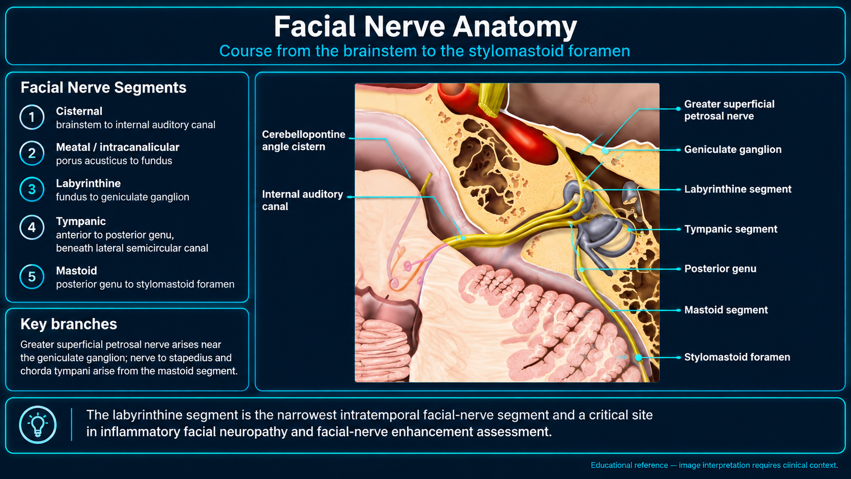
Facial nerve course from the brainstem to the stylomastoid foramen
Facial Nerve CourseClick to enlarge
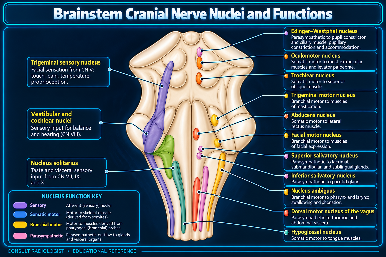
Brainstem cranial nerve nuclei and functions
Brainstem NucleiClick to enlarge
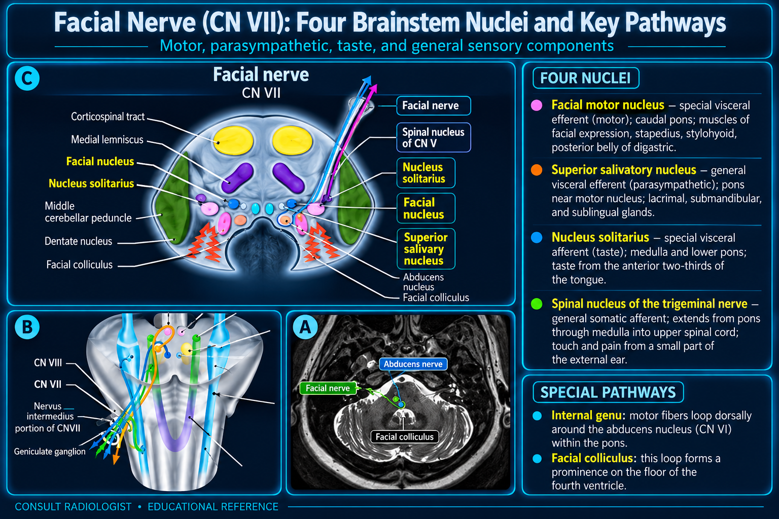
Four facial nerve nuclei and key pathways
CN VII Nuclei and PathwaysClick to enlarge
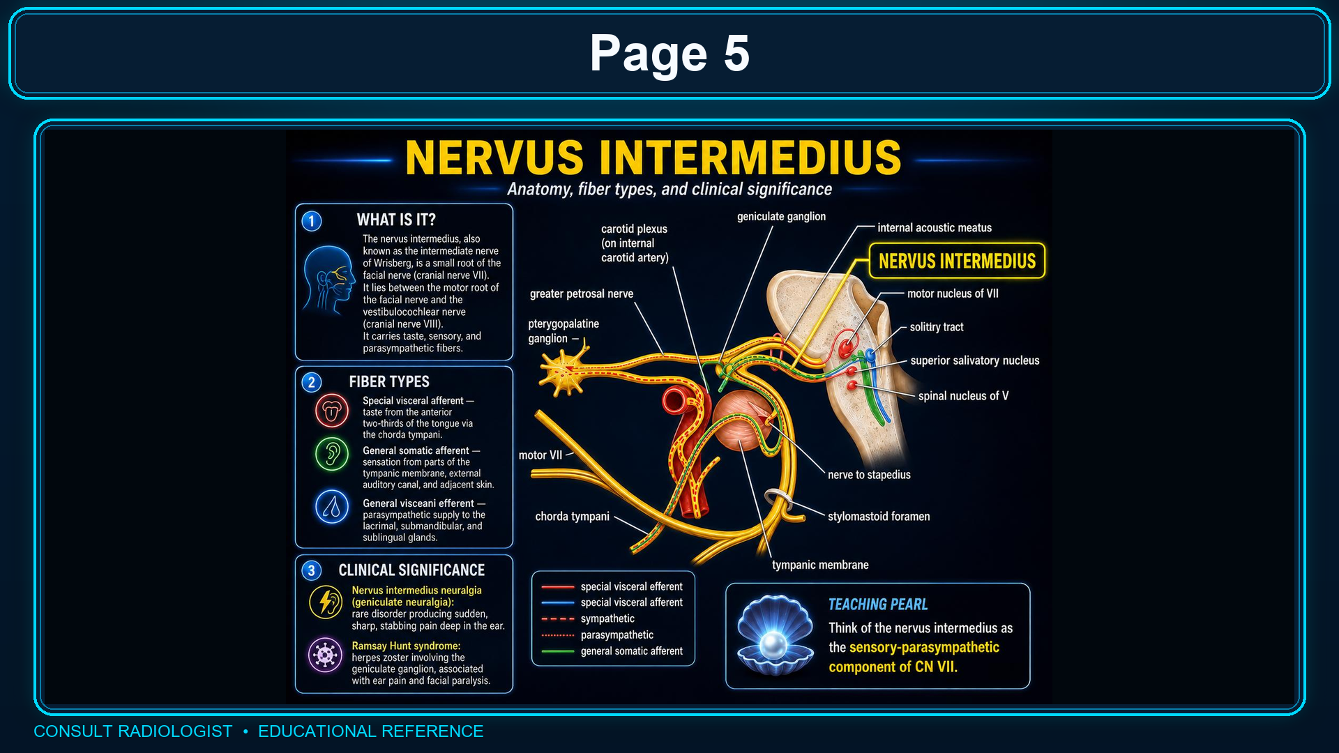
Nervus intermedius anatomy and fiber types
Nervus IntermediusClick to enlarge
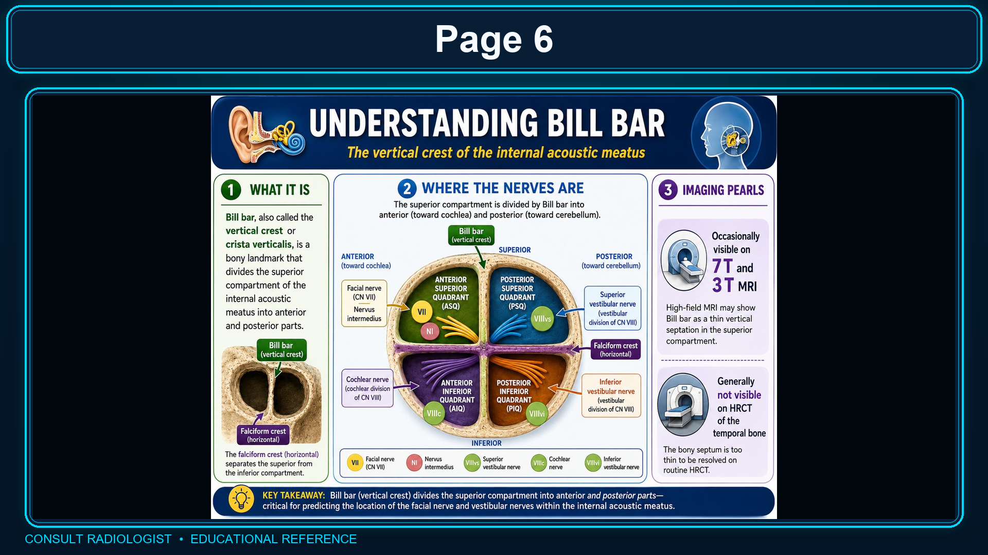
Bill bar and internal auditory canal fundus quadrants
Bill Bar and IAC FundusClick to enlarge
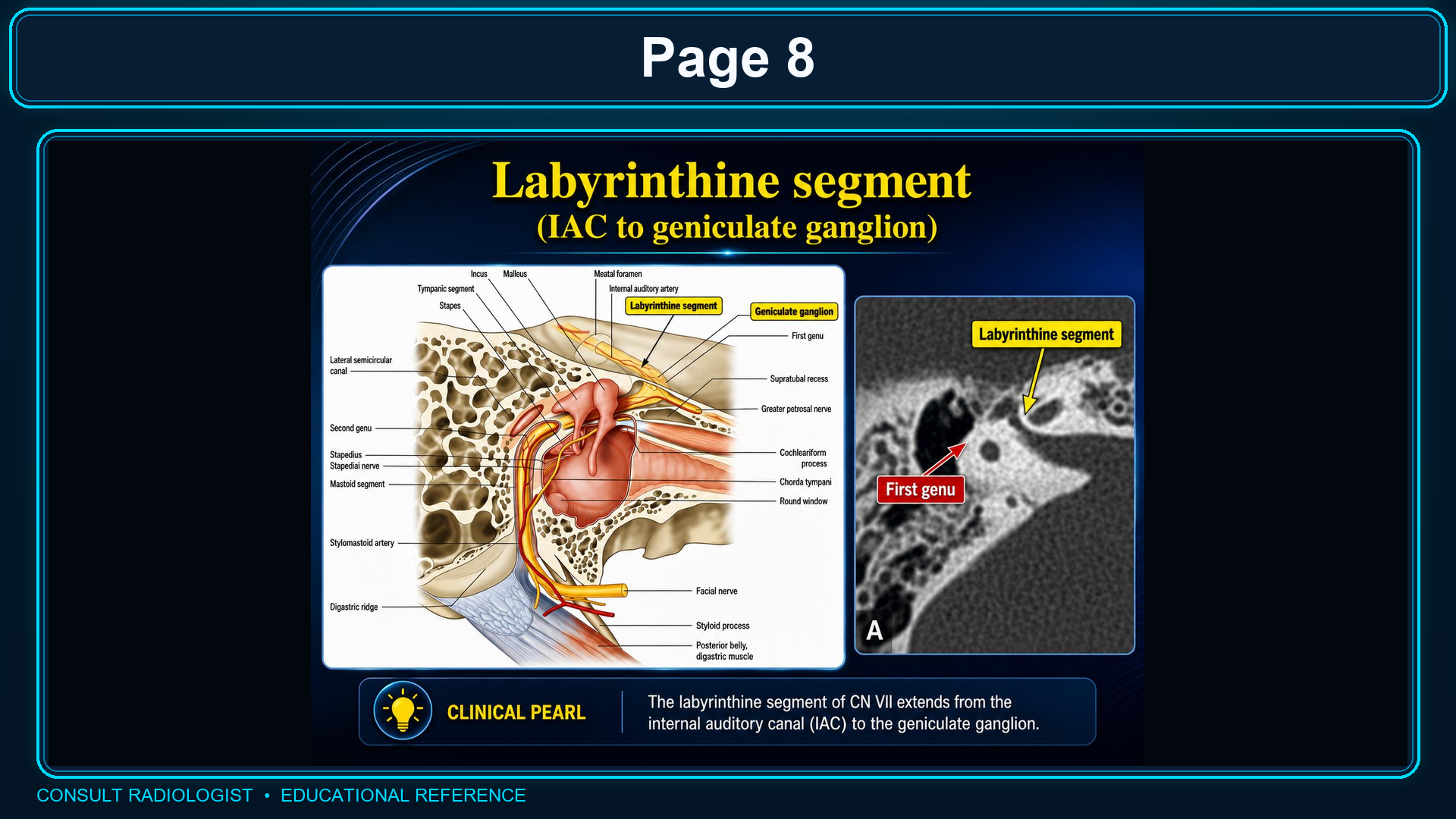
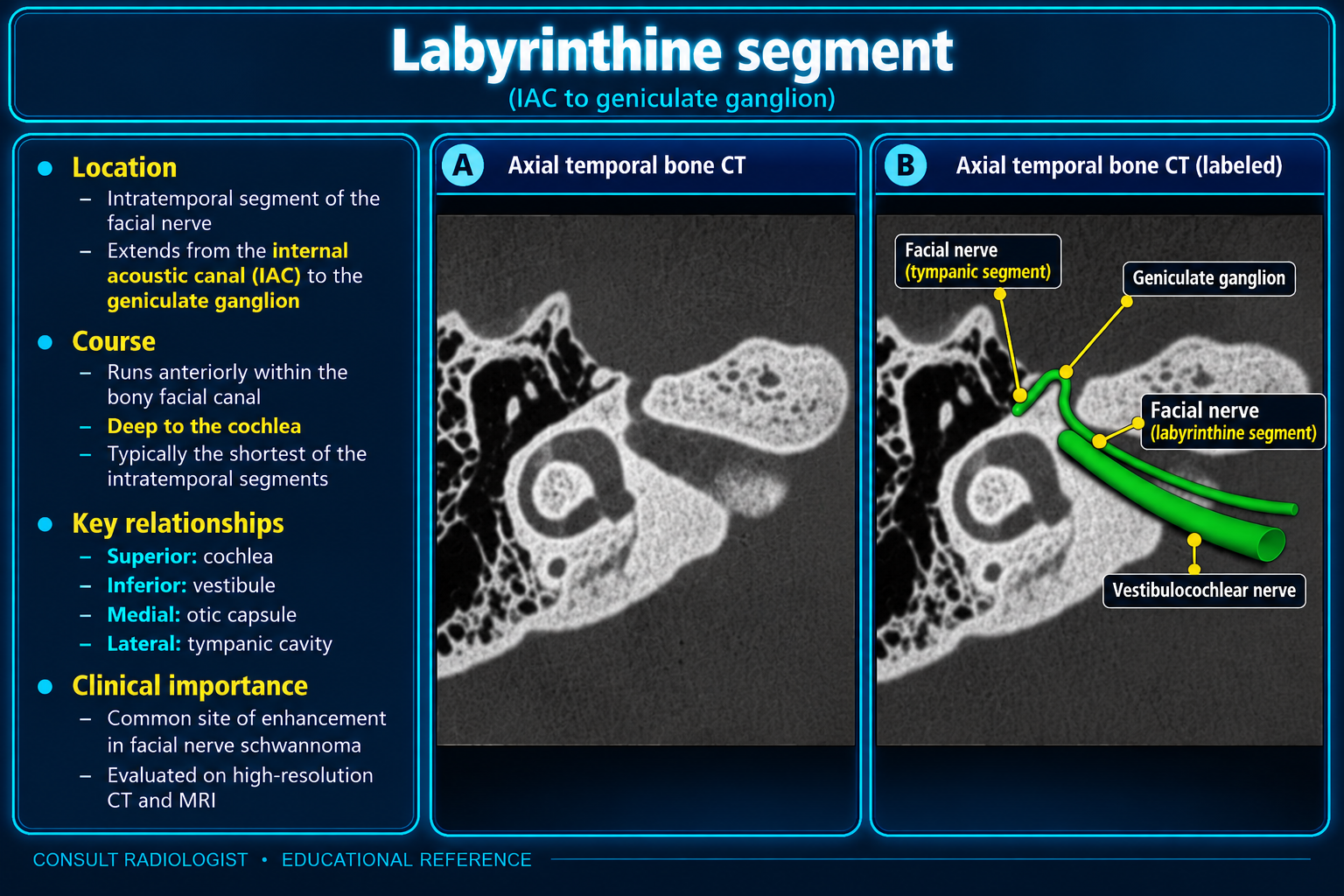
Labyrinthine facial nerve segment from the IAC to the geniculate ganglion
Labyrinthine Segment OverviewClick to enlarge
Axial temporal bone CT of the labyrinthine facial nerve segment
Labyrinthine Segment CTClick to enlarge
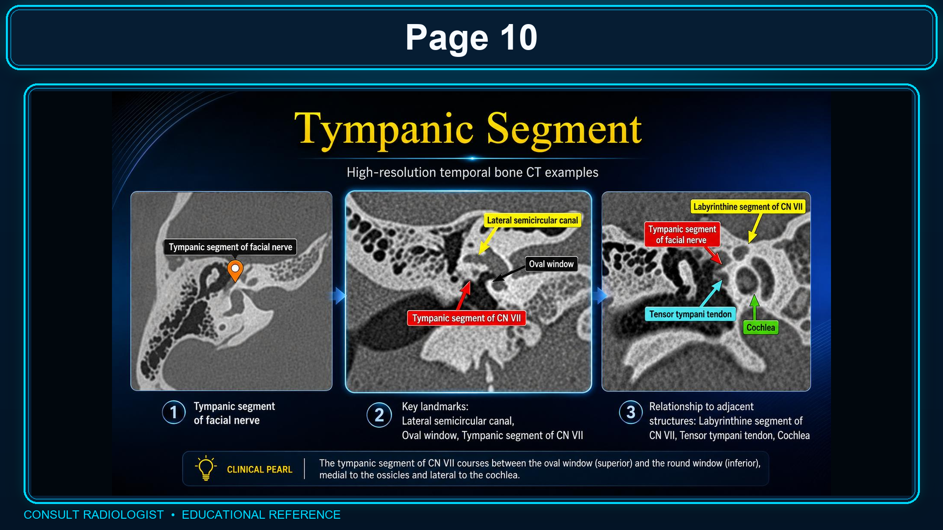
High-resolution CT examples of the tympanic facial nerve segment
Tympanic Segment CTClick to enlarge
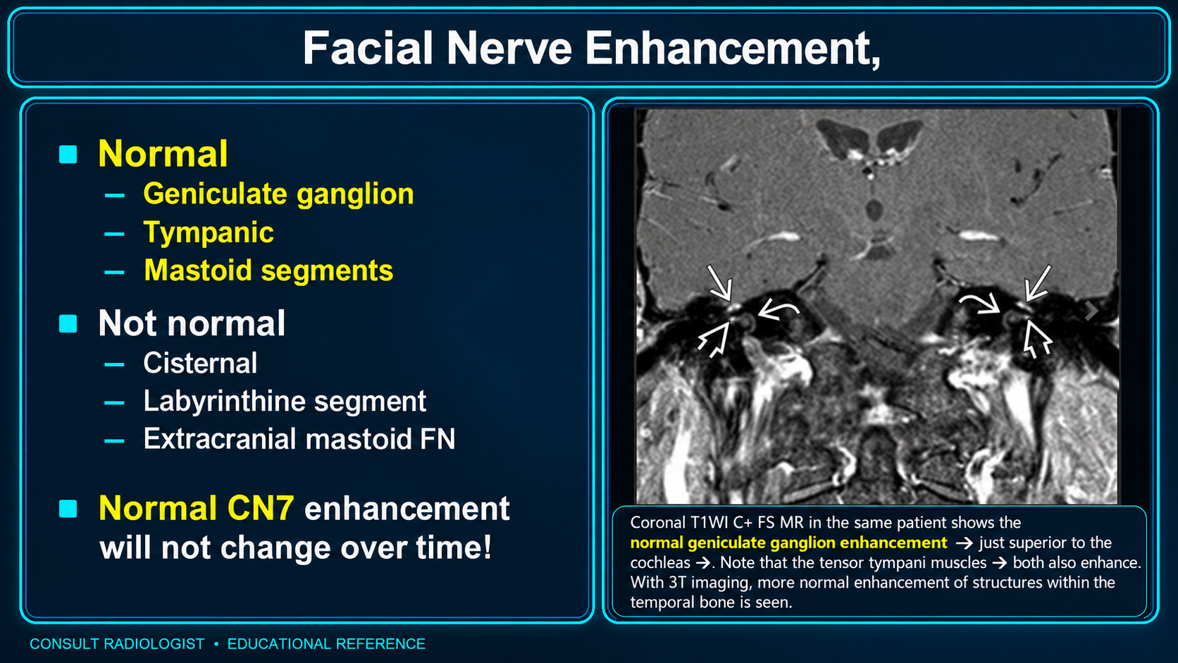
Normal and abnormal facial nerve enhancement patterns
Facial Nerve EnhancementClick to enlarge
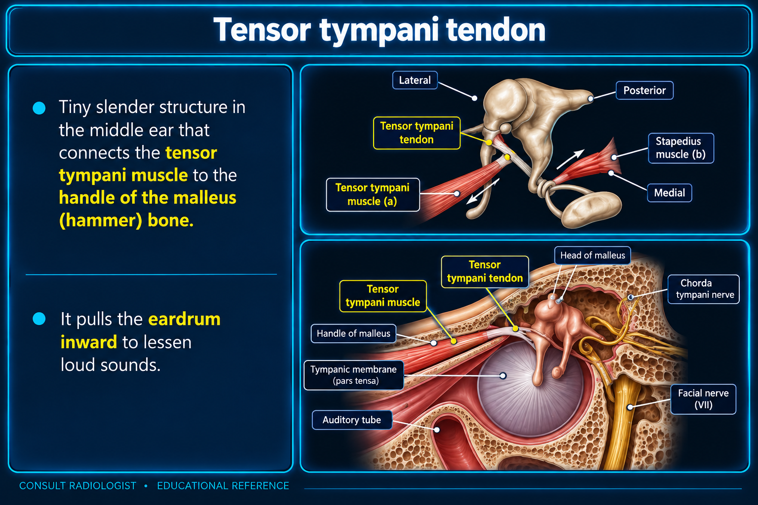
Tensor tympani tendon and adjacent facial nerve anatomy
Middle Ear RelationshipsClick to enlarge

Function and Central Anatomy

Mixed cranial nerveCN VII carries branchial motor, parasympathetic, taste, and a small somatic sensory component.

Four functional systems

  • Facial motor nucleus: facial expression, stapedius, stylohyoid, posterior digastric.
  • Superior salivatory nucleus: lacrimal, nasal/palatal, submandibular, and sublingual secretion.
  • Nucleus solitarius: taste from the anterior two-thirds of the tongue.
  • Spinal trigeminal nucleus: small sensory contribution from the external ear.

Pontine course

Motor fibers loop around the abducens nucleus, forming the internal genu and contributing to the facial colliculus. A dorsal pontine lesion can combine ipsilateral facial weakness with abducens or horizontal gaze dysfunction.

UMN versus LMN

Supranuclear lesions usually spare the forehead because the upper face receives bilateral corticobulbar input. Nuclear, fascicular, or peripheral lesions weaken the ipsilateral upper and lower face.

Course and Segments

From brainstem to facePontomedullary junction → CPA → IAC → labyrinthine segment → geniculate ganglion → tympanic segment → second genu → mastoid segment → stylomastoid foramen.

IAC fundus

CN VII is anterosuperior. The cochlear nerve is anteroinferior; superior and inferior vestibular nerves occupy the posterior quadrants. Bill bar separates CN VII from the superior vestibular nerve.

Intratemporal landmarks

  • Labyrinthine: shortest and narrowest segment, from fundus to geniculate ganglion.
  • Tympanic: below the lateral semicircular canal and above the oval window.
  • Second genu: turn near the pyramidal eminence and facial recess.
  • Mastoid: descends to the stylomastoid foramen.

Key branches

Greater petrosal nerve arises near the geniculate ganglion. Nerve to stapedius and chorda tympani arise from the mastoid segment.

Imaging and Enhancement

Normal enhancement pitfallMild symmetric geniculate, tympanic, and mastoid enhancement may be normal. Cisternal, canalicular, and labyrinthine enhancement is more suspicious.

MRI

  • Thin axial and coronal precontrast T1.
  • Thin fat-suppressed postcontrast T1.
  • Heavily T2 CISS, FIESTA, or SPACE for CPA/IAC anatomy.
  • Extend through parotid and neck when extracranial disease or perineural spread is suspected.

CT

High-resolution temporal bone CT evaluates facial canal caliber, dehiscence, fracture, cholesteatoma erosion, congenital aberrant course, geniculate fossa expansion, and the relationship to the oval window and ossicles.

Abnormal features

Asymmetry, nerve thickening, nodularity, masslike enhancement, facial canal expansion, foraminal fat loss, or progressive proximal extension.

Clinical Localization

FindingLesion proximal to
Decreased lacrimationGreater petrosal nerve / geniculate ganglion
HyperacusisNerve to stapedius
Anterior tongue taste lossChorda tympani
Facial weakness onlyDistal chorda level or extracranial CN VII
Parotid mass plus weaknessMalignancy/perineural spread until proven otherwise

Nervus intermedius

Carries taste, parasympathetic, and small somatic sensory fibers between the motor root of CN VII and CN VIII in the CPA/IAC.

Common Pathology

  • Bell palsy: smooth linear enhancement, often greatest in the distal canalicular, labyrinthine, and geniculate regions.
  • Ramsay Hunt: facial neuritis with possible CN VIII and inner-ear enhancement.
  • Facial nerve schwannoma: fusiform enhancing mass with smooth facial canal expansion.
  • Geniculate venous malformation: small symptomatic lesion with irregular honeycomb or spiculated bone.
  • Perineural tumor spread: enhancement from peripheral branches or parotid toward the stylomastoid foramen and skull base.
  • Trauma/cholesteatoma: fracture, dehiscence, or erosion of the facial canal.

Radiology Search Pattern

Brainstem

Inspect the facial nucleus/fascicle, facial colliculus, diffusion, demyelination, and adjacent CN VI pathways.

CPA and IAC

Look for a mass, neuritis, leptomeningeal disease, inner-ear enhancement, and continuity into the labyrinthine facial canal.

Temporal bone

Assess the labyrinthine segment, geniculate fossa, tympanic canal, second genu, mastoid segment, and stylomastoid foramen.

Parotid and face

Search for parotid or cutaneous primary tumor, branch enhancement, foraminal fat loss, and facial muscle denervation.

Reporting Pearls

Facial neuritis: “Smooth asymmetric enhancement of the facial nerve, greatest along the labyrinthine segment and geniculate ganglion, without nodular masslike enlargement.”
Schwannoma: “Fusiform enhancing lesion centered along CN VII with smooth expansion of the facial canal.”
Perineural spread: State the involved nerve, most proximal extent, skull-base and intracranial involvement, denervation, and visible primary or recurrent tumor.

High-yield distinction

Smooth linear enhancement favors neuritis. Nodular enlargement, bony expansion, foraminal obliteration, or progressive extension favors tumor, schwannoma, or perineural spread.
Secondary Links