Stafne bone cavity
Key Images
Look For First
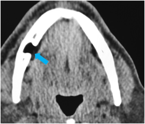
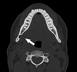
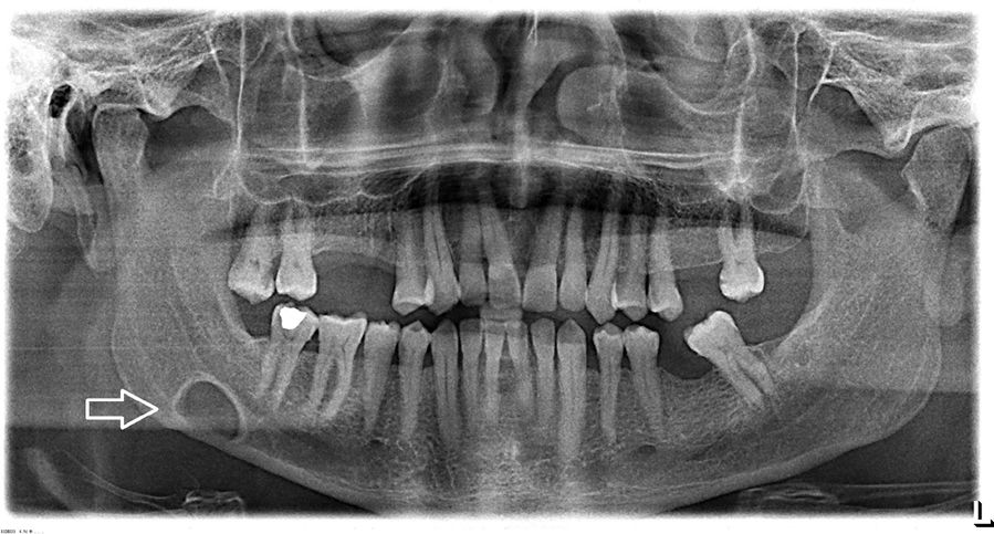
- Well-circumscribed, round radiolucent defect in the posterior mandible below the mandibular canal, between the molars and mandibular angle
- Medial cortical defect with preserved corticated rim on CT, with no enlargement on serial imaging
- Herniating submandibular gland tissue filling the mandibular defect on CT or MRI
Key Image Findings
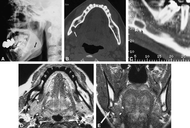
- Plain radiograph shows a unilocular, well-circumscribed radiolucent defect measuring 1–3 cm, located between the inferior alveolar canal and the inferior border of the mandible, typically between the molars and the angle of the jaw.
- CT reveals a shallow cortical defect through the medial cortex of the mandible with a well-defined corticated rim and no associated soft tissue abnormalities, distinguishing it from destructive or aggressive lesions.
- CT demonstrates herniation of submandibular gland tissue into the mandibular defect, confirming the benign nature of the finding.
- MRI shows continuation and herniation of submandibular gland signal (intermediate to high T2, similar to normal gland) into the mandibular defect.
- Sialography may demonstrate salivary gland tissue within the bone cavity, though this modality is less commonly used today.
- The defect remains static in size and radiographic appearance over time, with no progression on serial imaging—a key diagnostic feature.
- The radiolucent lesion may be superimposed on the lower anterior teeth on certain projections, potentially mimicking an odontogenic lesion.
- The defect may occasionally interrupt the inferior cortex of the mandible and may be palpable intraorally as an indentation.
Differential Diagnosis
- Odontogenic keratocyst: distinguished by larger size, aggressive behavior, association with impacted teeth, and multilocular or unilocular appearance; lacks gland herniation on CT/MRI.
- Ameloblastoma: typically larger, more aggressive with cortical perforation and root resorption; located near impacted teeth; no salivary gland herniation.
- Dentigerous cyst: always associated with the crown of an impacted tooth and does not show salivary gland herniation into the lesion.
- Radicular cyst: located at the apex of a non-vital tooth; demonstrates no gland tissue on imaging.
- Aneurysmal bone cyst (ABC): shows multilocular appearance with fluid-fluid levels on MRI and more aggressive cortical destruction.
- Myxoma or central hemangioma: typically show multilocular pattern and greater size; lack the characteristic gland herniation and static appearance.
Discussion
Stafne bone cavities result from remodeling of mandibular bone by adjacent submandibular salivary gland tissue and have been documented to regress after nearby gland resection, confirming the mechanical process of bone resorption.
The pathognomonic feature is herniation of submandibular gland tissue into the medial mandibular cortical defect, visible on CT and MRI, which definitively distinguishes it from destructive or cystic lesions.
Prevalence in middle-aged men (0.10–0.48%) suggests male predominance, though the biologic or mechanical reason is not well understood.
The static nature of the lesion on serial radiographs is diagnostically important and helps avoid unnecessary intervention or biopsy.
CT or MRI can confidently establish the diagnosis by demonstrating the gland tissue herniation and the absence of any concerning soft tissue pathology.
No clinical intervention is required; reassurance and documentation of the incidental finding prevent patient anxiety and unnecessary surveillance.
Reporting Pearls
Report the finding as: "A cortical defect of the medial mandible near the angle, consistent with a Stafne bone cavity, with submandibular gland tissue herniation into the defect. This is a benign, asymptomatic developmental variant requiring no follow-up." Emphasize the static nature and gland herniation to definitively exclude aggressive pathology.
Pitfalls
- Mistaking the radiolucent defect on plain radiographs for an odontogenic cyst or pathology, especially when superimposed on lower anterior teeth—clinical correlation and additional imaging should clarify the diagnosis.
- Confusing Stafne cavity with other lytic mandibular lesions (keratocyst, ameloblastoma, dentigerous cyst) without demonstrating the characteristic gland herniation on CT or MRI.
- Performing unnecessary biopsy or recommending follow-up imaging for a lesion that is static and benign; recognizing the diagnosis on cross-sectional imaging avoids overtreatment.
- Overlooking the importance of demonstrating submandibular gland tissue within the defect on advanced imaging—this finding is the key to confirming diagnosis and excluding other pathology.