Medication-related osteonecrosis of the jaw
Key Images
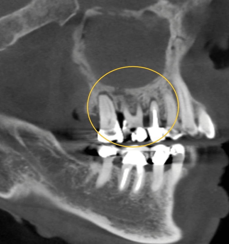
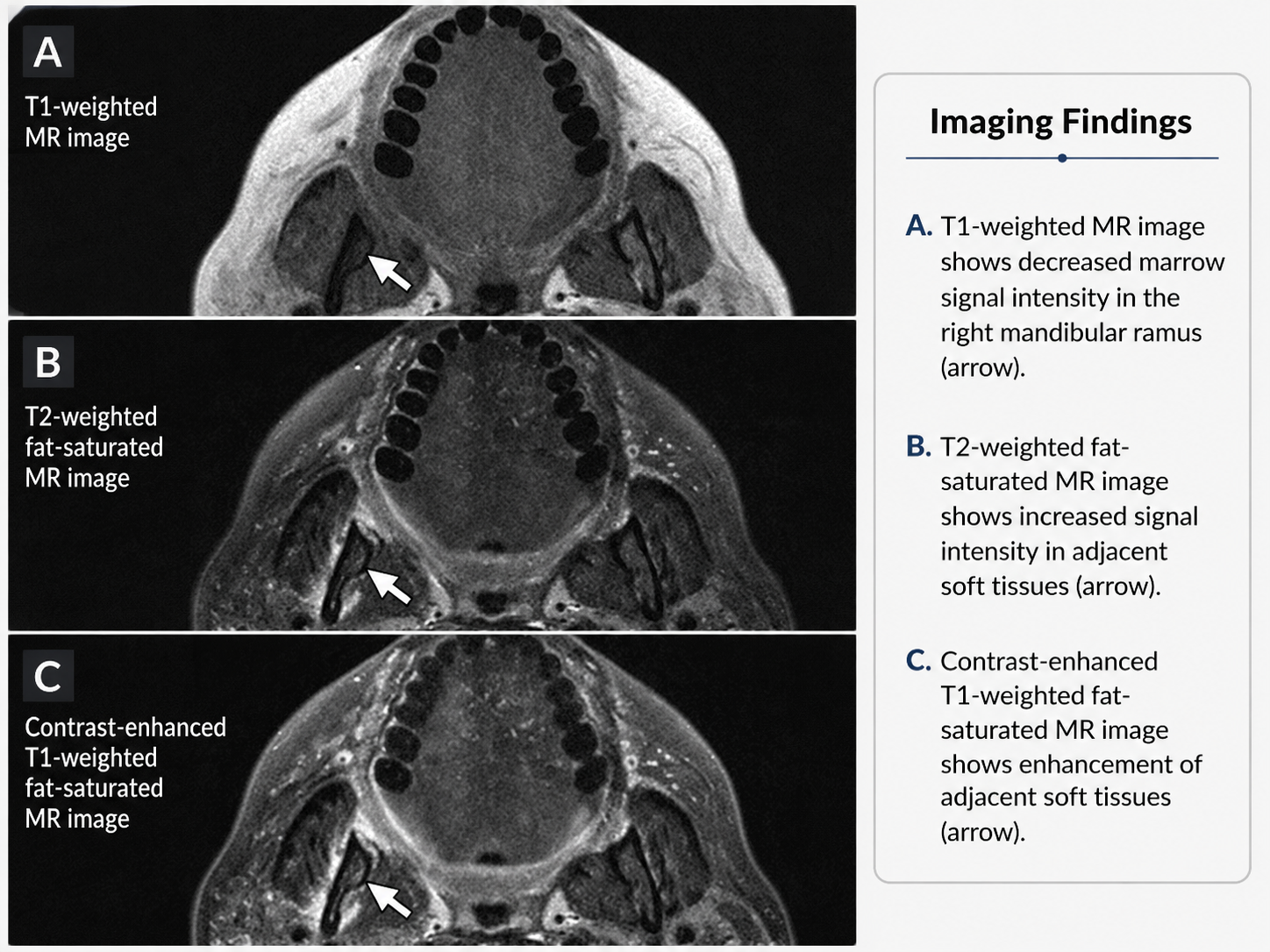
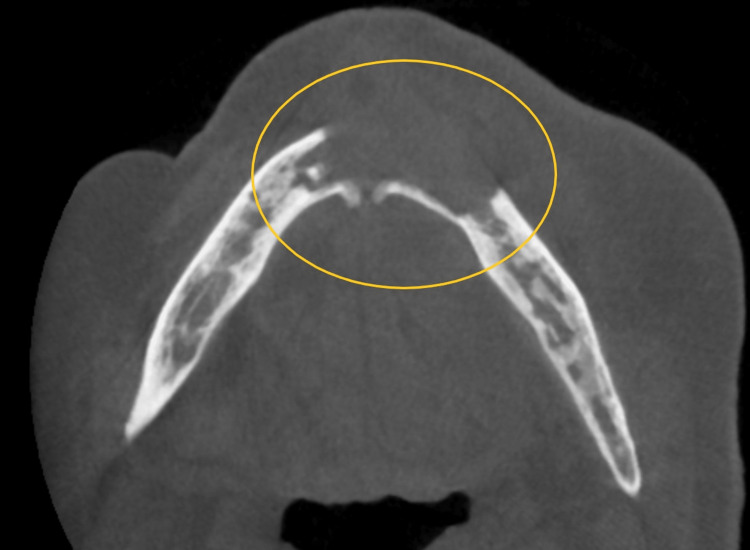
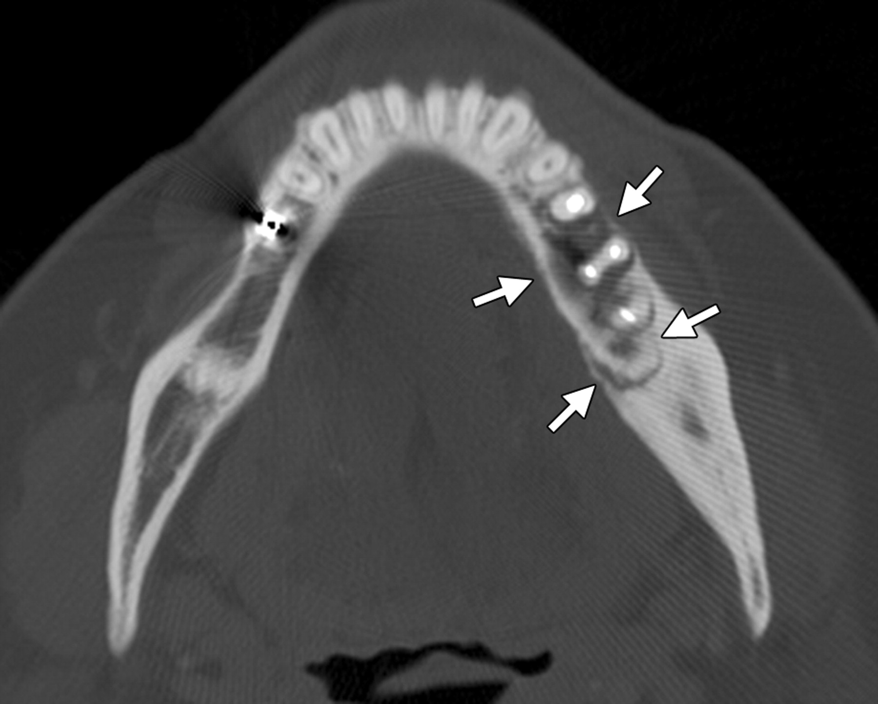
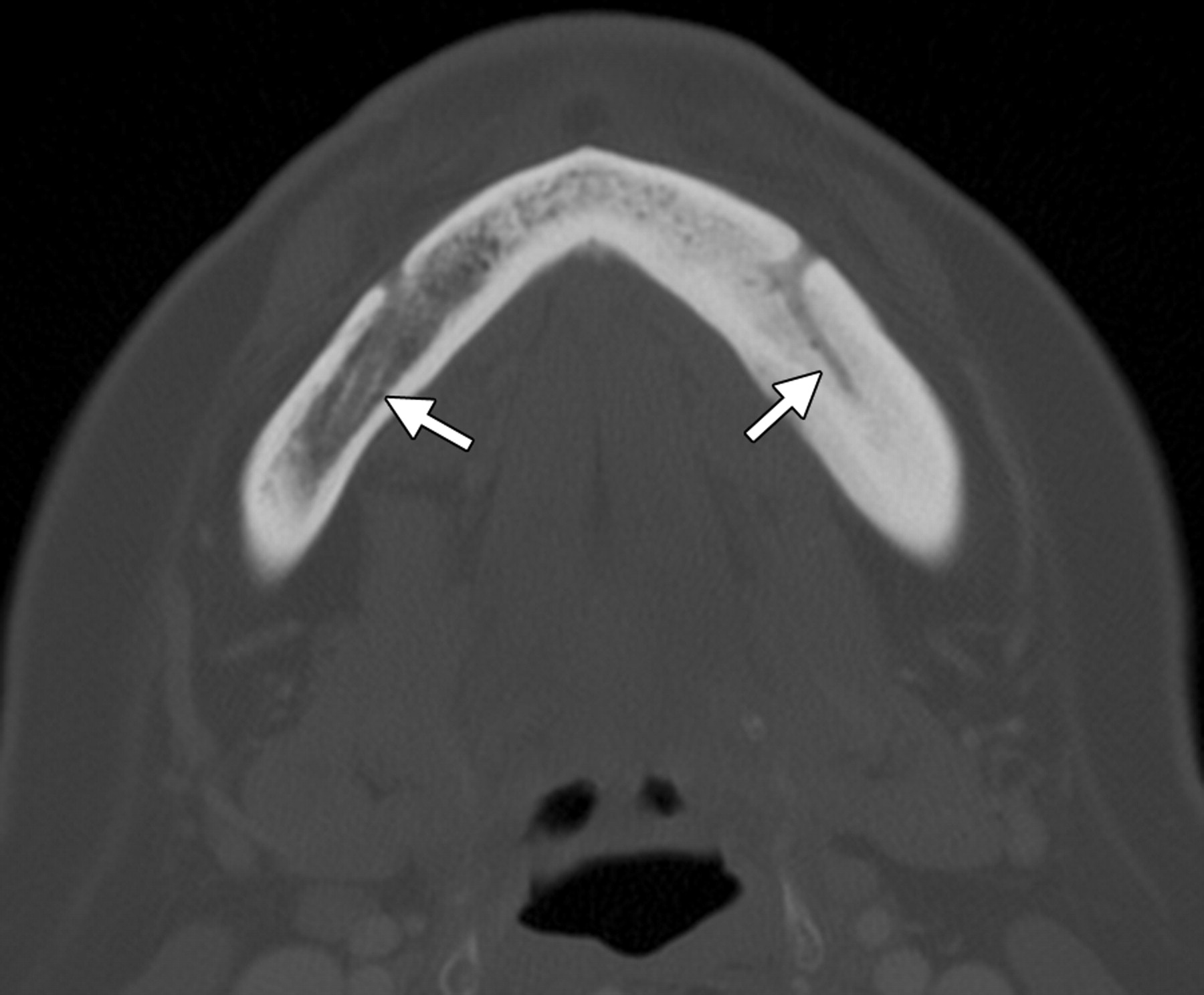
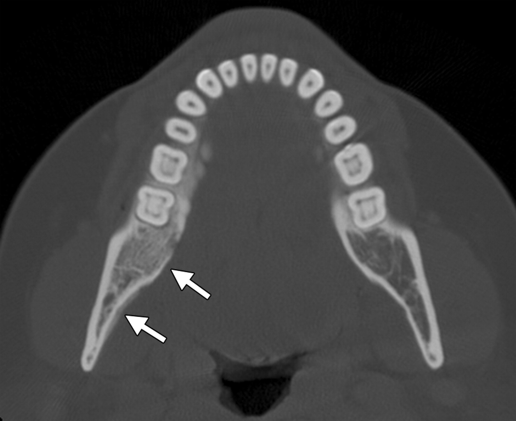
Look For First
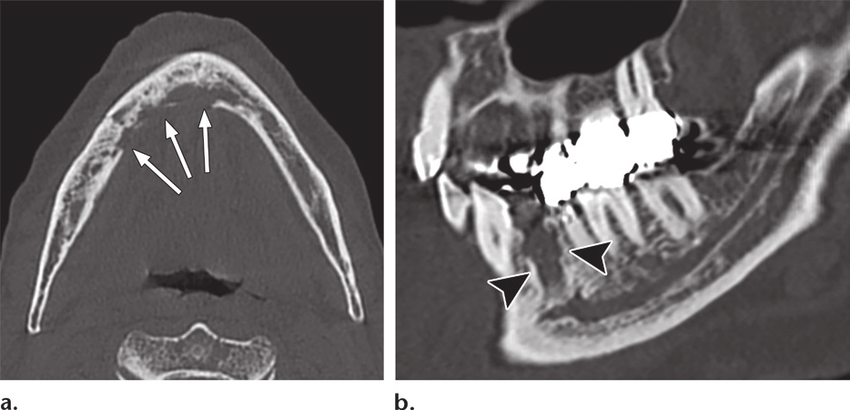
- Mandibular involvement (approximately twice as common as maxilla) with poorly defined lucency or mixed lytic-sclerotic bone change
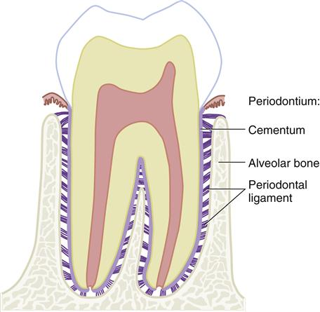
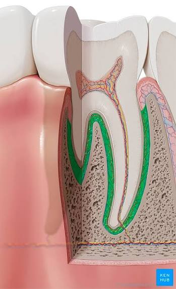
- Prominence or widening of the periodontal ligament space adjacent to a tooth root on CT—an early finding before extensive osseous destruction
- Focal sclerosis adjacent to tooth roots and bony sequestration with cortical destruction
- Clinical context of antiresorptive or antiangiogenic medication exposure without prior jaw radiation history
Key Image Findings
- Early MRONJ may be occult on conventional radiographs but CT is more sensitive, showing prominence or widening of the periodontal ligament and focal sclerosis adjacent to tooth roots before advanced changes develop.
- Mixed lytic-sclerotic bone change with poorly defined lucencies is characteristic; sequestration with exposed necrotic bone persisting for more than 8 weeks confirms clinical diagnosis in the appropriate medication context.
- Periosteal proliferation and new bone formation may occur around affected areas, though radiation-induced vascular damage in osteoradionecrosis typically limits new bone formation—an important distinguishing feature.
- Stage 0 MRONJ presents with imaging abnormalities (sclerosis, mixed change, periodontal ligament widening) and clinical symptoms before exposed bone becomes apparent, highlighting imaging's role in early detection.
- CT demonstrates cortical or adjacent structural destruction, helps establish full disease extent, and is superior to plain radiographs for assessing both early and advanced disease in treatment planning.
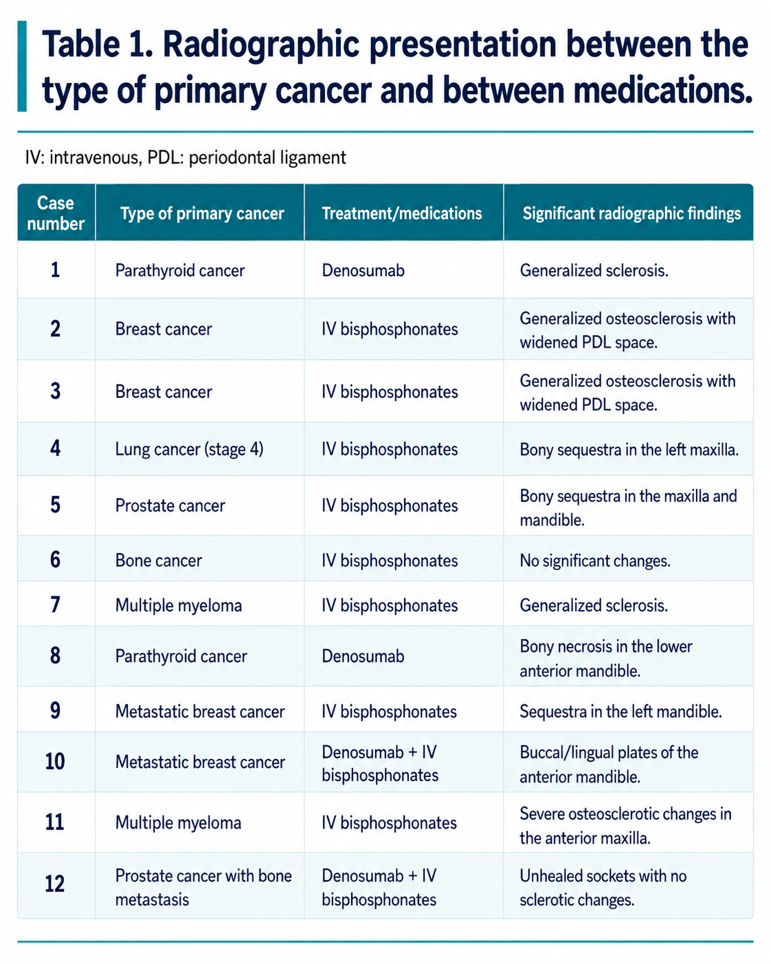
- Patients receiving high-dose or intravenous bisphosphonates demonstrate more severe osteonecrosis on imaging compared to oral bisphosphonate users, reflecting their substantially higher clinical risk.
- In advanced disease, extensive osseous destruction and superimposed infection obscure potentially distinguishing features between MRONJ and other osteonecrotic processes.
- The predilection for the alveolar portions of the jaws reflects their frequent exposure to dental trauma, tooth extraction, periodontal disease, and mechanical stress—the sites where MRONJ most commonly manifests.
Differential Diagnosis
- Osteoradionecrosis: History of therapeutic head-and-neck radiation is the critical distinguishing feature; both can appear very similar radiographically, but osteoradionecrosis typically shows limited periosteal new bone formation due to vascular damage.
- Chronic or bacterial osteomyelitis: Expected to show more periosteal new bone formation and less sclerosis than MRONJ; clinical presentation with fever and systemic infection helps differentiate.
- Recurrent or metastatic malignancy involving the jaw: Should demonstrate cortical destruction with expansile component and soft-tissue mass; obtain clinical history of primary cancer location and metastatic workup.
- Medication-induced or idiopathic osteonecrosis from other causes: Requires correlation with complete medication history documenting antiresorptive or antiangiogenic exposure.
Discussion
MRONJ pathophysiology involves suppression of normal bone remodeling, impaired bone healing, antiangiogenic effects, and local trauma/infection—leading to sequestrum formation and necrotic exposed bone.
Risk factors include IV bisphosphonate therapy (particularly high-dose), long-term exposure, treatment for malignancy rather than osteoporosis, and recent dental surgery with tooth extraction preceding MRONJ in approximately 65% of cases.
Clinical staging and diagnosis are primarily based on exposed or probeable necrotic bone persisting for more than 8 weeks with medication exposure and no jaw radiation history, but stage 0 MRONJ can present with imaging abnormalities and symptoms before exposed bone appears.
No characteristic imaging pattern reliably identifies either the causative medication or underlying malignancy; studies show substantial overlap in imaging appearance across different primary cancers and medication exposures.
High-dose or IV bisphosphonate therapy is associated with substantially greater risk of MRONJ compared to oral bisphosphonates, and when such therapy is planned, needed dental procedures should ideally be completed before treatment begins.
The mandible is affected approximately twice as often as the maxilla, and imaging plays an essential role in detecting early or stage 0 disease, determining extent, identifying complications, and guiding conservative or surgical management.
Reporting Pearls
When reporting MRONJ, clearly document the location (mandible vs. maxilla, specific tooth or region), extent of disease (focal vs. multifocal), presence and severity of mixed lytic-sclerotic change, periodontal ligament widening adjacent to tooth roots, sequestration, and any cortical perforation or adjacent structural involvement—then correlate findings with the patient's documented antiresorptive or antiangiogenic medication exposure and absence of prior jaw radiation to support the diagnosis.
Pitfalls
- Early MRONJ, particularly stage 0, may be occult on conventional radiography alone; CT should be obtained when clinical suspicion is high, as it detects early findings such as periodontal ligament widening and focal sclerosis before visible lucencies develop.
- Assuming imaging findings alone can identify the specific causative medication or underlying primary malignancy; no sufficiently specific imaging pattern exists, and clinical history of medication exposure is essential for diagnosis.
- Distinguishing MRONJ from osteoradionecrosis on imaging alone is unreliable because they appear very similar radiographically; the critical differentiating factor is the patient's treatment history—jaw irradiation favors osteoradionecrosis, antiresorptive/antiangiogenic exposure without radiation favors MRONJ.
- Overinterpreting periosteal new bone formation as evidence against MRONJ; periosteal proliferation can occur with MRONJ and should not be used in isolation to exclude the diagnosis—it is more commonly expected with bacterial osteomyelitis.