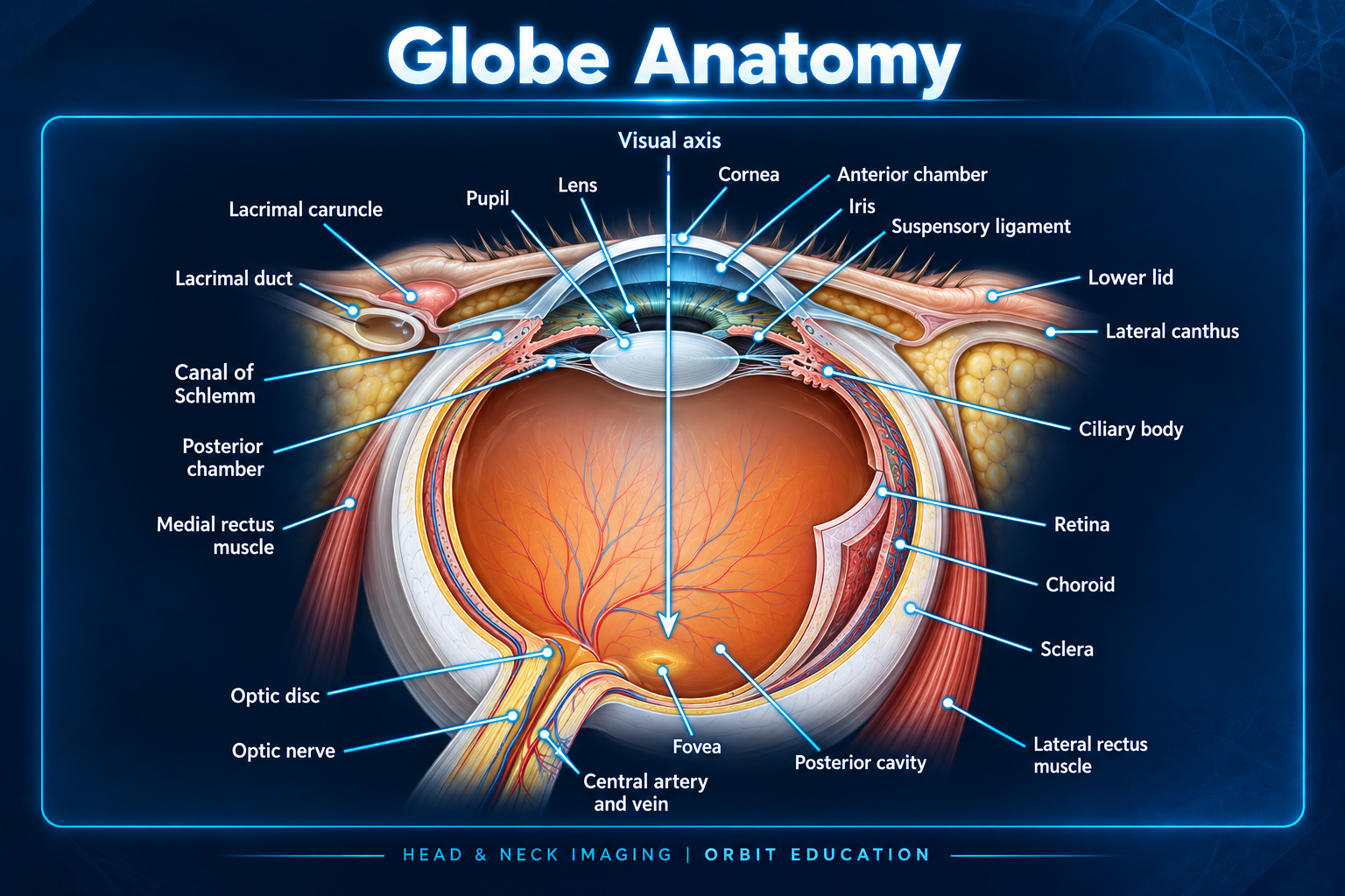
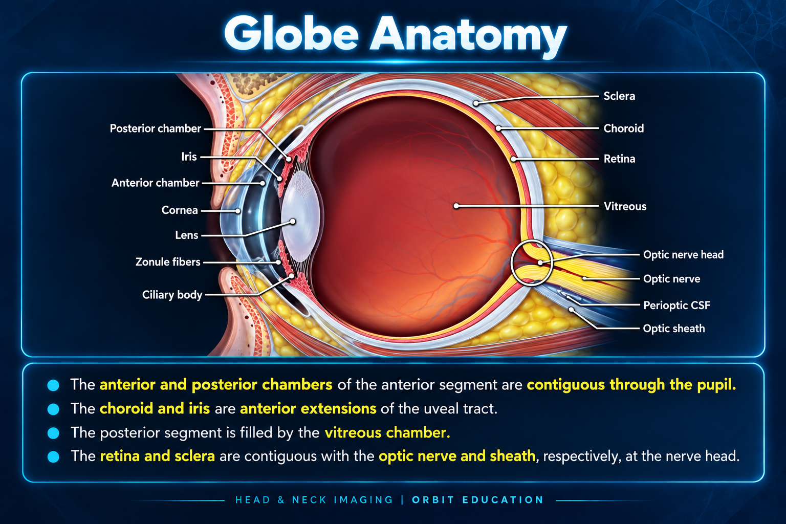
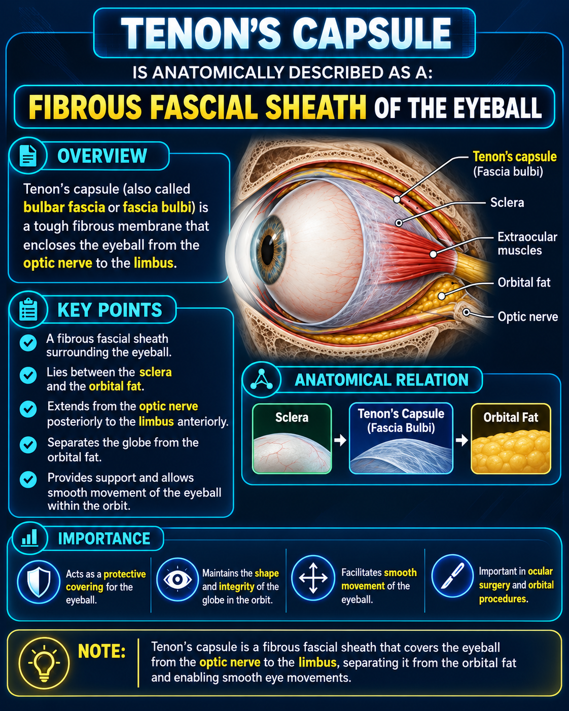
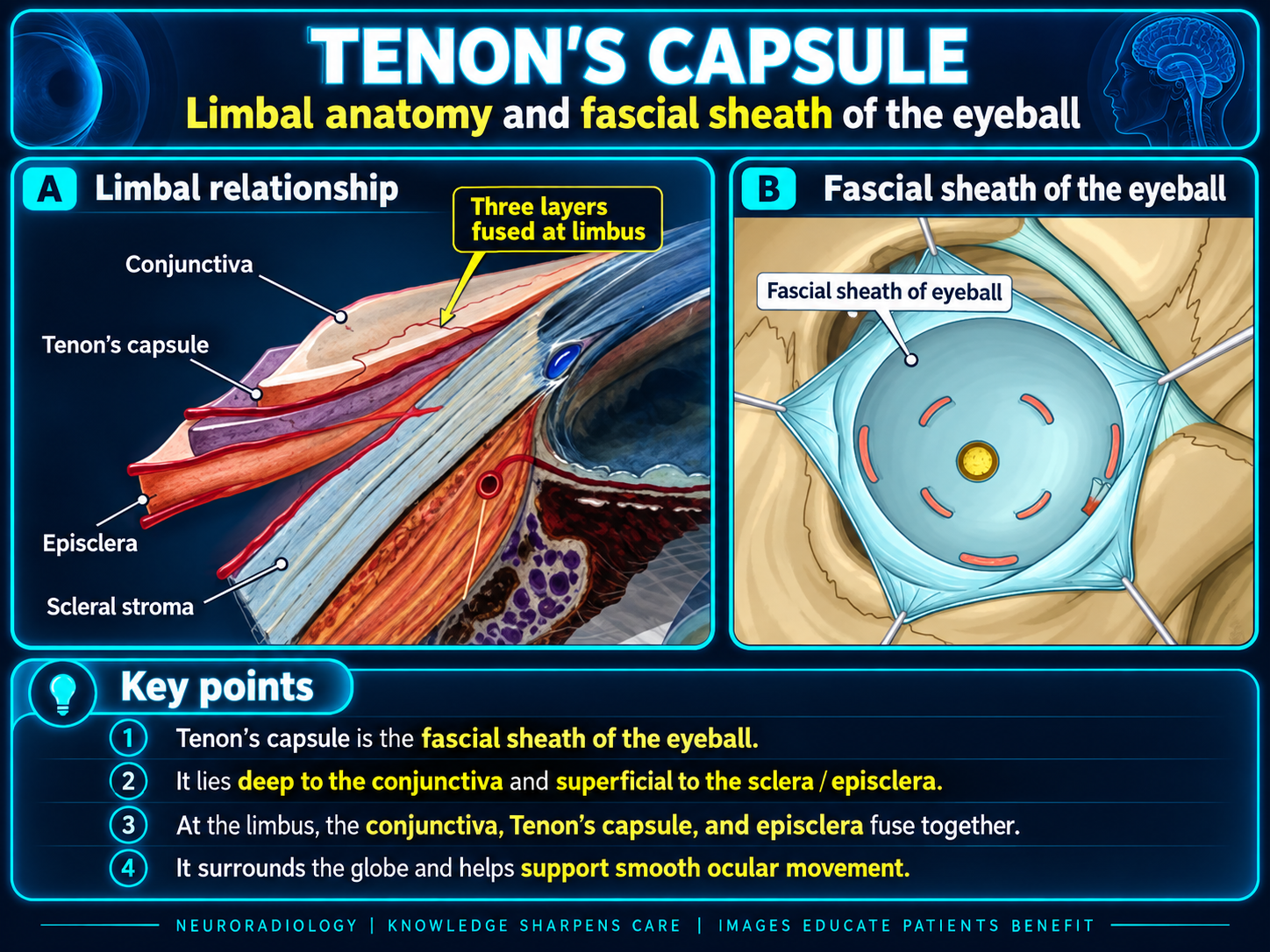
The globe is a slightly asymmetric sphere, approximately 24–25 mm in sagittal diameter, housed within the orbit and moved by six extraocular muscles attached to the sclera. It has three wall layers—fibrous, vascular, and neural—and three fluid-containing chambers: anterior, posterior, and vitreous.
Cornea → aqueous chambers → lens → vitreous → retina → optic nerve
Global Organization
- Globe: approximately 24 mm wide and 24–25 mm long; the cornea forms the visible anterior convexity.
- Limbus: junction of transparent cornea and opaque sclera.
- Equator: circumferential midpoint used to localize lesions, scleral buckles, and vortex veins.
- Anterior pole: central corneal apex.
- Posterior pole: posterior globe containing the macula and optic nerve head.
- Extraocular muscles: superior, inferior, medial, and lateral recti plus superior and inferior obliques.
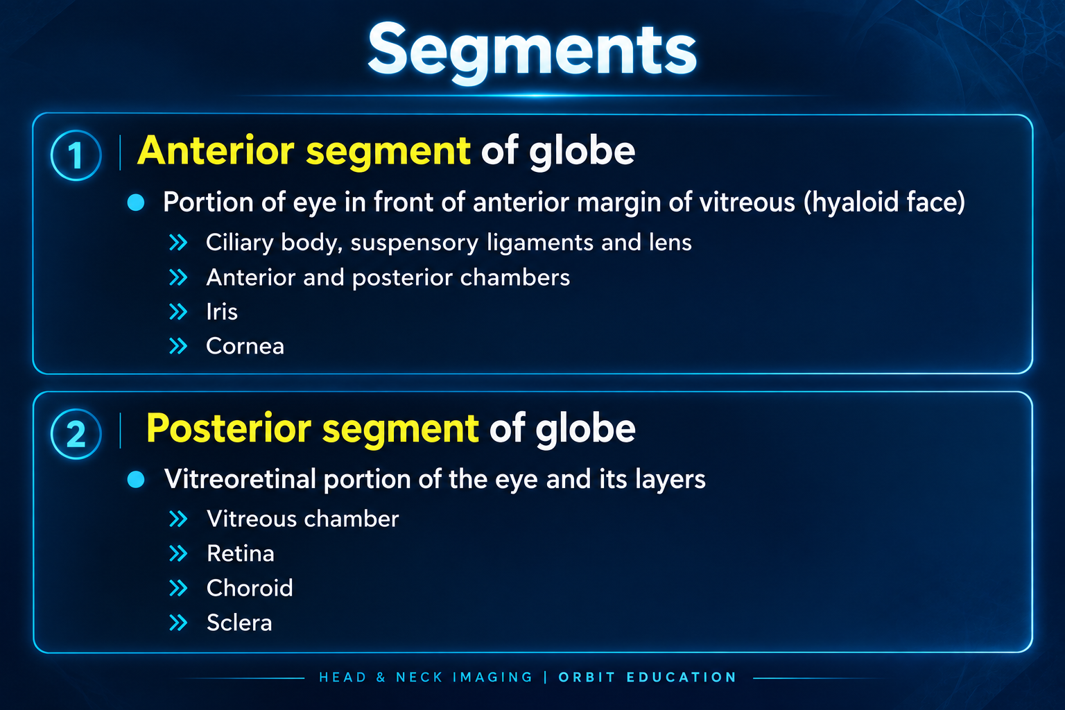
Segments
Anterior: cornea, anterior chamber, iris, posterior chamber, ciliary body, zonules, and lens.
Posterior: vitreous, retina, choroid, optic nerve head, and posterior sclera.
Globe Wall
| Layer | Components | Role |
|---|
| Fibrous | Cornea, sclera | Structural shell and major refractive surface |
| Uveal / vascular | Iris, ciliary body, choroid | Light control, aqueous production, accommodation, and outer retinal support |
| Neural | Retina | Visual processing; ganglion-cell axons form the optic nerve |
Fibrous tunic
- Cornea: transparent, avascular anterior one-sixth of the fibrous coat.
- Sclera: opaque collagenous coat extending from limbus to optic nerve.
- Conjunctiva: covers anterior sclera and reflects onto eyelids; it does not cover the cornea.
Uvea
- Iris: regulates light through the pupil.
- Ciliary body: produces aqueous humor and changes zonular tension for accommodation.
- Choroid: vascular pigmented layer supporting the RPE and outer retina.
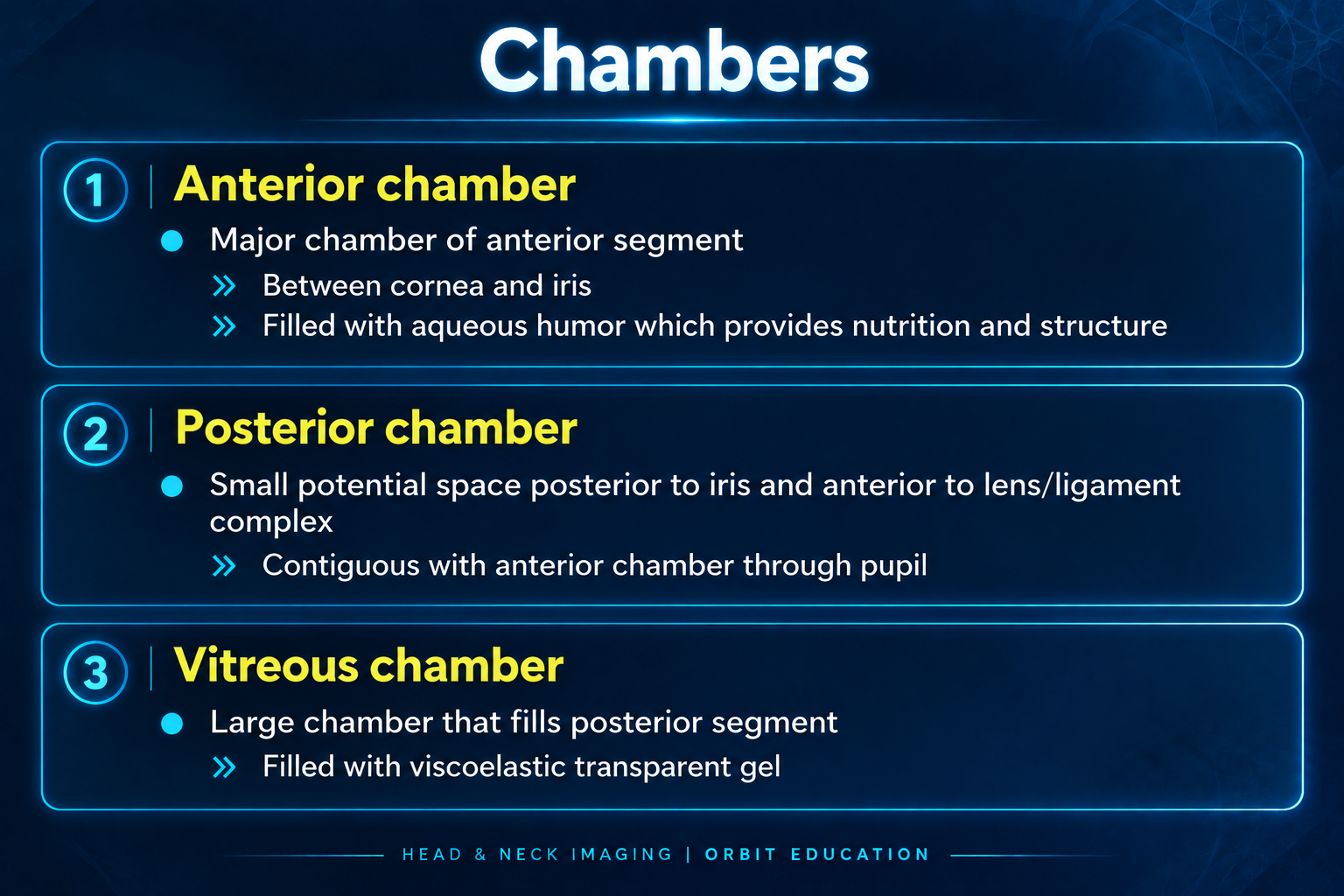
Chambers & Contents
| Chamber | Boundaries | Contents |
|---|
| Anterior | Posterior cornea to anterior iris | Aqueous humor |
| Posterior | Posterior iris to anterior lens, zonules, and ciliary body | Aqueous humor |
| Vitreous | Posterior lens to retina | Vitreous humor |
Aqueous pathway
- Produced by ciliary processes
- Posterior chamber
- Through the pupil
- Anterior chamber
- Trabecular meshwork
- Schlemm canal and episcleral venous drainage
Lens and vitreous
The avascular biconvex lens is suspended by zonules. The vitreous is a transparent gel with firm attachment at the vitreous base, optic disc, macula, and retinal vessels.
Retina & Posterior Pole
- RPE: supports photoreceptors, forms part of the outer blood-retinal barrier, and absorbs stray light.
- Neurosensory retina: contains photoreceptors, bipolar cells, interneurons, and ganglion cells.
- Rods: low-light and peripheral vision.
- Cones: color and high-acuity central vision.
- Macula and fovea: fine central vision and maximal visual acuity.
- Optic disc: axonal exit and central retinal vessel entry/exit; no photoreceptors.
- Optic nerve: ganglion-cell axons transmitting visual information as cranial nerve II.
Blood Supply
The ophthalmic artery, arising from the internal carotid artery, provides the principal arterial supply.
- Central retinal artery: inner retina.
- Posterior ciliary arteries: choroid, outer retina, ciliary body, and portions of the optic nerve head.
- Anterior ciliary arteries: anterior segment and episcleral circulation.
- Vortex veins: major choroidal venous drainage.
- Ophthalmic veins: orbital venous drainage.
Imaging Landmarks
On CT and routine MRI, the retina, choroid, and sclera are usually seen as one thin posterior wall complex rather than three separately resolved layers.
Identify routinely
- Cornea and anterior chamber
- Lens and vitreous
- Posterior wall complex
- Optic nerve head and optic nerve
- Extraocular muscles and orbital fat
The retina/choroid/sclera complex normally lies smoothly against the globe. Separation of these layers underlies retinal, choroidal, and hemorrhagic detachments.