Endophthalmitis
Key Images
Look For First
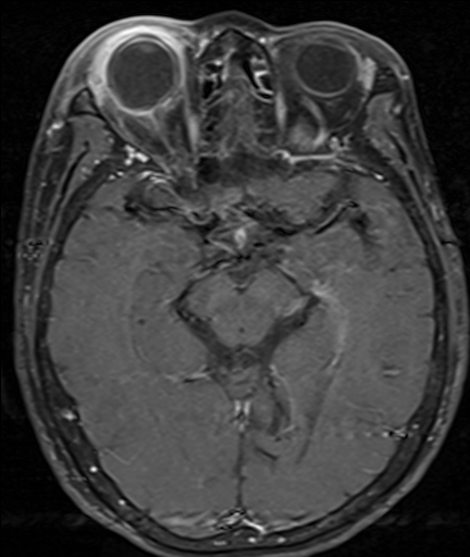
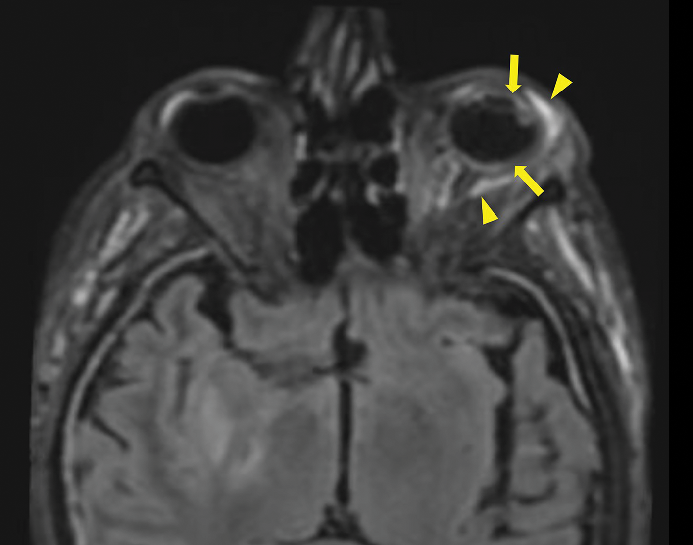
- High FLAIR signal in the vitreous humor on MRI
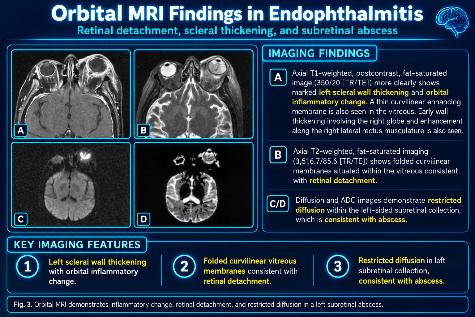
- Restricted diffusion (high DWI signal) in the vitreous resembling an abscess
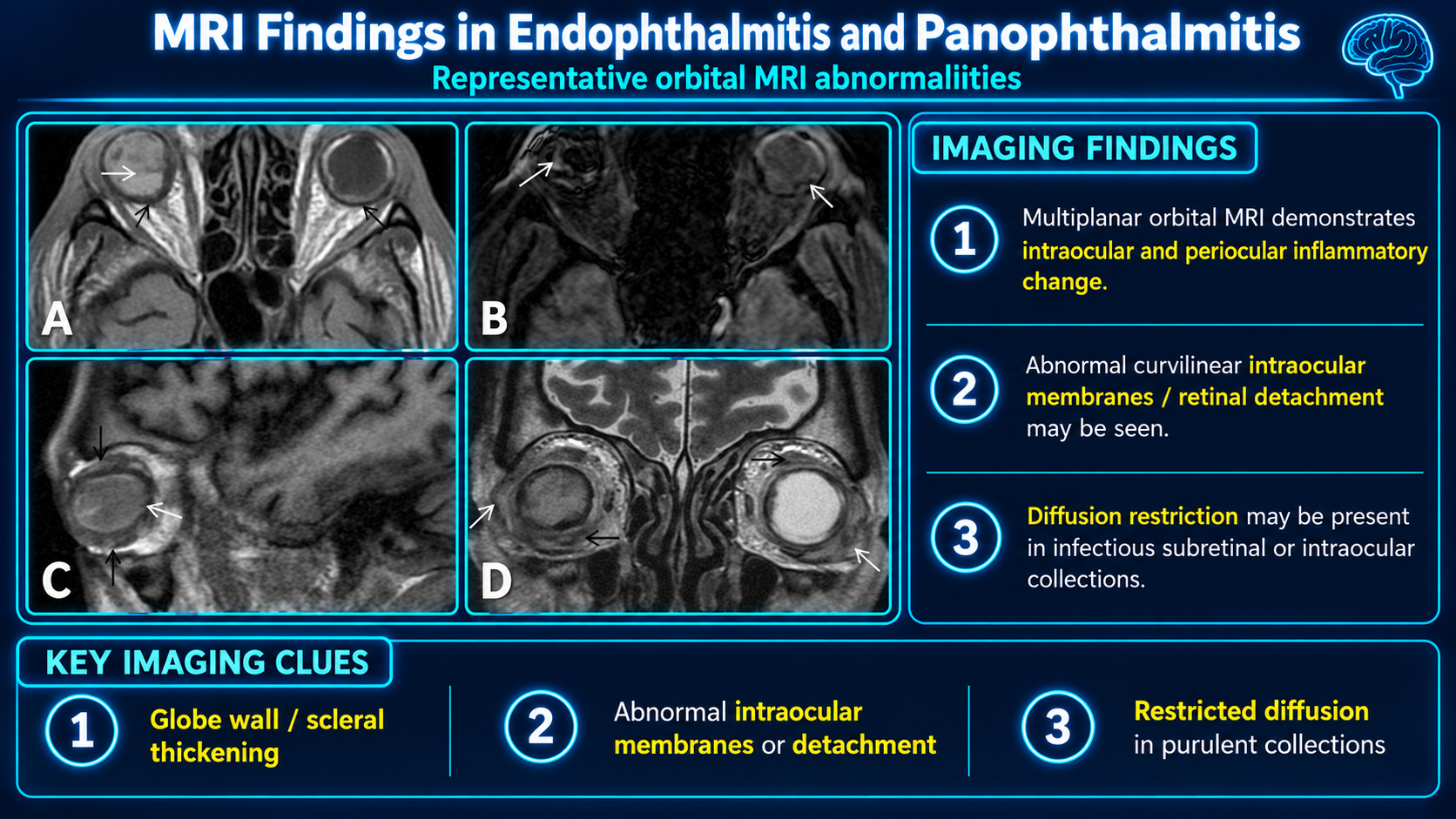
- Smooth or irregular thickening and enhancement of the uveoscleral layer
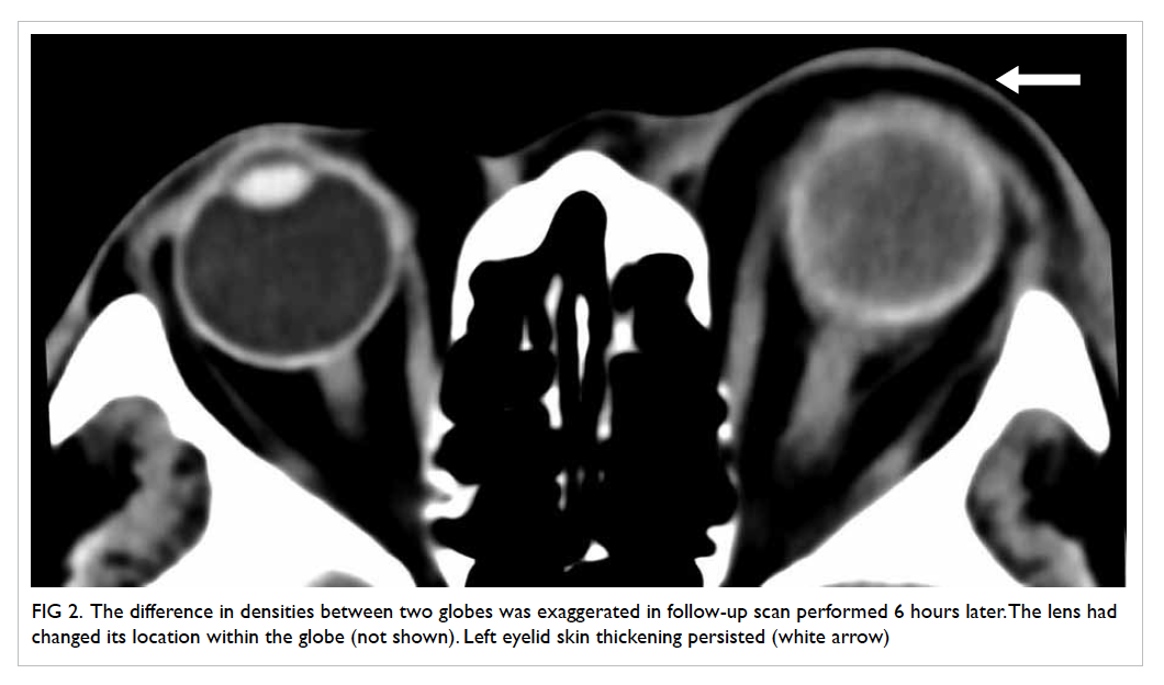
- Hyperdensity or echogenic debris within the vitreous on CT or ultrasound
Key Image Findings
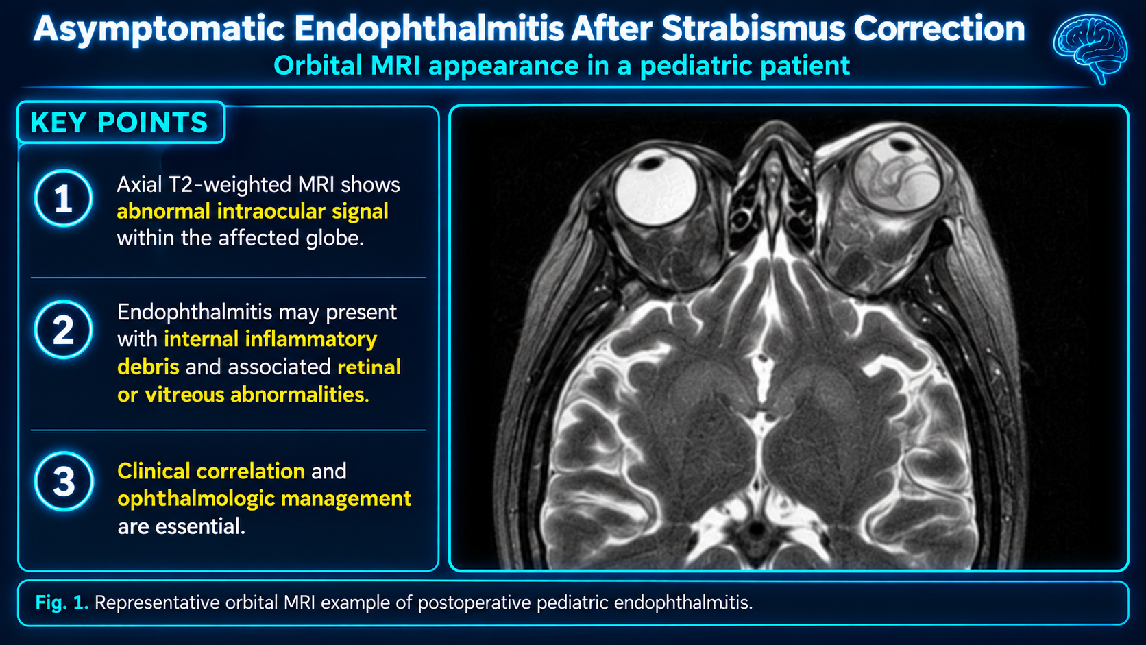
- MRI shows areas of high FLAIR signal within the vitreous humor, which is a key feature reflecting the proteinaceous inflammatory exudate.
- Restricted diffusion on diffusion-weighted imaging (DWI) of the vitreous is virtually diagnostic of endophthalmitis, appearing similar to an abscess with high signal intensity.
- T1-weighted MRI demonstrates isointensity or hyperintensity of the vitreous depending on proteinaceous content and hemorrhage.
- CT imaging shows hyperdensity of the vitreous humor, with choroidal enhancement on post-contrast imaging often evident in early disease.
- Ultrasound reveals intraocular echogenic debris, membranes, and increased echogenicity of the vitreous humor, sometimes with chorioretinal thickening or retinal/choroidal detachments.
- Scleral thickening and smooth or irregular thickening with enhancement of the uveoscleral layer are characteristic findings across imaging modalities.
- Proptosis and intraorbital fat stranding may be present, indicating raised intraorbital pressure and orbital involvement.
- Early imaging may show an unremarkable globe, making diagnosis challenging in the initial disease phase; progression manifests with the imaging features described above.
Differential Diagnosis
- Panophthalmitis—distinguished by extension beyond the sclera with edema or exudates in the sub-Tenon's space, lacrimal gland enhancement, and subconjunctival abscess.
- Orbital cellulitis—presents with pre and post-septal stranding and more extensive extraocular inflammation; endophthalmitis is intraocular inflammation.
- Vitreous hemorrhage—lacks the restricted diffusion pattern and inflammatory enhancement; blood appears hyperintense on T1 but lacks the abscess-like DWI restriction.
- Endophthalmitis versus early abscess—both show restricted diffusion, but clinical and laboratory correlation helps distinguish endophthalmitis from other intraocular infections.
- Tractional retinal detachment—can mimic retinal detachment seen in endophthalmitis but lacks the intraocular inflammation and echogenic debris.
- Posterior uveitis—may show similar inflammation but is typically less fulminant and lacks the hyperdensity and restricted diffusion pattern typical of endophthalmitis.
Discussion
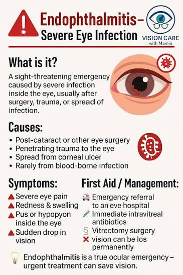
Endophthalmitis is a medical emergency requiring prompt imaging to differentiate it from other causes of intraocular inflammation and guide immediate intravitreal antibiotic therapy.
Restricted diffusion on DWI is virtually diagnostic of endophthalmitis, reflecting the dense proteinaceous inflammatory exudate and cellular material within the vitreous, similar to an abscess.
Decreased diffusion restriction on follow-up MRI is thought to correlate positively with treatment response and can be used to monitor therapeutic efficacy.
The distinction between endophthalmitis and panophthalmitis is critical clinically: endophthalmitis remains intraocular, while panophthalmitis extends beyond the sclera and indicates more severe orbital involvement.
Exogenous causes (ocular surgery, penetrating injury, orbital cellulitis spread) account for approximately 93-98% of endophthalmitis cases, while endogenous hematogenous seeding is less common at 2-7%.
Early in disease, imaging may be unremarkable, making clinical diagnosis essential before imaging changes become apparent; this underscores the importance of clinical correlation.
Reporting Pearls
Report the restricted diffusion in the vitreous as "DWI hyperintensity with low ADC values in the vitreous humor, consistent with abscess-like endophthalmitis" and always specify the presence or absence of extraocular extension (panophthalmitis features) to guide clinical management and distinguish endophthalmitis from more extensive orbital involvement.
Pitfalls
- Early endophthalmitis may present with a deceptively unremarkable globe on imaging; clinical suspicion and DWI sequences are essential for diagnosis even when conventional imaging appears normal.
- Confusing endophthalmitis with panophthalmitis: failing to assess for sub-Tenon's space edema, lacrimal gland enhancement, and pre/post-septal stranding can result in underestimating disease severity.
- Misinterpreting vitreous hemorrhage as endophthalmitis: blood may be hyperdense or hyperintense on T1, but it lacks the restricted diffusion pattern and inflammatory enhancement characteristic of infection.
- Overlooking orbital cellulitis as the source in exogenous cases: careful assessment of pre and post-septal stranding and spread from periocular infection is necessary to identify the primary inciting event.