Nasopharynx Anatomy

Imaging orientation: The nasopharynx is the superior pharynx, extending from the posterior choanae to the upper surface of the soft palate. Evaluate mucosal symmetry, both fossae of Rosenmüller, the Eustachian tube orifices, skull base, parapharyngeal spaces, retropharyngeal nodes, and pathways through the pharyngobasilar fascia.

Visual Reference

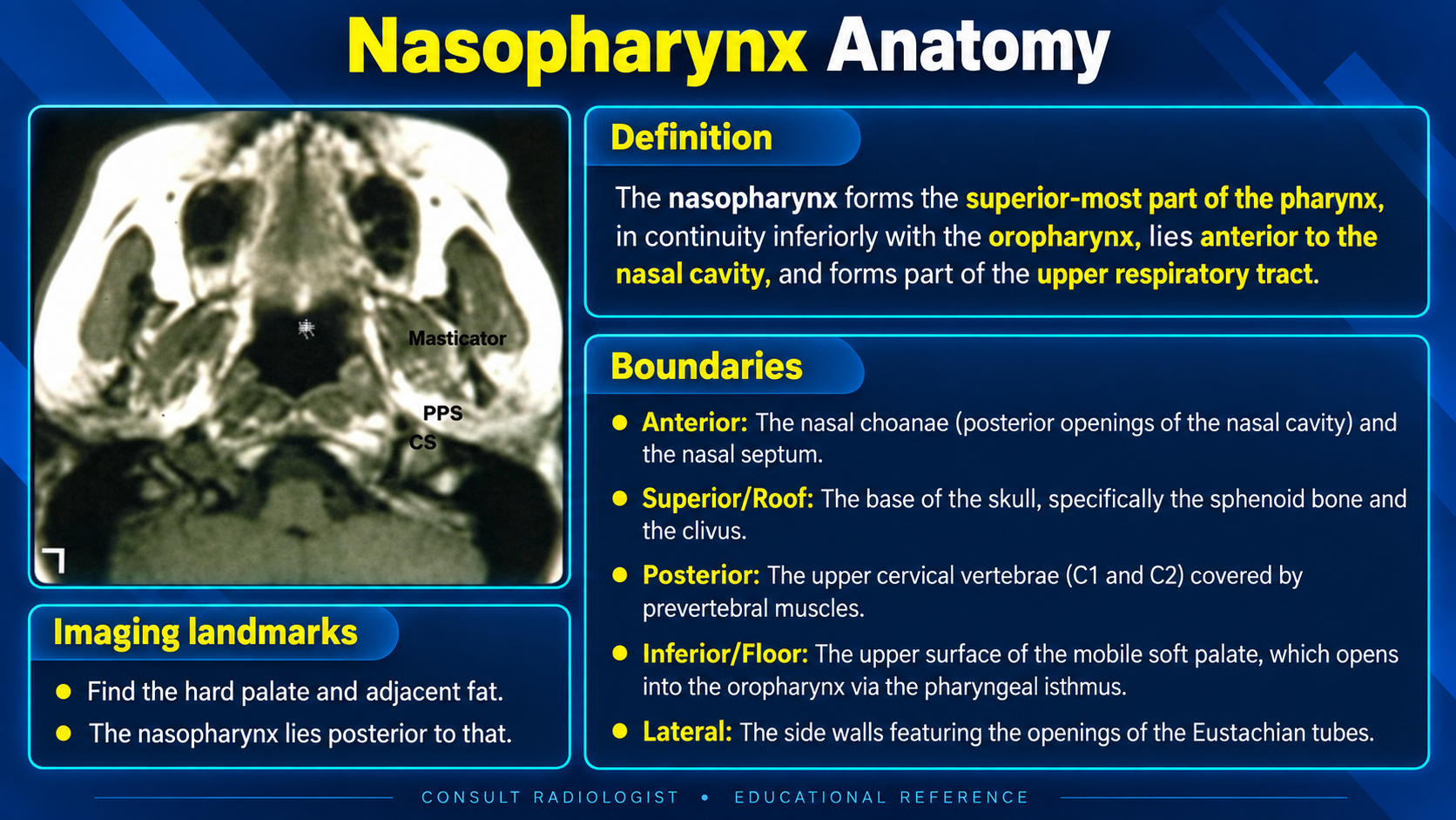
Nasopharynx boundaries and axial imaging landmarks
Boundaries and OrientationClick to enlarge
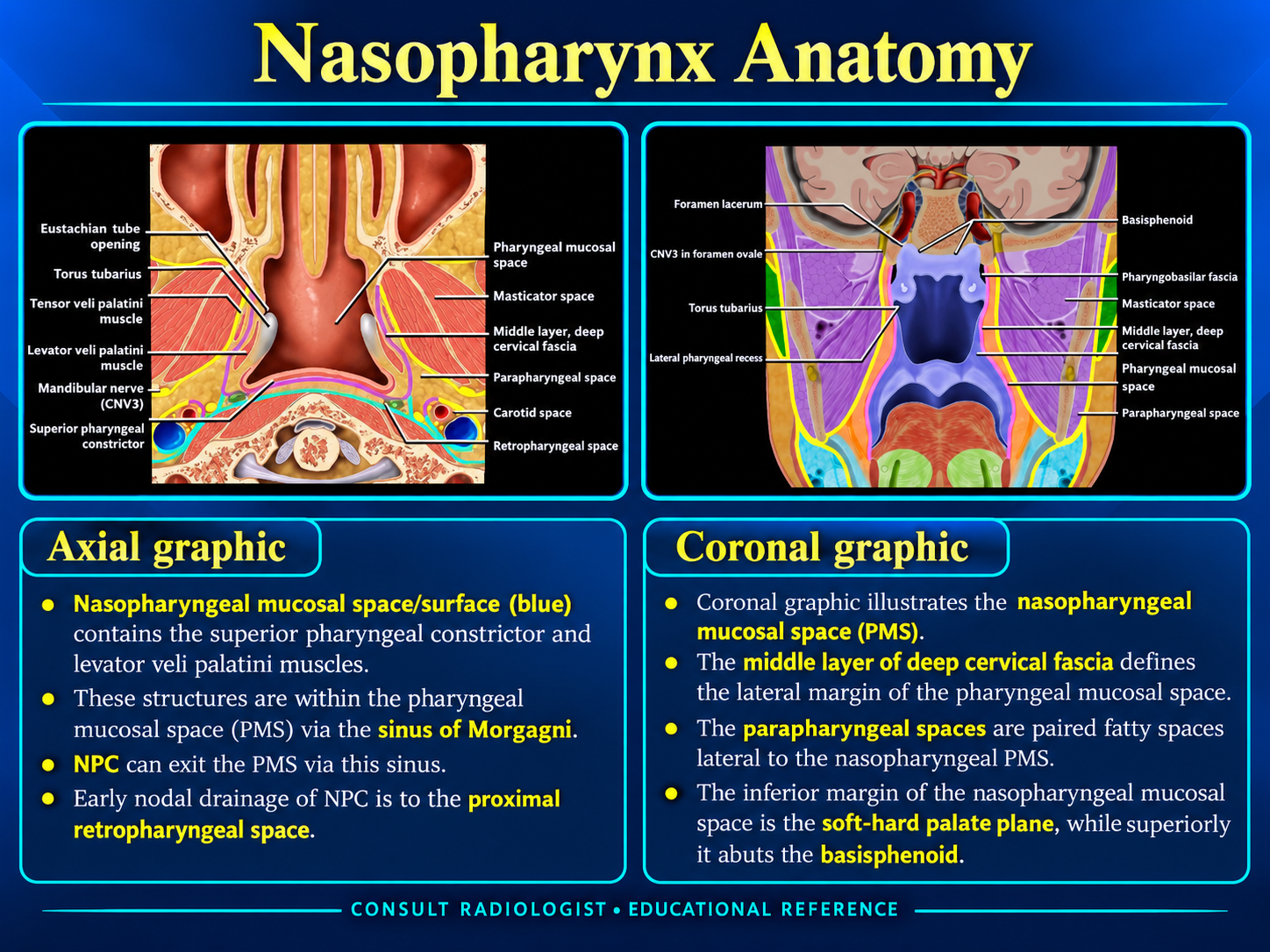
Axial and coronal nasopharyngeal anatomy
Axial and Coronal AnatomyClick to enlarge
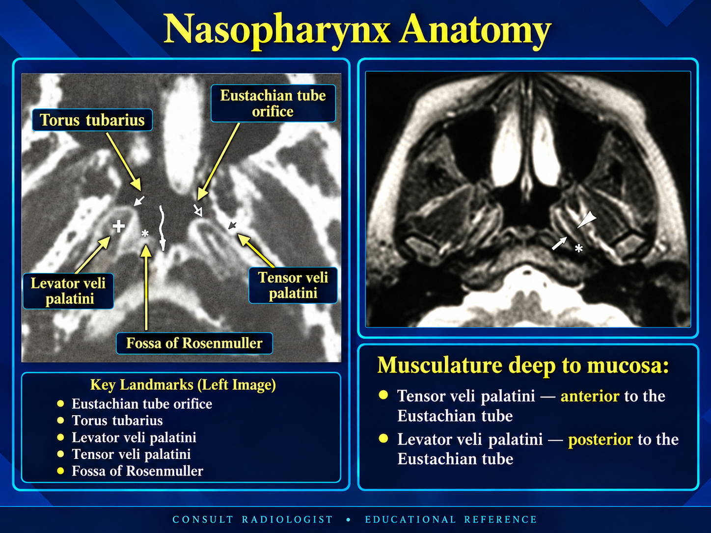
Nasopharyngeal lateral wall and Eustachian tube landmarks
Lateral Wall LandmarksClick to enlarge
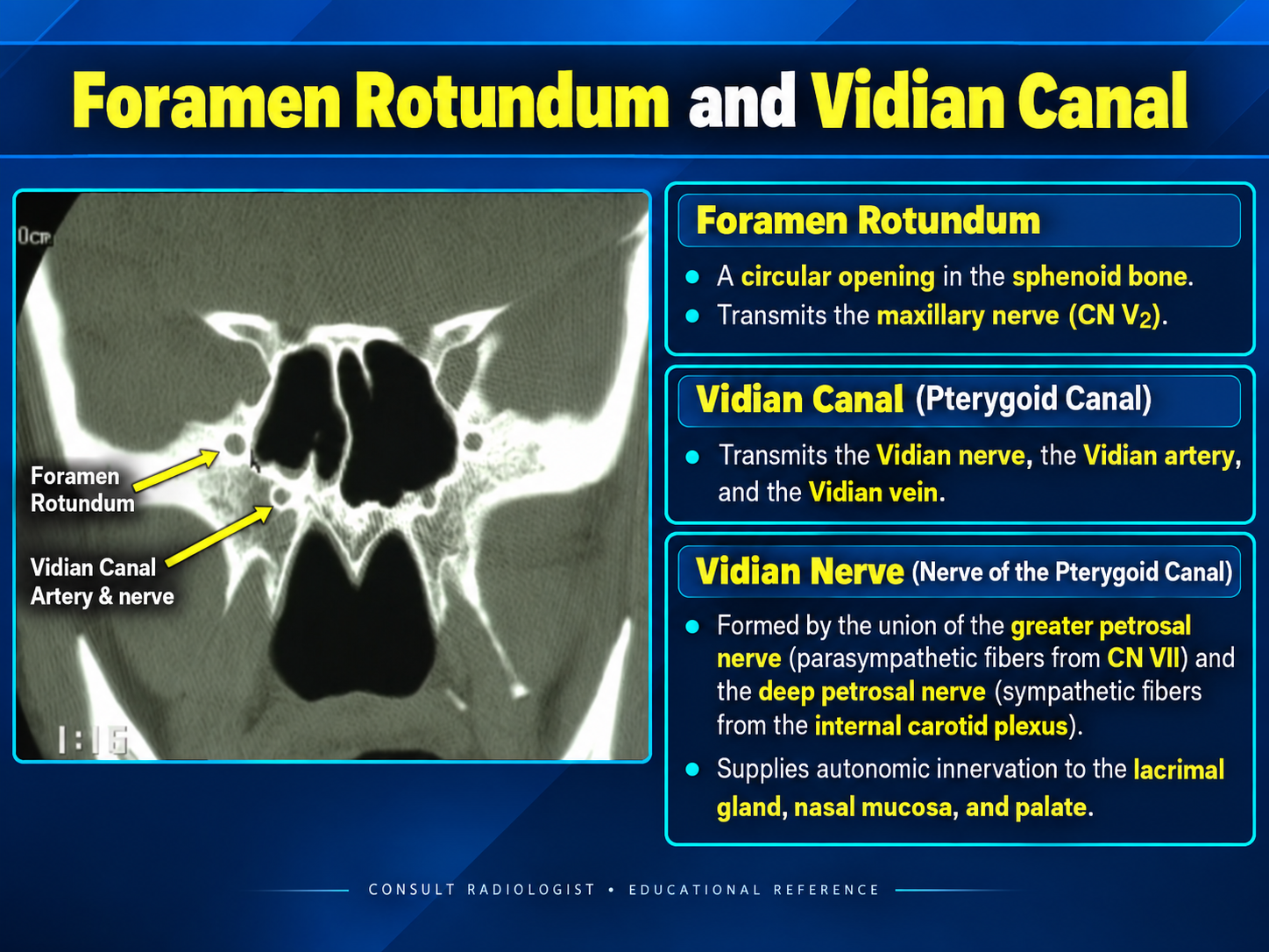
Foramen rotundum and vidian canal anatomy
Skull-Base PathwaysClick to enlarge

Boundaries

Superior pharyngeal compartmentThe nasopharynx lies posterior to the nasal cavity and above the soft palate.
  • Anterior: posterior choanae and posterior nasal septum/vomer.
  • Superior: sphenoid floor, clivus, and pharyngeal vault.
  • Posterior: prevertebral muscles over C1 and C2.
  • Inferior: hard–soft palate plane and mobile superior surface of the soft palate.
  • Lateral: Eustachian tube orifices, torus tubarius, and fossae of Rosenmüller.

Adenoid tissue normally occupies the roof and posterior wall, particularly in children and young adults.

Lateral Wall Landmarks

High-yield siteThe fossa of Rosenmüller is the most common origin of nasopharyngeal carcinoma and must be compared side to side.
  • Eustachian tube orifice: central on the lateral wall.
  • Torus tubarius: mucosal ridge over the cartilaginous tube.
  • Fossa of Rosenmüller: recess posterior to the torus.
  • Tensor veli palatini: anterior to the Eustachian tube.
  • Levator veli palatini: posterior and medial to the tube.
  • Sinus of Morgagni: superior fascial gap transmitting the Eustachian tube and levator muscle.

Adjacent Spaces and Spread

Read beyond the mucosaNasopharyngeal disease can cross the pharyngobasilar fascia and spread along predictable deep-space and skull-base pathways.
  • Lateral spread into the parapharyngeal and carotid spaces.
  • Posterior spread to prevertebral muscles and upper cervical spine.
  • Superior spread into the sphenoid sinus, clivus, and skull base.
  • Anterior spread toward the nasal cavity and pterygopalatine fossa.
  • Perineural spread along V2 through foramen rotundum or along the vidian canal.

Imaging Search Pattern

RegionWhat to assess
MucosaSymmetry, mass, ulceration, adenoid contour
Lateral wallRosenmüller recess, torus, tube orifice
Deep spacesParapharyngeal, carotid, retropharyngeal
Skull baseClivus, sphenoid, foramina, marrow signal
Neural pathwaysV2, foramen rotundum, vidian canal, cavernous sinus
NodesRetropharyngeal and bilateral level II–V chains

Lymphatic Drainage

Lateral pathway

Lymphatics traverse the superior constrictor and drain to lateral retropharyngeal, upper deep cervical, and posterior-triangle nodes.

Medial pathway

The roof and posterior wall drain toward median retropharyngeal nodes.

Imaging pearl: Retropharyngeal nodes are an early and common site of spread. Nasopharyngeal carcinoma may also present with bilateral level II nodal disease.

Systemic drainage

Deep cervical nodal chains ultimately drain to the jugular lymphatic trunks.

Vessels and Innervation

Arterial supply

  • Ascending pharyngeal artery.
  • Ascending palatine branch of the facial artery.
  • Ascending cervical artery.
  • Branches of the maxillary artery.

Venous drainage

Pharyngeal and pterygoid venous plexuses drain toward the internal jugular system.

Innervation

Sensation is supplied by the pharyngeal branch of V2 and CN IX. Motor supply travels through the pharyngeal plexus, principally via CN X with contributions associated with CN IX and sympathetic fibers.

Reporting Checklist

Primary site: side, subsite, craniocaudal extent, and fossa of Rosenmüller involvement.
Local extension: nasal cavity, oropharynx, parapharyngeal space, carotid space, prevertebral muscles, clivus, sphenoid sinus, and intracranial compartment.
Perineural spread: V2/foramen rotundum, vidian canal, PPF, Meckel cave, and cavernous sinus.
Nodes: retropharyngeal and bilateral cervical nodal stations, including necrosis and extranodal extension.

Symmetry caveat

Normal lymphoid tissue can be prominent, especially in younger patients, but focal asymmetric mucosal thickening, deep infiltration, skull-base change, or nodal disease warrants concern.
Secondary Links