Lymph Node Levels of the Neck

Head and neck imaging approach: Assign every suspicious node a side and level, then describe size, morphology, necrosis or cystic change, extranodal extension, and relationship to vessels and the primary tumor. Use levels I–VI for the standard cervical compartments and name retropharyngeal, parotid, facial, occipital, or mediastinal nodes specifically.

Visual Reference

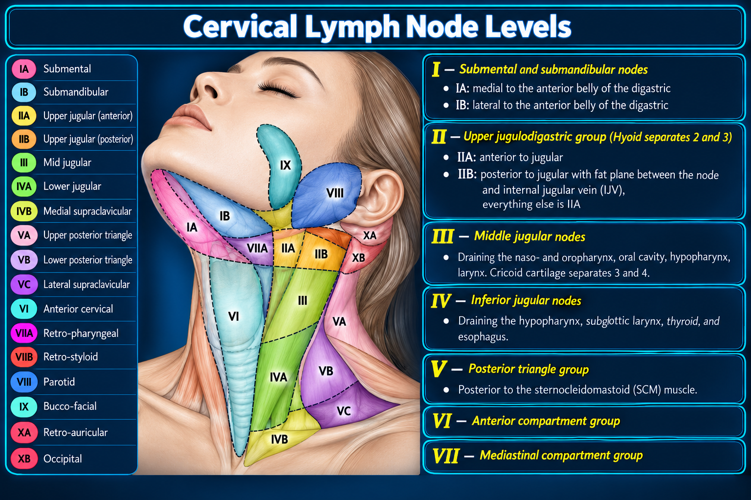
Illustrated cervical lymph node levels
Cervical Nodal MapClick to enlarge
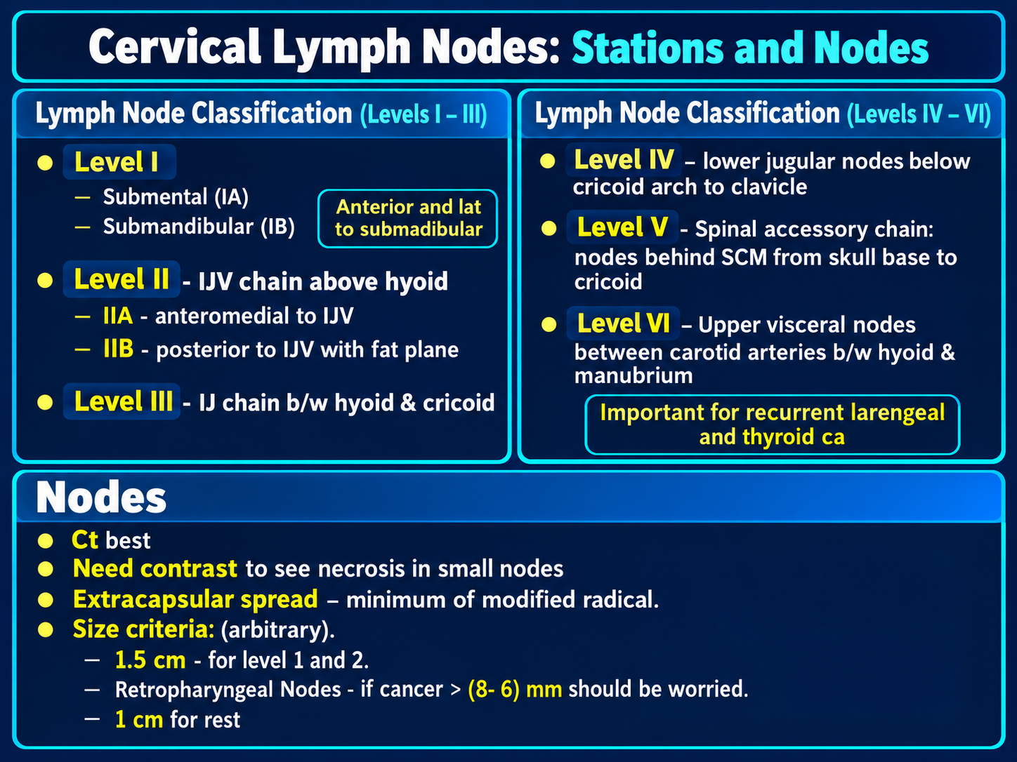
Cervical lymph node stations and imaging criteria
Levels I–VI OverviewClick to enlarge
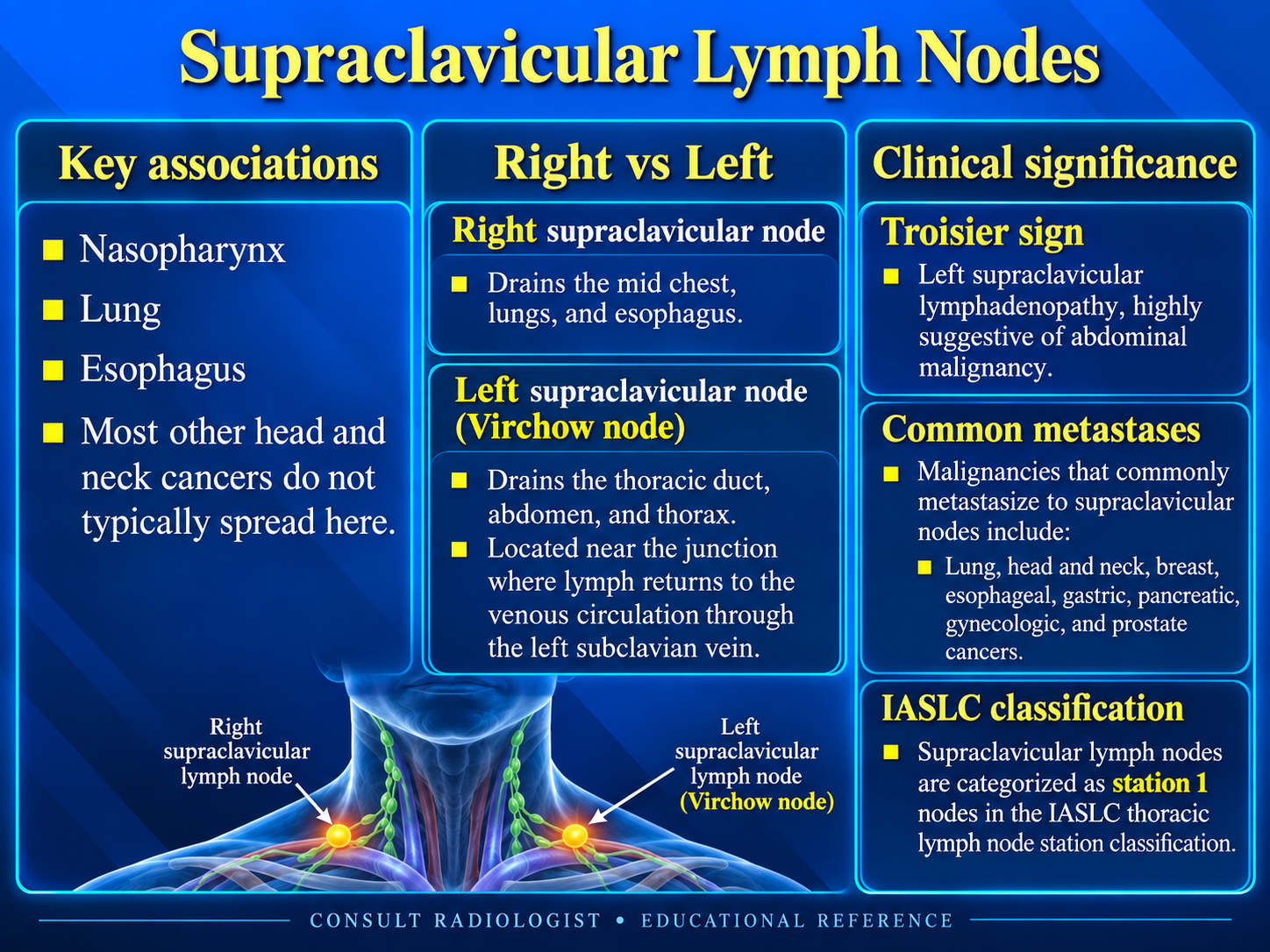
Supraclavicular lymph node drainage and significance
Supraclavicular NodesClick to enlarge
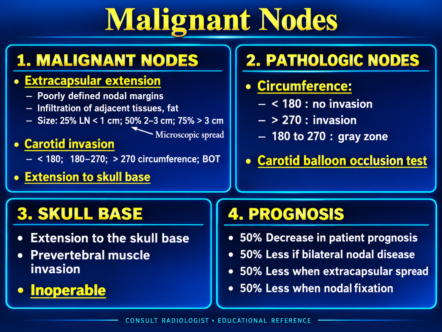
Imaging features of malignant cervical lymph nodes
Malignant Nodal FeaturesClick to enlarge
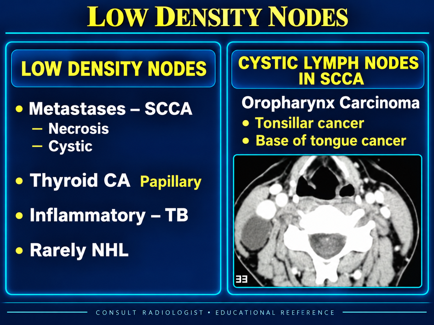
Differential diagnosis for low density cervical nodes
Low-Density NodesClick to enlarge

Levels I and II

Suprahyoid nodal groupsLevel I surrounds the submandibular and submental spaces. Level II follows the upper internal jugular chain from the skull base to the hyoid.

Level I

  • IA: between the anterior digastric bellies, above the hyoid and below the mylohyoid.
  • IB: posterolateral to the anterior digastric belly in the submandibular triangle.

Level II

  • IIA: anterior to, lateral to, or inseparable from the internal jugular vein.
  • IIB: posterior to the vein with an intervening fat plane.

Common drainage includes the oral cavity, oropharynx, nasopharynx, parotid region, and upper aerodigestive tract.

Levels III and IV

Middle and lower jugular chainThe hyoid separates levels II and III; the cricoid arch separates levels III and IV.

Level III

Extends from the hyoid to the cricoid, between the anterior and posterior margins of the sternocleidomastoid, lateral to the carotid space.

Level IV

Extends from the cricoid to the clavicle along the lower internal jugular chain. Medial supraclavicular nodes are included.

Drainage pattern

Levels III and IV receive drainage from the pharynx, larynx, cervical esophagus, thyroid, and other upper aerodigestive sites. Lower left-sided nodes may also reflect thoracic or abdominal malignancy.

Levels V and VI

Posterior triangle and central compartmentLevel V is posterior to the sternocleidomastoid. Level VI lies between the carotid arteries in the anterior visceral compartment.

Level V

  • VA: above the cricoid, including the spinal accessory chain.
  • VB: below the cricoid, including lateral supraclavicular nodes.

Level VI

From hyoid to manubrium, medial to the common carotid arteries; includes prelaryngeal (Delphian), pretracheal, paratracheal, and perithyroidal nodes.

Typical associations

Level V: nasopharyngeal, cutaneous, and thyroid primaries. Level VI: thyroid, subglottic larynx, cervical esophagus, and trachea.

Boundary Quick Reference

LevelSuperior → inferiorKey landmark
IMandible/mylohyoid → hyoidAnterior digastric separates IA and IB
IISkull base → hyoidIJV relationship separates IIA and IIB
IIIHyoid → cricoidMiddle jugular chain
IVCricoid → clavicleLower jugular chain
VSkull base → claviclePosterior to SCM; cricoid separates VA/VB
VIHyoid → manubriumBetween common carotids

Nodes Outside Levels I–VI

Name these groups directly rather than forcing them into the six-level scheme:

  • Retropharyngeal
  • Parotid and preauricular
  • Facial, buccal, and mandibular
  • Occipital and posterior auricular
  • Superior mediastinal
Nomenclature: Some surgical systems call superior mediastinal nodes “level VII,” but usage is inconsistent. A precise anatomic name is clearer in an imaging report.

Assessing a Suspicious Node

  • Morphology: round shape, loss of fatty hilum, irregular margin, heterogeneous enhancement.
  • Necrosis or cystic change: highly suspicious in the appropriate clinical setting; in an adult, consider HPV-associated oropharyngeal carcinoma and papillary thyroid carcinoma.
  • Extranodal extension: indistinct capsule, infiltration of adjacent fat or muscle, matted nodes, or invasion of nearby structures.
  • Vascular relationship: describe narrowing, occlusion, or circumferential contact with the carotid artery and internal jugular vein.

Size thresholds vary by nodal group and clinical context; morphology and interval change can outweigh short-axis diameter.

Reporting Checklist

Location: side, level, and nodal group; report the largest or most suspicious node in each relevant station.
Morphology: short-axis size, cystic/necrotic component, calcification, enhancement, and extranodal extension.
Extent: adjacent vessel, muscle, skin, prevertebral, skull-base, and mediastinal involvement; note bilateral or contralateral disease.
Primary search: correlate the drainage pattern with mucosal, thyroid, salivary, cutaneous, thoracic, and abdominal primary sites.

Supraclavicular pearl

Right-sided nodes predominantly receive right thoracic drainage. Left supraclavicular (Virchow) nodes receive thoracic-duct drainage and may reflect thoracic or abdominopelvic malignancy.

Secondary Links