Epiglottis and Pre-epiglottic Space
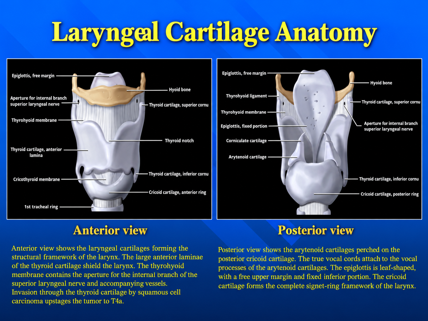
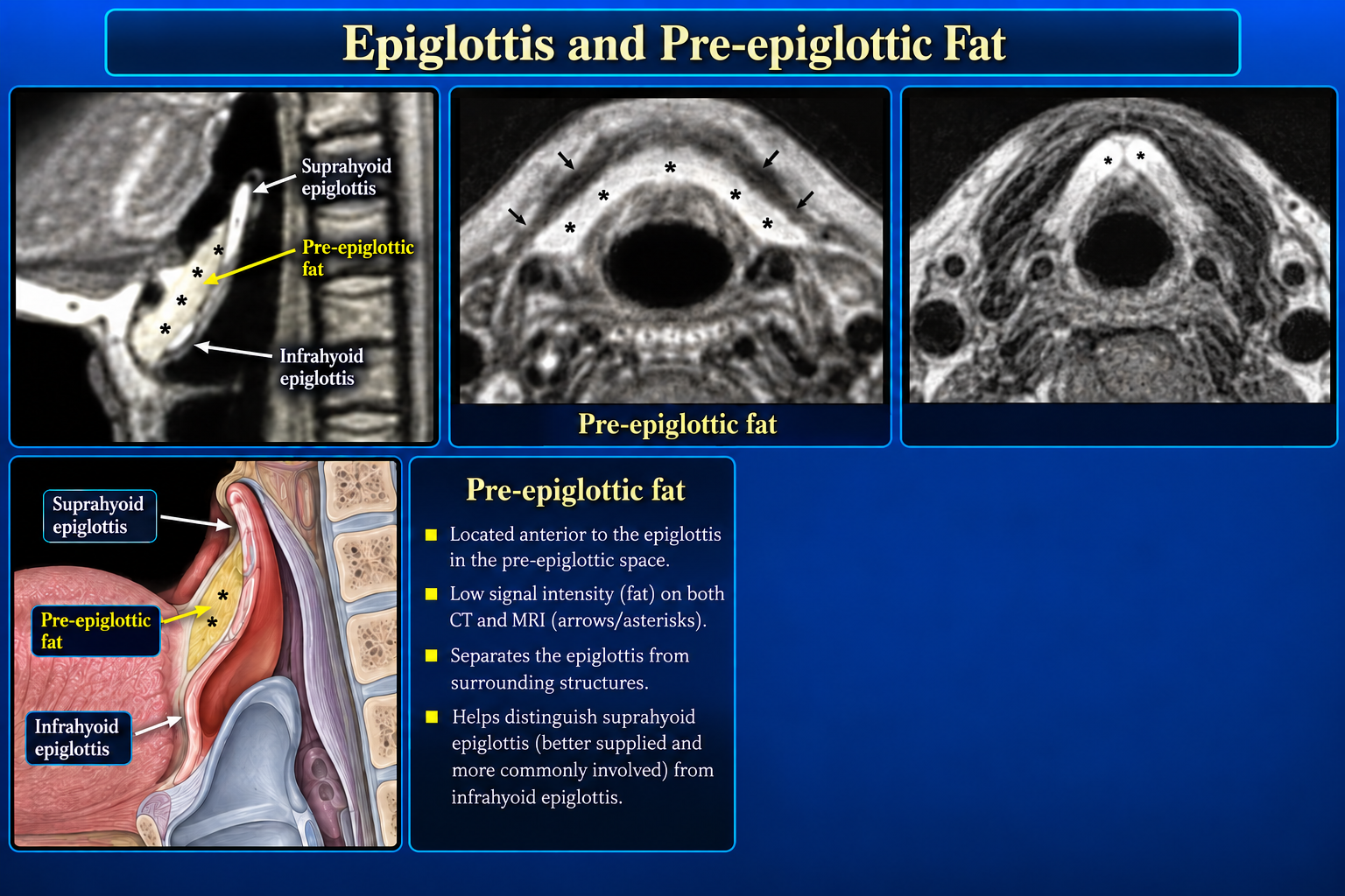
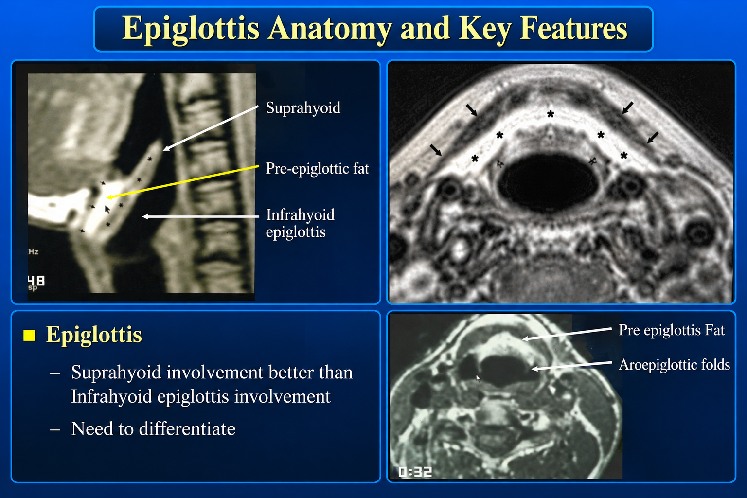
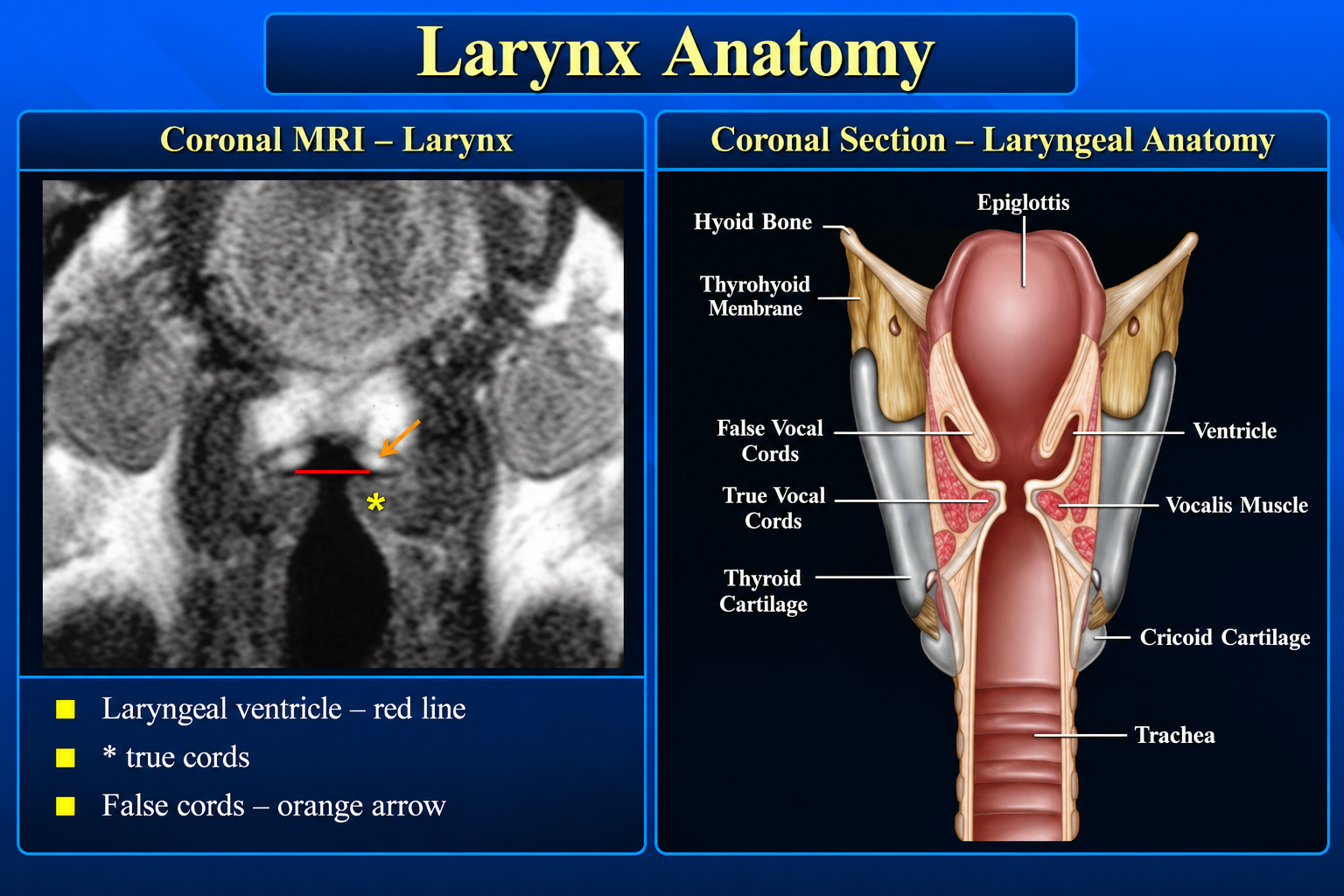
The hyoid divides the epiglottis into suprahyoid and infrahyoid portions. The suprahyoid segment forms the free upper margin; the infrahyoid segment attaches inferiorly through the thyroepiglottic ligament.
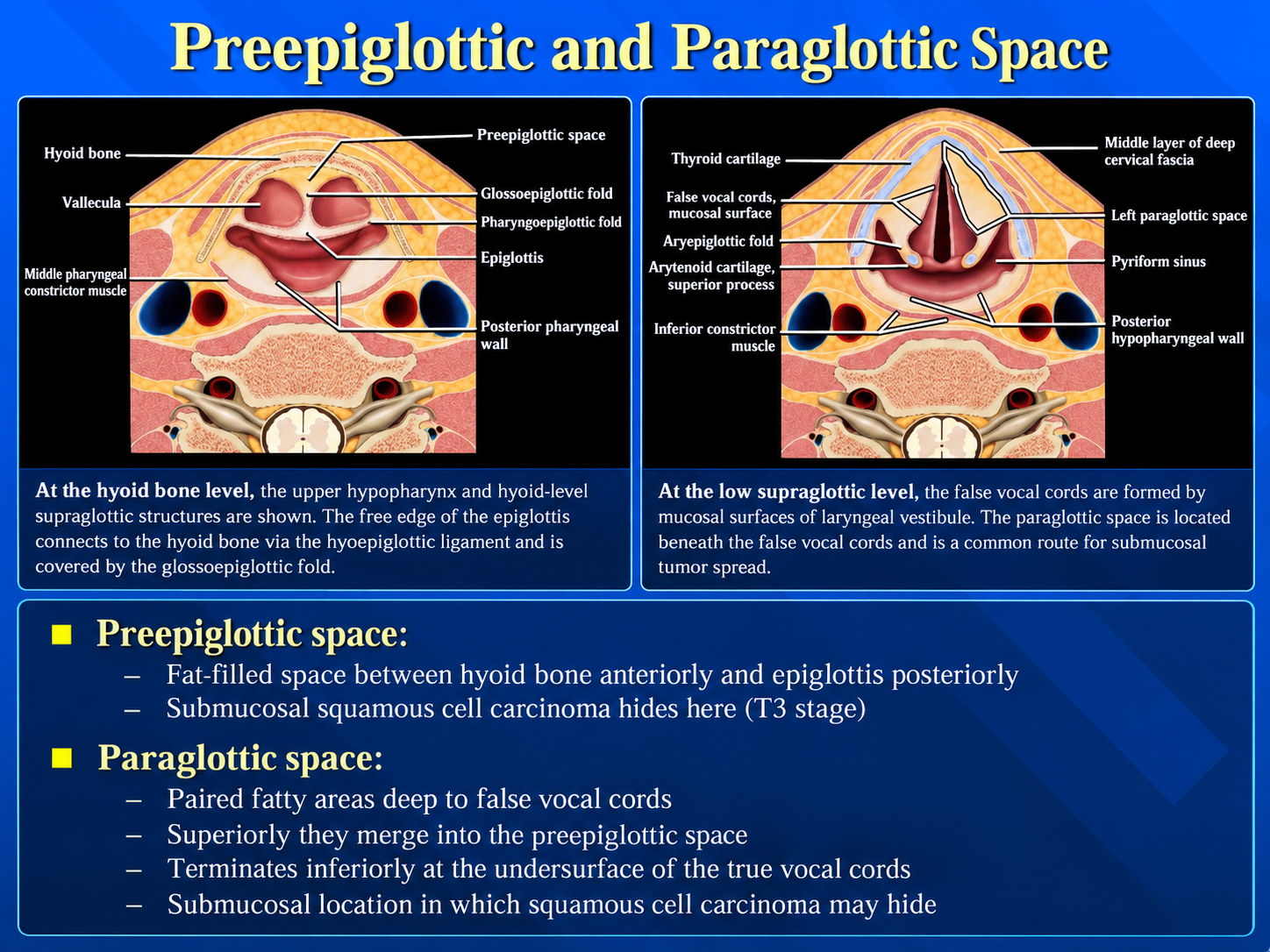
Pre-epiglottic boundaries
- Anterior: hyoid, thyrohyoid membrane, and upper thyroid cartilage.
- Posterior: epiglottis.
- Superior: hyoepiglottic ligament.
- Inferolateral: paraglottic spaces.
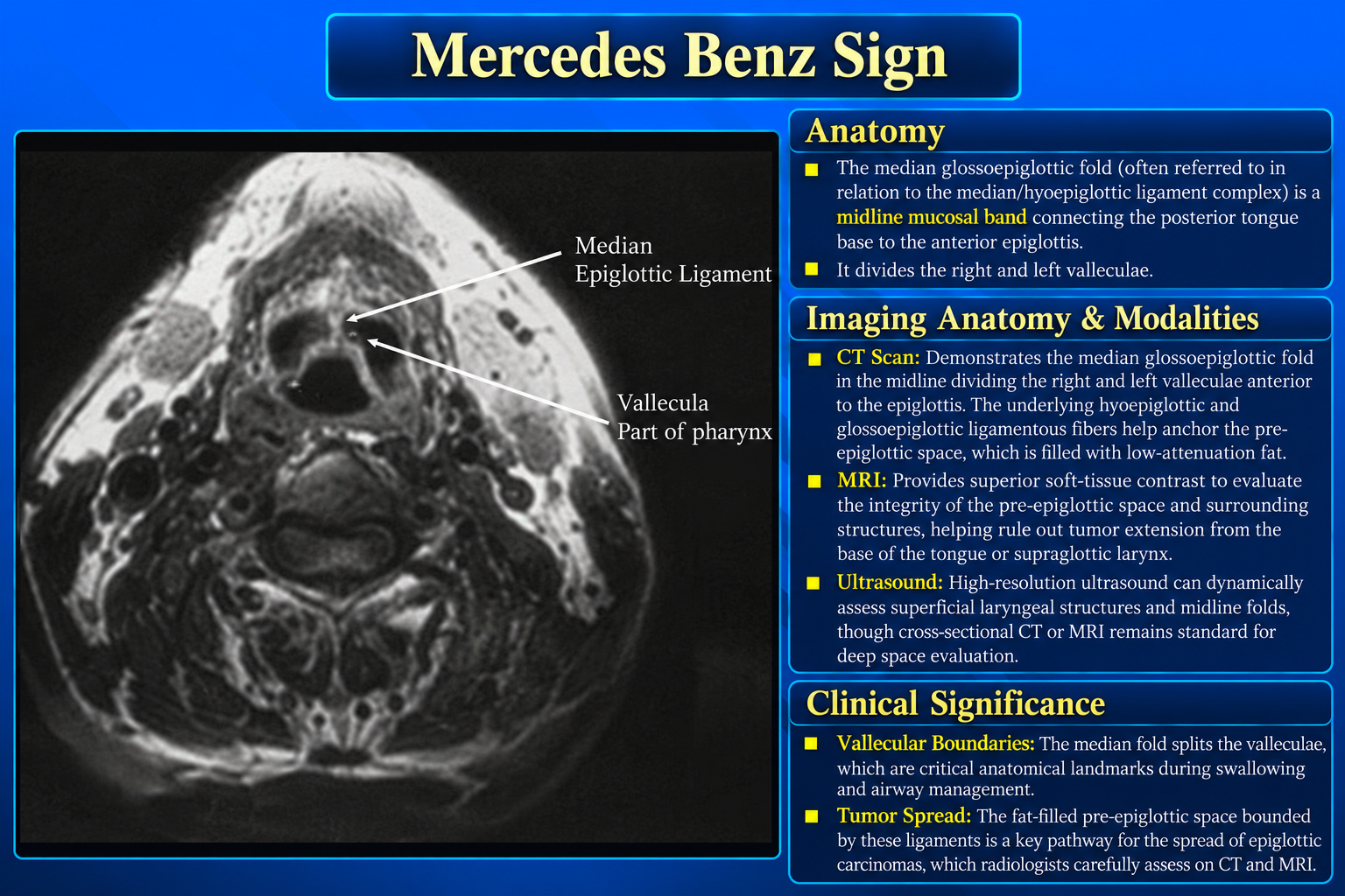
Vallecular landmark: The median glossoepiglottic fold separates the paired valleculae, producing the classic Mercedes-Benz configuration on axial images.
Suggested Reporting Language
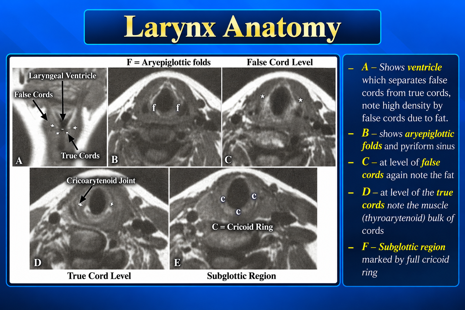
The larynx is symmetric without focal mucosal mass. The pre-epiglottic and paraglottic fat planes are preserved. The vocal cords are symmetric in position. No cartilage destruction or extralaryngeal extension is identified.
When abnormal
State the primary subsite, cranio-caudal extent, cord mobility if known, commissural or deep-space involvement, cartilage invasion, extralaryngeal spread, and nodal disease.
Technique note
Thin-section contrast-enhanced CT is commonly used for mucosal and cartilage assessment; MRI is complementary for deep-space, marrow, and perineural evaluation.