Venous extradural (epidural) hematomas
Key Images
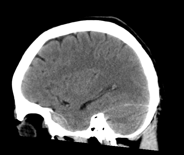
Look For First
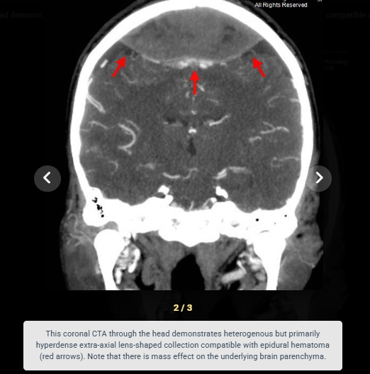
- Biconvex lentiform hyperdensity at atypical EDH locations (vertex, anterior temporal, posterior fossa)
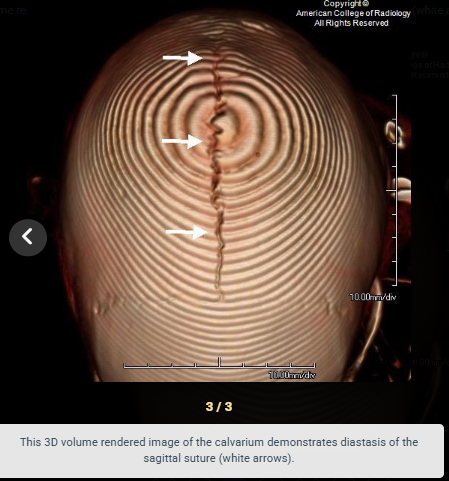
- Hematoma crossing a suture line, particularly at the sagittal suture (distinguishes from typical arterial EDH)
- Associated skull fracture or suture diastasis, especially sagittal suture widening
- Focal hypo- or isodensity (swirl sign) within the hyperdense collection indicating active venous bleeding
Key Image Findings
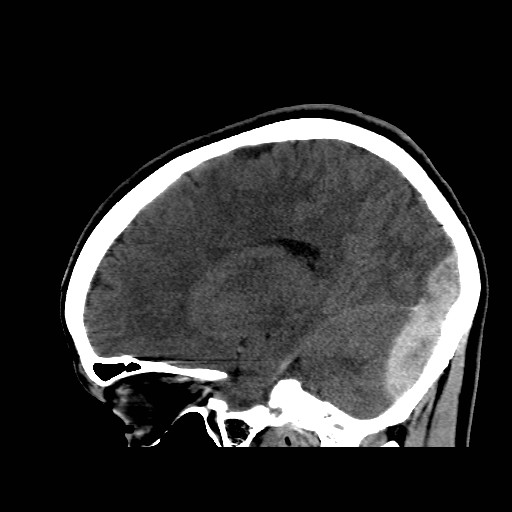
- Biconvex or lentiform extra-axial hyperdensity on CT that is sharply marginated from adjacent brain and skull.
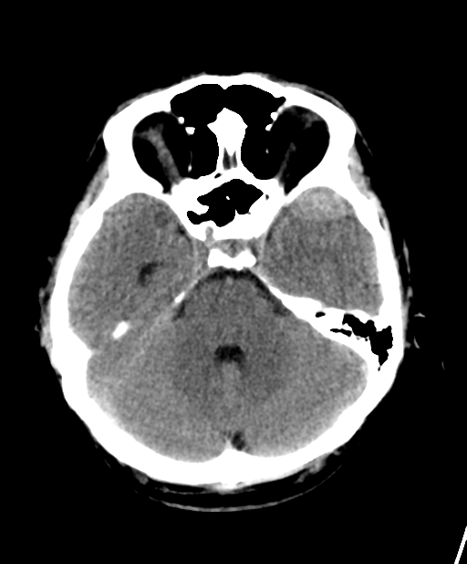
- Venous EDHs preferentially occur at three classic locations: vertex (superior sagittal sinus), anterior middle cranial fossa (sphenoparietal sinus), and occipital/posterior fossa (transverse sinus).
- Unlike typical arterial EDHs, venous EDHs can cross suture lines due to the linear anatomic distribution of the involved dural venous sinuses.
- Swirl sign—focal areas of hypo- or isodensity within the predominantly hyperdense hematoma—indicates active bleeding and suggests venous source.
- The displaced dural venous sinus may be elevated away from the inner table of skull, creating a characteristic appearance on coronal reformats.
- In the chronic phase, venous EDHs become increasingly heterogeneous with areas of lower density reflecting clot organization and resorption.
- Associated skull fractures (especially sagittal, occipital) or suture diastasis are commonly present on bone windows.
- Venous EDHs typically produce smaller volume bleeds and less mass effect than arterial EDHs, often without significant midline shift or herniation.
Differential Diagnosis
- Arterial epidural hematoma: biconvex collection, but typically stops at suture lines and occurs in classic middle meningeal distribution over convexity; venous EDHs cross sutures and favor atypical locations.
- Subdural hematoma: located in the subdural space (between dura and arachnoid), crescent-shaped rather than lentiform, follows brain contours more closely, and extends across hemispheres.
- Subarachnoid hemorrhage: blood within the subarachnoid space, fills sulci and surrounds vessels, not a focal extra-axial mass.
- Intracerebral hemorrhage: located within the brain parenchyma with edema and mass effect directed inward, not a peripheral extra-axial collection.
- Dural metastasis or dural-based tumor: enhances uniformly with contrast and has irregular margins; lacks the acute hyperdensity of fresh blood.
- Epidural abscess: gradual onset, lacks the focal sharp margins of acute EDH, and typically has associated clinical signs of infection.
Discussion
Venous EDHs have a more benign clinical course than arterial EDHs because dural venous sinuses have lower pressure, resulting in slower bleeding and smaller hematoma volumes.
The ability of venous EDHs to cross suture lines is a crucial distinguishing feature; this occurs because the involved venous sinus runs along the dural attachments (e.g., superior sagittal sinus along the falx).
Younger patients are at higher risk; after age 60, dural adhesions become tighter and venous EDHs become rare, making them an important consideration in geriatric head trauma.
Conservative management is often appropriate for venous EDHs with small volume, stable neurologic status (GCS > 8), and no mass effect, requiring only close monitoring and serial imaging.
Vertex location with sagittal suture involvement is the most common presentation; displacement of the superior sagittal sinus inferiorly away from bone is a key imaging sign.
The swirl sign in venous EDH indicates active ongoing bleeding from the opened sinus and may predict hematomas that require intervention rather than observation.
Reporting Pearls
Describe venous EDHs by location (vertex, anterior temporal, or posterior fossa), note whether they cross suture lines, document associated fractures or diastasis, comment on the presence of swirl sign, and report the absolute volume and degree of mass effect; specifically state that the hematoma appears to be of venous rather than arterial origin based on location and morphology.
Pitfalls
- Mistaking a venous EDH for a subdural hematoma because both can occur at the vertex; look for the lentiform shape and extradural location (outside dura) of EDH versus the crescent shape of subdural.
- Overestimating the severity of venous EDHs; even when large, these hematomas typically have better clinical prognosis than arterial EDHs and may be managed conservatively with imaging surveillance.
- Assuming all EDHs are arterial in origin and expecting them to respect suture lines; venous EDHs commonly cross sutures, especially at the sagittal suture, which should prompt investigation for sinus involvement.
- Missing the swirl sign or subtle active bleeding within the collection; careful examination at multiple density windows can reveal hypo/isodense foci that indicate ongoing venous bleeding and guide management decisions.