Time Table & Treatment Guidlines

Acute Ischemic Stroke Treatment Flowchart

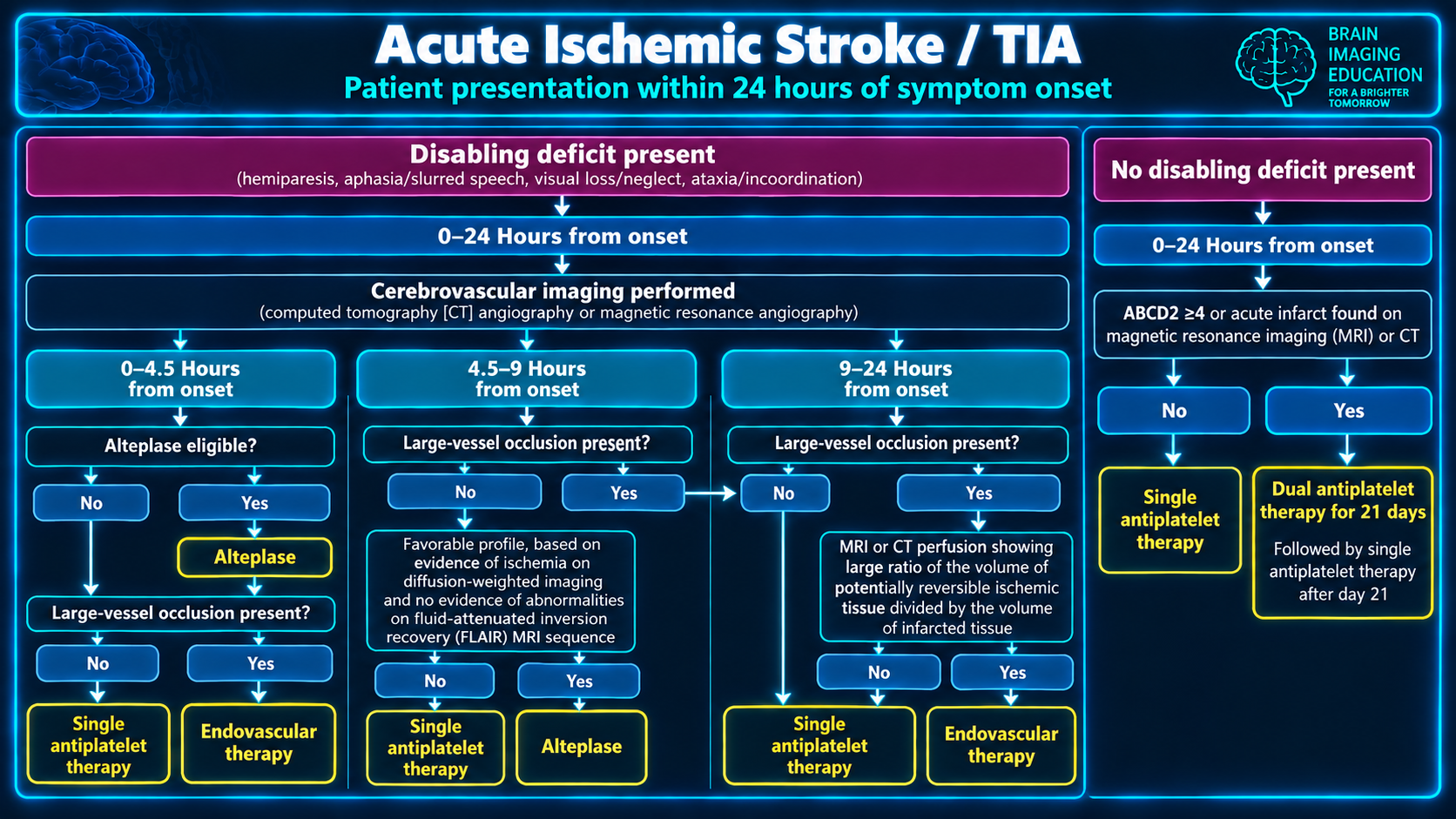
Acute ischemic stroke and TIA treatment flowchart for patients presenting within 24 hours
Acute Ischemic Stroke / TIAClick to enlarge

Time zero is the last-known-well (LKW) time. Reperfusion benefit declines continuously with delay, so imaging, eligibility assessment, thrombolysis, and thrombectomy workflow should occur in parallel whenever possible. The 2026 AHA/ASA guideline is the current comprehensive U.S. reference and includes broader late-window thrombolysis and EVT eligibility than prior versions. ahajournals

Consolidated Acute Ischemic Stroke Timeline

Time from LKWIV thrombolysisEVTKey operational point
Prehospital / arrivalIdentify potential disabling deficit and exclude hypoglycemia.Identify probable LVO and route to an EVT-capable pathway when system design supports it.Establish accurate LKW, premorbid mRS, anticoagulant history, glucose, and BP. Obtain NCCT immediately; add CTA head/neck for suspected LVO without delaying lysis. ahajournals
0–4.5 hClass I: eligible patients with disabling AIS should receive either alteplase 0.9 mg/kg (maximum 90 mg) or tenecteplase 0.25 mg/kg promptly. Benefit is greatest in the first 3 hours.Class I EVT for eligible anterior-circulation LVO; do not withhold IVT because EVT is planned.Target door-to-needle is 60 minutes or less. Give IVT first when indicated, then proceed directly to angiography—do not await clinical response.
0–6 hStandard IVT eligibility still depends on the 4.5-hour window.EVT is indicated for eligible ICA or M1 occlusion and now includes selected large-core infarcts, including ASPECTS 3–5.Avoid requiring CT perfusion in otherwise eligible early-window LVO; NCCT and CTA are usually sufficient for fast triage. ahajournals
4.5–9 h or wake-up/unknown onsetClass IIa: IVT is reasonable with a favorable advanced-imaging profile: DWI-FLAIR mismatch or CT/MR perfusion mismatch demonstrating potentially salvageable tissue.If LVO is present, assess independently for EVT eligibility.This is an imaging-selected thrombolysis window, not simply a clock-based extension.
6–16 hIVT may be considered only in selected late presenters, especially when EVT is unavailable or unsuitable and favorable imaging is present.Class I: EVT for appropriately selected anterior-circulation LVO patients, traditionally based on DAWN/DEFUSE-3-type clinical-core or perfusion-core mismatch.Advanced imaging is central to late-window selection.
16–24 hClass IIb: IVT may benefit a narrow, imaging-selected LVO group; evidence is most relevant when EVT cannot be performed, rather than as routine bridging before EVT.EVT remains reasonable in selected patients; original evidence was strongest with DAWN-type selection.Assess core size, penumbra, collateral context, disability baseline, occlusion site, and feasibility of rapid reperfusion.
Up to 24 h: posterior circulationNo routine late IVT pathway without appropriate imaging-based selection.The 2026 guidance supports EVT for selected basilar artery occlusion patients, particularly with NIHSS at least 10 and without extensive established infarction. ahajournalsPosterior-circulation imaging assessment should emphasize established brainstem injury and vascular confirmation, not NIHSS alone.

Eligibility and Workflow Details

  • Historical 3–4.5-hour “relative exclusions”—age over 80, NIHSS over 25, prior stroke plus diabetes, and oral anticoagulant use—should not be used as simplistic automatic exclusions. Anticoagulant decisions require agent-specific timing and laboratory/medication information, particularly for DOAC exposure.
  • For IVT/EVT candidates, maintain blood pressure below 185/110 mm Hg before thrombolysis and below 180/105 mm Hg after reperfusion treatment. ahajournals
  • Core workflow targets include arrival-to-groin puncture within 90 minutes for direct arrivals and within 60 minutes for transferred EVT candidates. Minimize nonessential imaging and avoid repeat scanning at the receiving EVT center if adequate source imaging is available.
  • The practical radiology contribution is a rapid, explicit report: hemorrhage status, ASPECTS/established core, occlusion location, tandem lesion, collateral appearance when assessed, perfusion core/penumbra and mismatch when obtained, and posterior-fossa infarct burden for basilar occlusion.

Antiplatelet and Early Medical Therapy

  • For minor noncardioembolic, nondisabling ischemic stroke or high-risk TIA not treated with IVT/EVT, start short-course DAPT—typically aspirin plus clopidogrel—for 21 days when appropriate, then transition to single antiplatelet therapy.
  • Aspirin within 24–48 hours is appropriate for most ischemic-stroke patients not receiving thrombolysis. After IV alteplase or tenecteplase, defer antiplatelet/anticoagulant treatment until follow-up brain imaging at approximately 24 hours excludes hemorrhage.
  • DAPT is not a substitute for reperfusion in a disabling stroke and should not delay vascular imaging or transfer for possible EVT.

A practical way to remember the framework is: early window = treat rapidly on clinical and CT/CTA grounds; late/unknown window = treat selectively using tissue viability imaging. What would you include in a one-line CTA/CTP impression to make the late-window EVT decision immediately actionable for the stroke team?

Secondary Links