Hemorrhagic Transformation

Reference Images

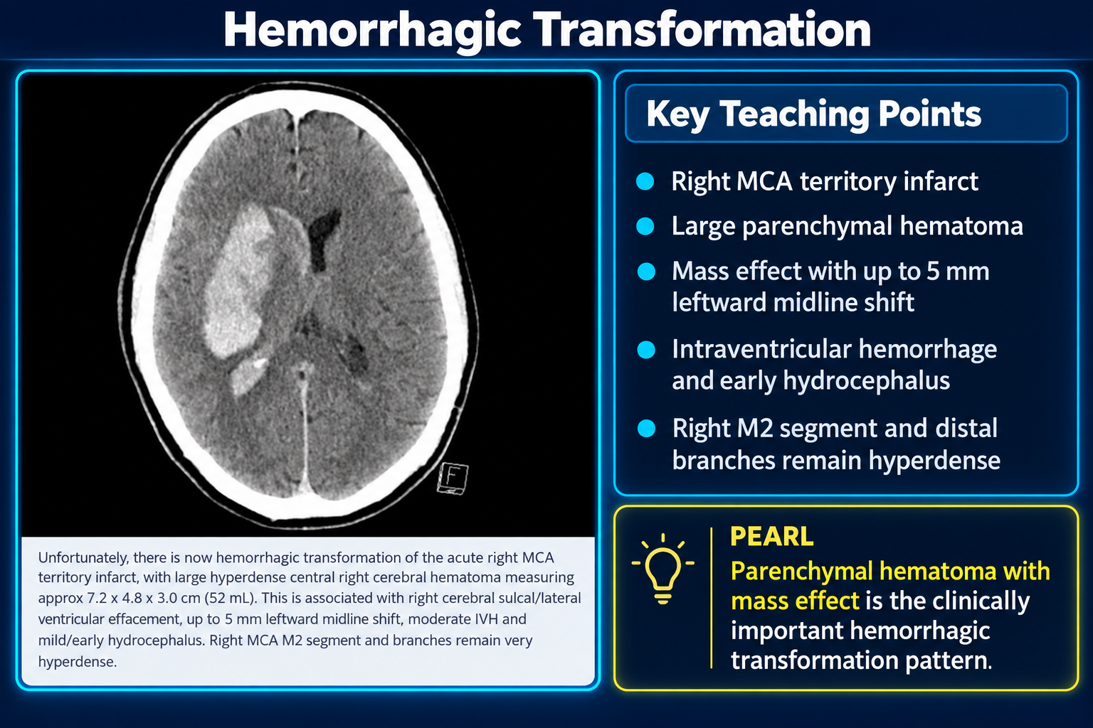
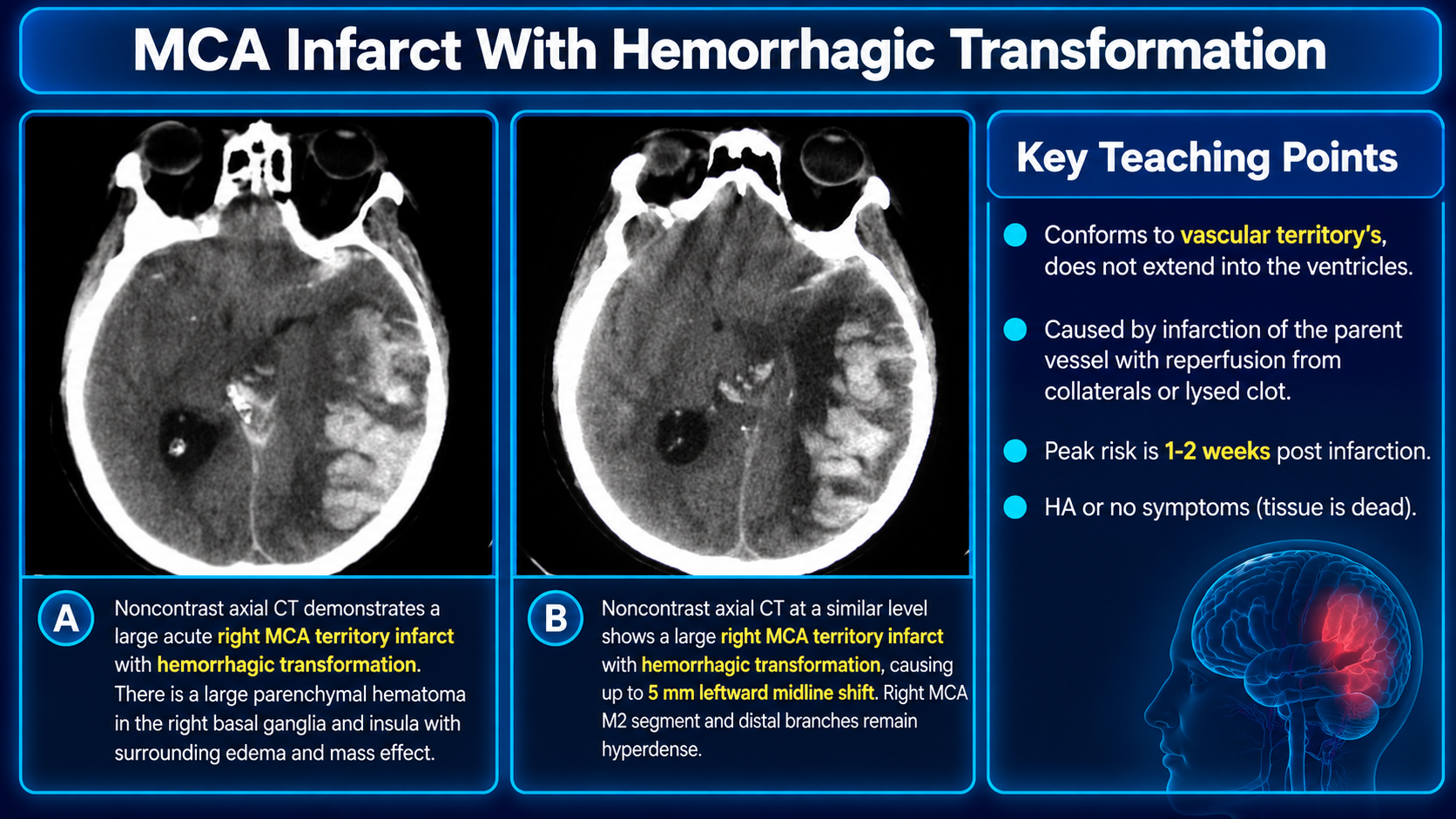
Teaching graphic showing hemorrhagic transformation of a right MCA infarct with a large parenchymal hematoma, mass effect, intraventricular hemorrhage, and early hydrocephalus
Hemorrhagic Transformation Teaching SummaryClick to enlarge
Teaching graphic showing hemorrhagic transformation of a right MCA infarct with a large parenchymal hematoma, mass effect, intraventricular hemorrhage, and early hydrocephalus
Hemorrhagic Transformation Quick NotesClick to enlarge

Hemorrhagic transformation is a complication of cerebral ischemic stroke and can significantly worsen prognosis. It includes two processes with different imaging appearances and prognostic implications: hemorrhagic infarction (petechial hemorrhage) and parenchymal hematoma.

When reporting hemorrhagic transformation, specify which pattern is present, its extent, associated mass effect, and any intraventricular or extra-axial extension.

Clinical Overview

More than half of cerebral infarcts develop some hemorrhagic component at some stage. Most hemorrhagic transformation is petechial hemorrhage (89%); a minority is hematoma (11%). A 2024 series of almost 1400 patients found HI1 to be the most common subtype, followed by HI2 and subarachnoid hemorrhage, while PH2 represented approximately 3%.

Many petechial hemorrhages are asymptomatic. Parenchymal hematomas are the subtype consistently associated with neurological deterioration and worse long-term outcomes.

  • Older age and larger stroke size
  • Cardioembolic stroke etiology
  • Anticoagulant use, thrombolytic therapy, or other recanalization
  • Fever, hyperglycemia, and low serum cholesterol
  • Elevated systolic blood pressure in the acute setting

After intravenous thrombolysis, predictors include NIHSS >14, proximal MCA occlusion, CT hypodensity affecting more than one-third of the MCA territory, delayed recanalization beyond 6 hours, and absent collateral flow.

Significant hemorrhagic transformation usually manifests as rapid, often profound clinical deterioration. In untreated patients it rarely occurs during the first 6 hours and is usually seen during the first few days, with most cases occurring within 4 days. Following thrombolysis or thrombectomy, most hemorrhage occurs within 24 hours of treatment.

Hemorrhagic transformation is attributed to collateral perfusion of infarcted tissue or reperfusion of tissue containing damaged vessels, with blood extravasation or diapedesis.

Classification and Imaging

SubtypeImaging definition
HI1Petechial hemorrhages at the infarct margins
HI2Confluent petechial hemorrhages throughout the infarcted tissue, without hemorrhage-related mass effect
PH1Hematoma involving no more than 30% of the infarcted area, with minimal mass effect
PH2Hematoma involving more than 30% of the infarcted area, with significant mass effect

This system is incorporated into the Heidelberg bleeding classification.

Petechial hemorrhage produces tiny punctate foci or regional increased attenuation, often most pronounced in gray matter. It should not be confused with the fogging phenomenon, which occurs 2-3 weeks after infarction.

Secondary hematoma combines features of ischemic stroke and cerebral hemorrhage. Rarely, CTA may show a spot sign.

MRI demonstrates infarct-related restricted diffusion on DWI/ADC. Blood-sensitive sequences, especially SWI, are more sensitive than CT for early blood products.

Postcontrast FLAIR sulcal hyperintensity after reperfusion, known as HARM, and several perfusion and diffusion markers are associated with greater risk.

Reporting and Pitfalls

  • Infarct territory and extent
  • HI1, HI2, PH1, or PH2 pattern when applicable
  • Hematoma dimensions and estimated volume
  • Mass effect, sulcal or ventricular effacement, and midline shift
  • Intraventricular, subarachnoid, or other extra-axial extension
  • Hydrocephalus or herniation
  • Residual hyperdense vessel or persistent large-vessel occlusion when visible

Petechial hemorrhage usually does not alter prognosis or treatment. Large secondary hematomas can substantially worsen morbidity and survival and may offset the benefit gained from successful reperfusion.

Contrast staining after contrast administration, including after endovascular clot retrieval, may mimic hemorrhage. Follow-up CT at 19-24 hours is most specific: persistent hyperdensity favors hemorrhage, whereas contrast staining decreases in density over time.

The original two-slide teaching file remains available as a read-only source: Hemorrhagic Transformation PDF.

The PDF emphasizes an MCA-territory distribution, reperfusion through collaterals or a lysed clot, and the possibility of headache or no symptoms.

Educational source content and links supplied by the user; original teaching PDF preserved.

Secondary Links