Middle Cerebral Artery

The middle cerebral artery (MCA) is one of the three major paired arteries supplying the brain. It arises from the internal carotid artery as the larger of its two principal terminal branches, the other being the anterior cerebral artery. The MCA courses laterally into the lateral sulcus, where its branches supply much of the lateral cerebral cortex.

Reference Images from the Teaching PDF

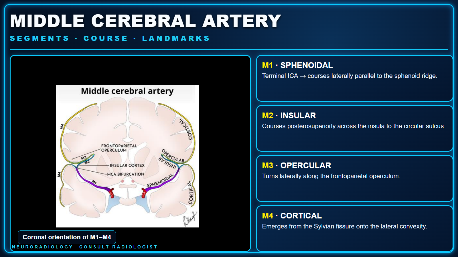
Themed teaching card showing a preserved coronal MCA diagram beside concise descriptions of the M1 through M4 segments
MCA Segments and CourseClick to enlarge
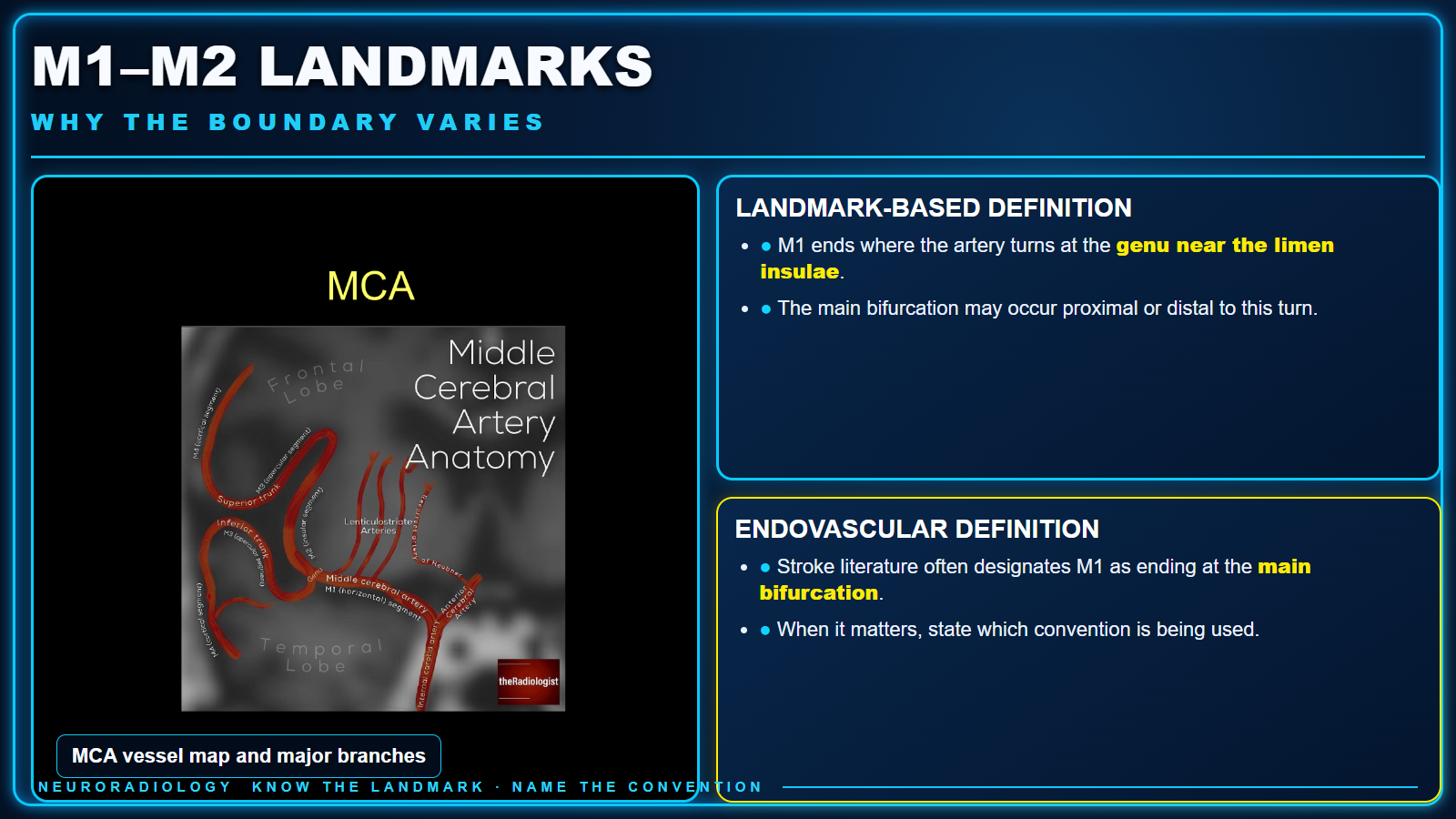
Themed teaching card comparing landmark-based and endovascular definitions of the M1 to M2 boundary
M1–M2 Landmark DefinitionsClick to enlarge
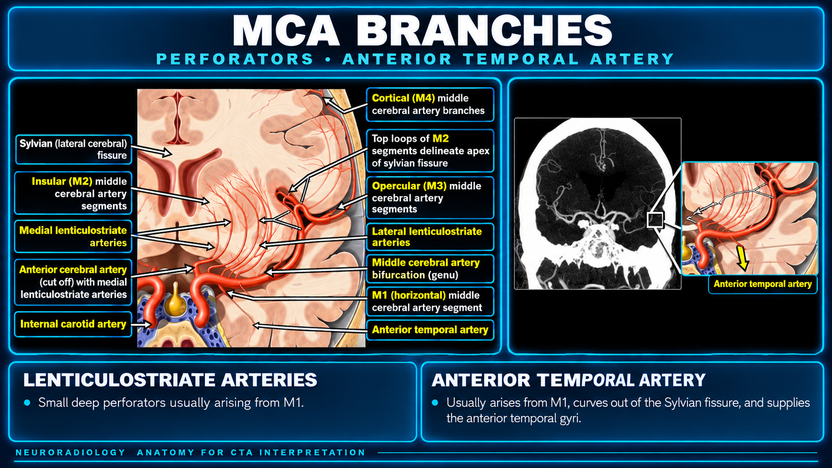
Themed teaching card preserving the labeled branch diagram and CTA example for the lenticulostriate and anterior temporal arteries
MCA Branches and CTA AnatomyClick to enlarge

MCA Segments

M1 · Sphenoidal / horizontal

  • Begins at the terminal ICA bifurcation.
  • Courses laterally, approximately parallel to the sphenoid ridge.
  • Ends at the genu beside the limen insulae or at the main bifurcation, depending on the convention used.

M2 · Insular

  • Begins at the genu/limen insulae or the main bifurcation.
  • Courses posterosuperiorly in the insular cleft.
  • Ends at the circular sulcus, where the vessel makes a right-angle to hairpin turn.

M3 · Opercular

  • Begins at the circular sulcus of the insula.
  • Courses laterally along the frontoparietal operculum.
  • Ends at the external or superior surface of the Sylvian fissure.

M4 · Cortical

  • Begins as branches exit the Sylvian fissure.
  • Courses over the lateral cerebral convexity.
  • Terminates within the final cortical territories.

Landmarks and Nomenclature

The M1–M2 boundary is not universally defined. Fischer’s landmark-based description places the transition where the artery turns at the genu near the limen insulae. Because the main bifurcation may occur proximal or distal to that turn, M1 can include the bifurcation in this system.

Endovascular stroke literature often defines M1 as ending at the main bifurcation. When the distinction matters, state which convention is being used rather than assuming a universal boundary.

M2 branches ramify across the insular surface within the Sylvian fissure. At the circular sulcus, they turn outward and become opercular M3 branches. After emerging onto the cerebral surface, they are designated cortical M4 branches.

The original seven-slide teaching file remains available as a read-only reference: Middle Cerebral Artery PDF.
Branches and Reading Pearls

Small perforators usually arise from M1 and course medially to supply deep gray nuclei and adjacent white matter. Their origin helps distinguish the proximal horizontal segment from the more distal insular branches.

The anterior temporal artery is usually an M1 branch. It curves out of the Sylvian fissure across the temporal lobe and supplies the anterior portions of the superior, middle, and inferior temporal gyri.

For an occlusion, name the side, segment, and the landmark used when anatomy is variant or the M1–M2 transition is ambiguous. A segment label alone can conceal a proximal or distal bifurcation pattern.

Text supplied by the user; diagrams preserved from the original MCA teaching PDF.

Secondary Links