Anterior temporal lobe perivascular spaces
Key Images
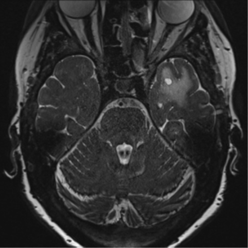
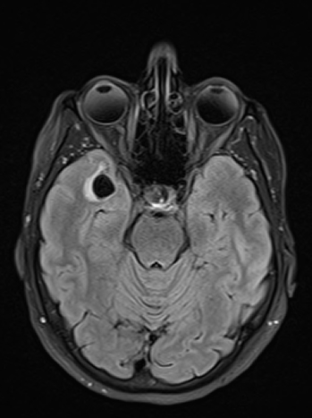
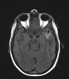
Look For First
- CSF-signal cyst in anterior temporal lobe or frontal operculum with surrounding T2/FLAIR hyperintense edema in ~80% of cases
- Vascular loop of middle cerebral artery (MCA branch) indenting the cortex directly adjacent to the cyst on MRA/high-resolution imaging
- Focal cortical thinning at the site of cyst contact
- No enhancement on post-contrast T1-weighted imaging despite presence of edema
Key Image Findings
- Internal CSF signal intensity on all sequences (hypointense T1, hyperintense T2, suppressed on FLAIR) with facilitated diffusion on ADC identical to cerebrospinal fluid
- Surrounding edema/gliosis appears as hypointense signal on T1 and hyperintense signal on T2/FLAIR that does not suppress on FLAIR, distinguishing it from pure CSF
- Associated hypointense diffusion (reduced signal on DWI) and facilitated diffusion in the surrounding edema/gliosis zone
- MCA branch or vascular loop ('knuckle' appearance) directly indenting the adjacent cortex, visible on high-resolution T2 sequences and confirmed on MRA
- Focal region of cortical thinning at the site of vascular contact and cyst indentation (~90% of cases have MCA contact)
- No enhancement on post-contrast T1-weighted imaging, which is the key feature distinguishing these from enhancing tumors
- No susceptibility artifact, blooming, or calcification on SWI/GRE sequences
- Cyst size remains stable over time; surrounding edema may fluctuate in conspicuity in a small percentage of cases but size invariably stable
Differential Diagnosis
- Cystic tumor with surrounding edema: distinguished by presence of enhancement on post-contrast imaging, unstable cyst size, and absence of directly contacting vascular loop indenting cortex
- Other perivascular spaces (Virchow-Robin spaces): anterior temporal lobe variants differ by frequently having surrounding edema (~80%), whereas typical perivascular spaces elsewhere rarely have significant edema
- Arachnoid cyst: lacks the characteristic MCA vascular loop indent, cortical thinning, and surrounding edema that define anterior temporal lobe perivascular spaces
- Porencephalic cyst: distinguished by larger size, different location (not specifically in anterior temporal lobe), and lack of associated vasculature
- Intracystic hemorrhage or tumor: SWI/GRE shows no susceptibility artifact or blood products to suggest hemorrhage or necrotic tumor
- IDH wild-type minimally hypercellular astrocytoma on histology: distinguished by normal brain cell composition (IDH wild-type reactive astrocytes) rather than true neoplastic cells
Discussion
The etiology of anterior temporal lobe perivascular spaces remains uncertain but is likely related to vascular compression of small communicating channels by a looped MCA branch, resulting in focal CSF accumulation and abnormal local CSF dynamics.
Unlike typical perivascular (Virchow-Robin) spaces elsewhere in the brain, anterior temporal lobe variants frequently have associated T2/FLAIR hyperintense edema (~80% of cases), which ranges from mild to extensive and can evolve over time.
The presence of surrounding edema can create a concerning appearance mimicking a cystic tumor with surrounding vasogenic edema, leading to unnecessary workup, biopsy, and potential over-treatment if the entity is not recognized.
If histology is obtained, reactive astrocytosis surrounding the cyst (normal brain response to the lesion) can be misinterpreted as a minimally hypercellular astrocytic tumor; IDH wild-type status confirms these are normal cells, not neoplastic.
Recognition of this entity is critical to avoid unnecessary surgical intervention, additional treatment, and patient morbidity through informed conservative follow-up imaging.
High-resolution T2-weighted sequences, post-contrast T1-weighted imaging, and MRA are essential for diagnosis and demonstrating the key distinguishing features (vascular loop, no enhancement, cortical thinning).
Reporting Pearls
Describe as: "CSF-signal lesion in [anterior temporal lobe/frontal operculum] with associated T2/FLAIR hyperintense edema and focal cortical thinning at the site of adjacent MCA loop indentation; no enhancement; imaging features consistent with opercular perivascular space rather than neoplasm. Recommend follow-up to confirm stability."
Pitfalls
- Misdiagnosing as a cystic tumor or mass due to the presence of surrounding edema, which is atypical for most perivascular spaces and can appear concerning on initial review.
- Failing to identify and report the adjacent MCA vascular loop on MRA and high-resolution imaging, which is the key distinguishing feature that confirms the diagnosis.
- Pursuing unnecessary biopsy or surgery based on imaging concern, which then yields histology showing only reactive astrocytosis; subsequent misinterpretation of normal reactive cells as IDH wild-type glioblastoma can lead to over-treatment.
- Failing to recognize that the cyst size is invariably stable whereas tumor size would change; stable imaging over follow-up imaging is reassuring and supports benign diagnosis.