Wilson disease
Key Images
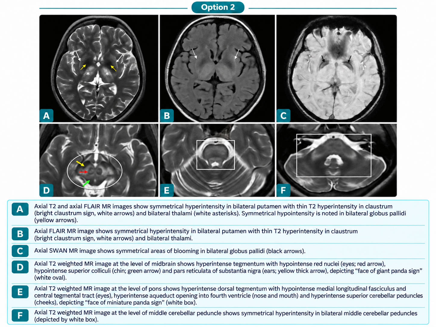
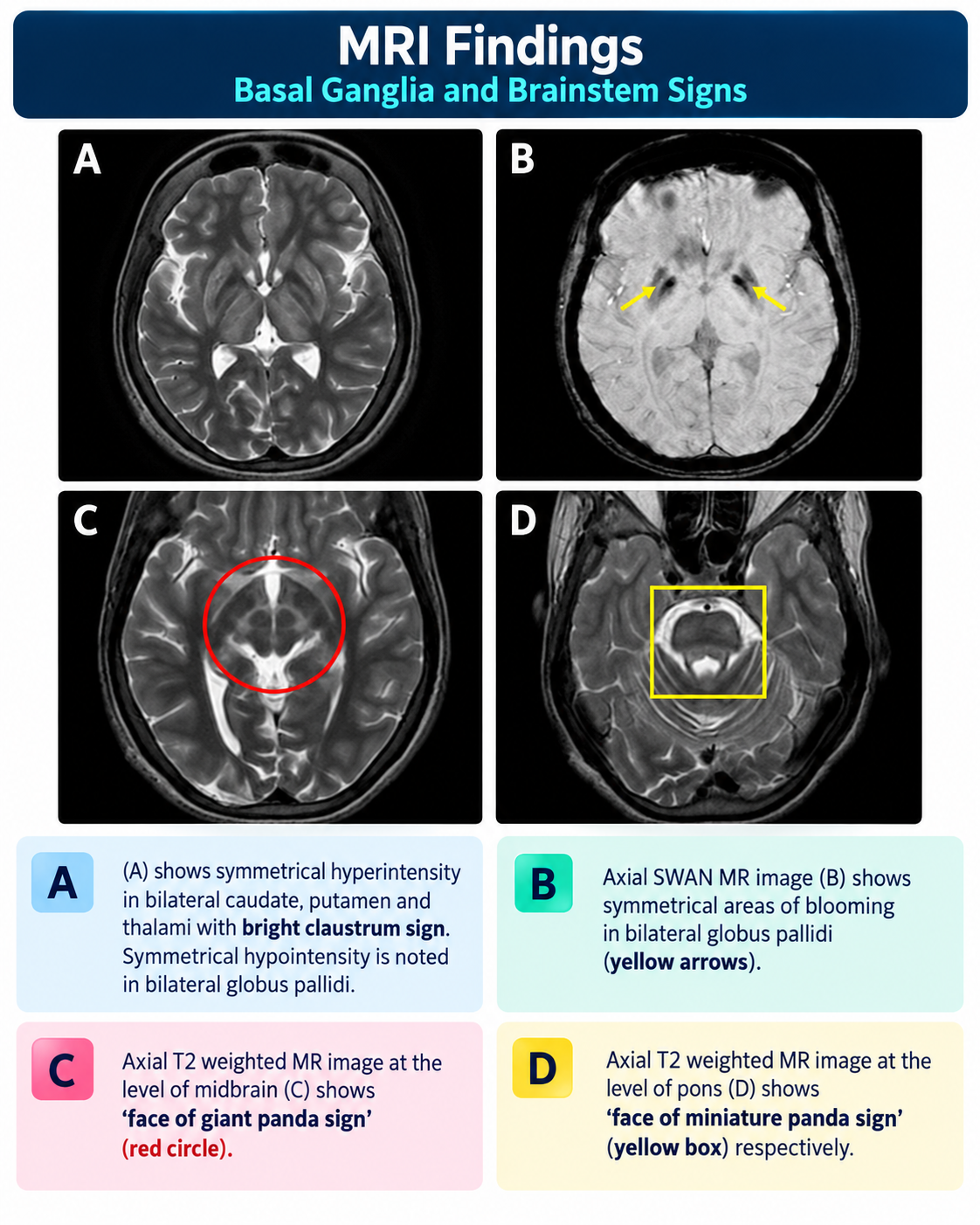
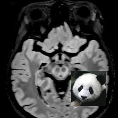
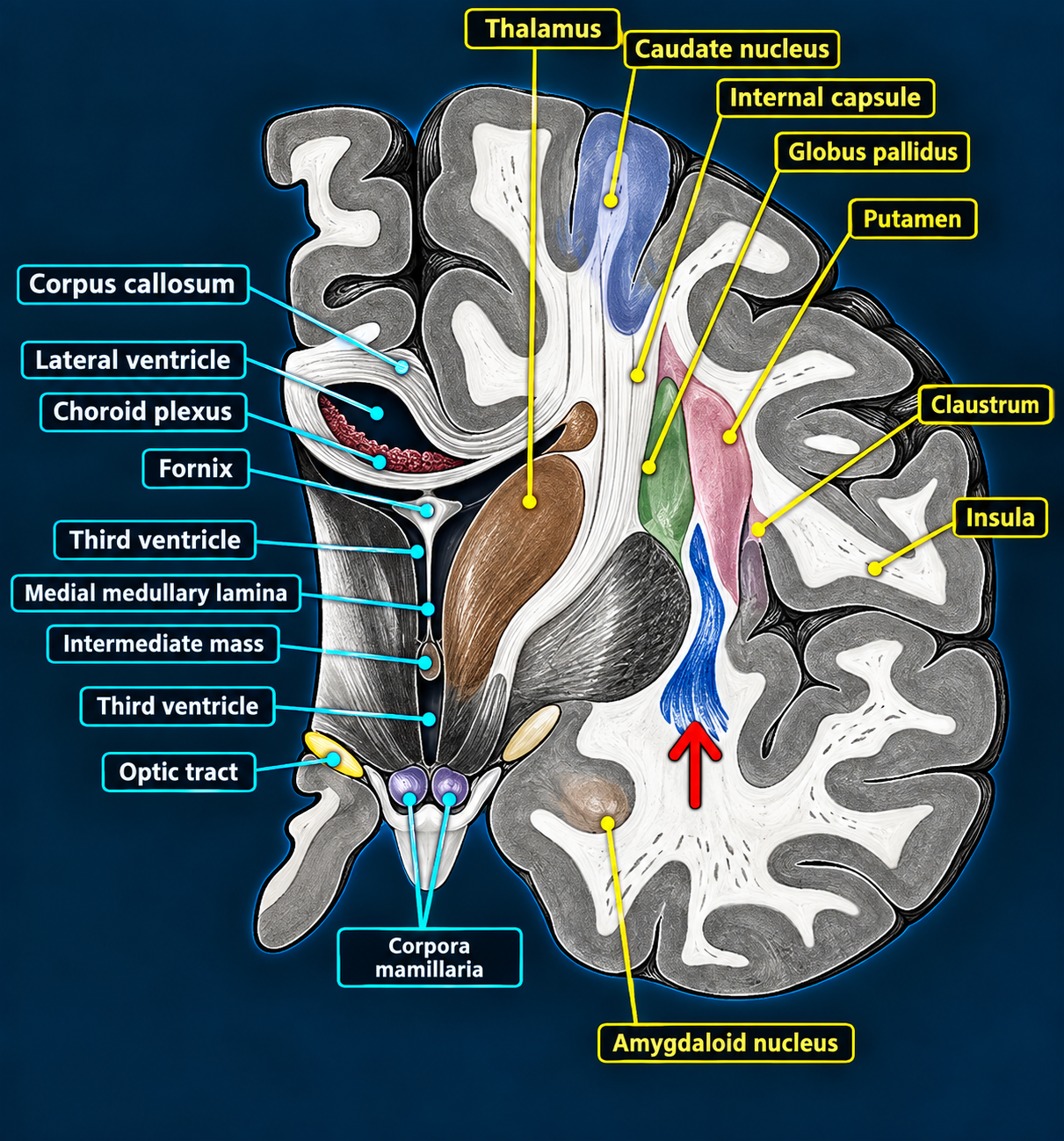
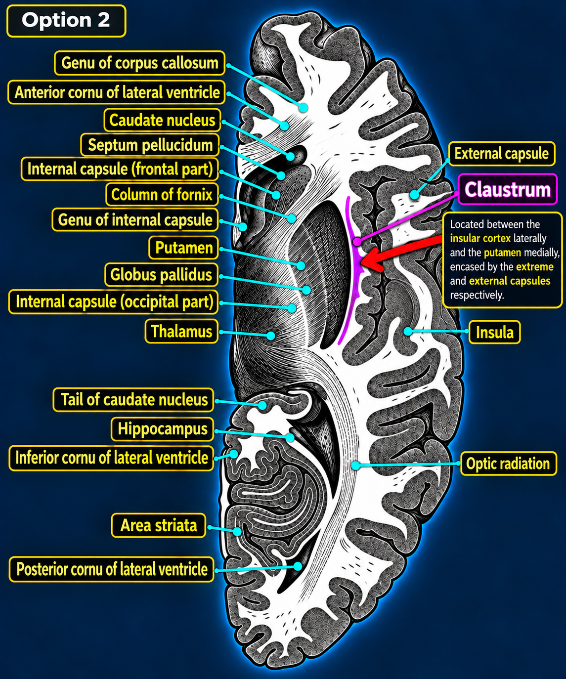
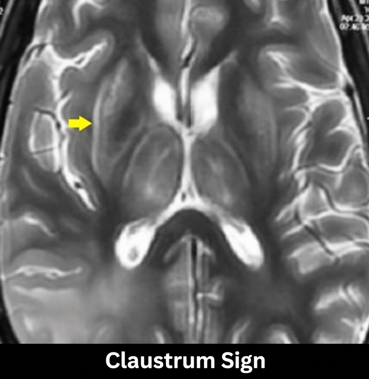
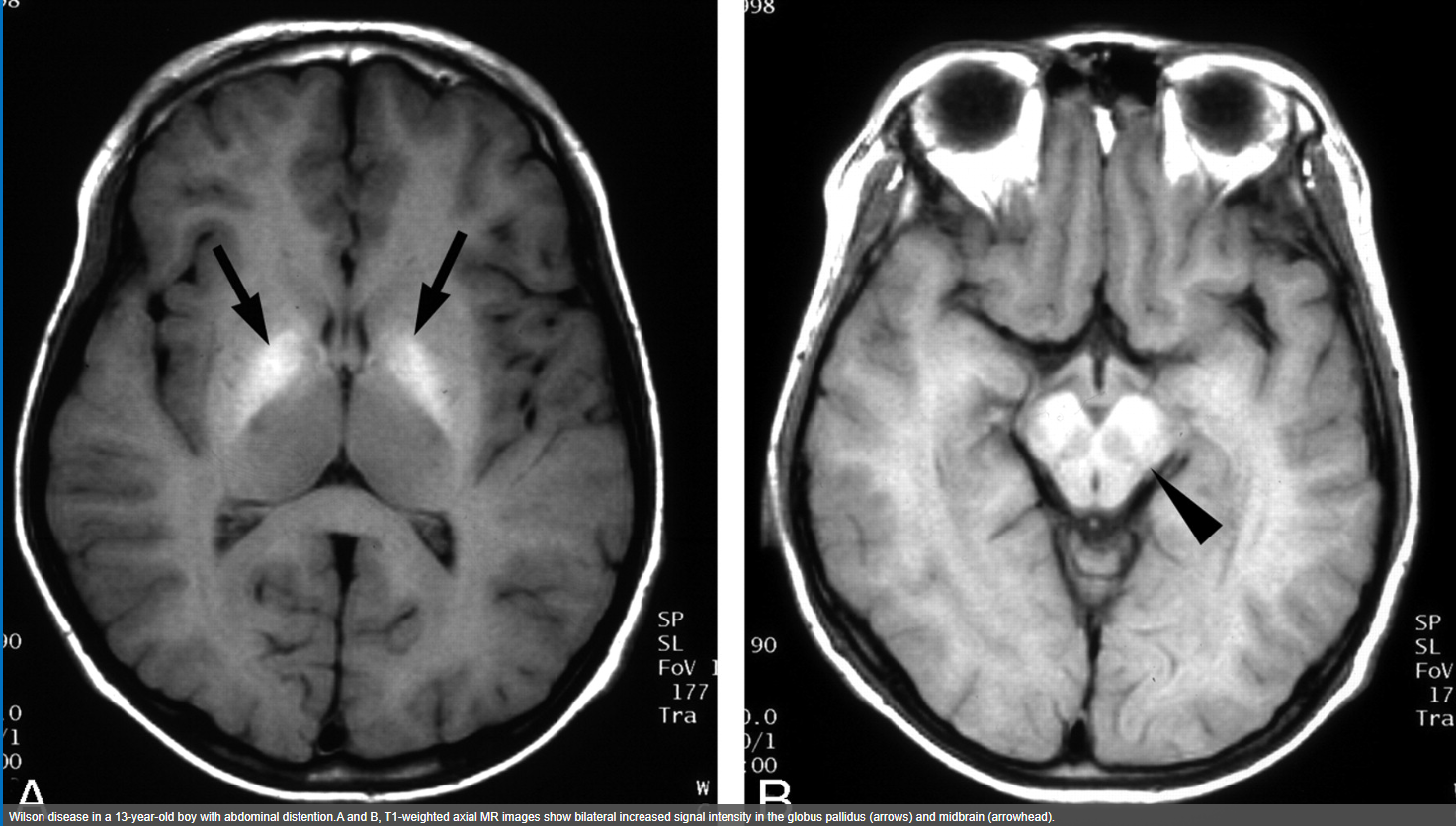
Look For First
- Basal ganglia signal abnormalities on T2/FLAIR MRI indicating free copper deposition and oxidative damage in the CNS
- Cirrhosis and portal hypertension on liver imaging reflecting progressive hepatic copper accumulation
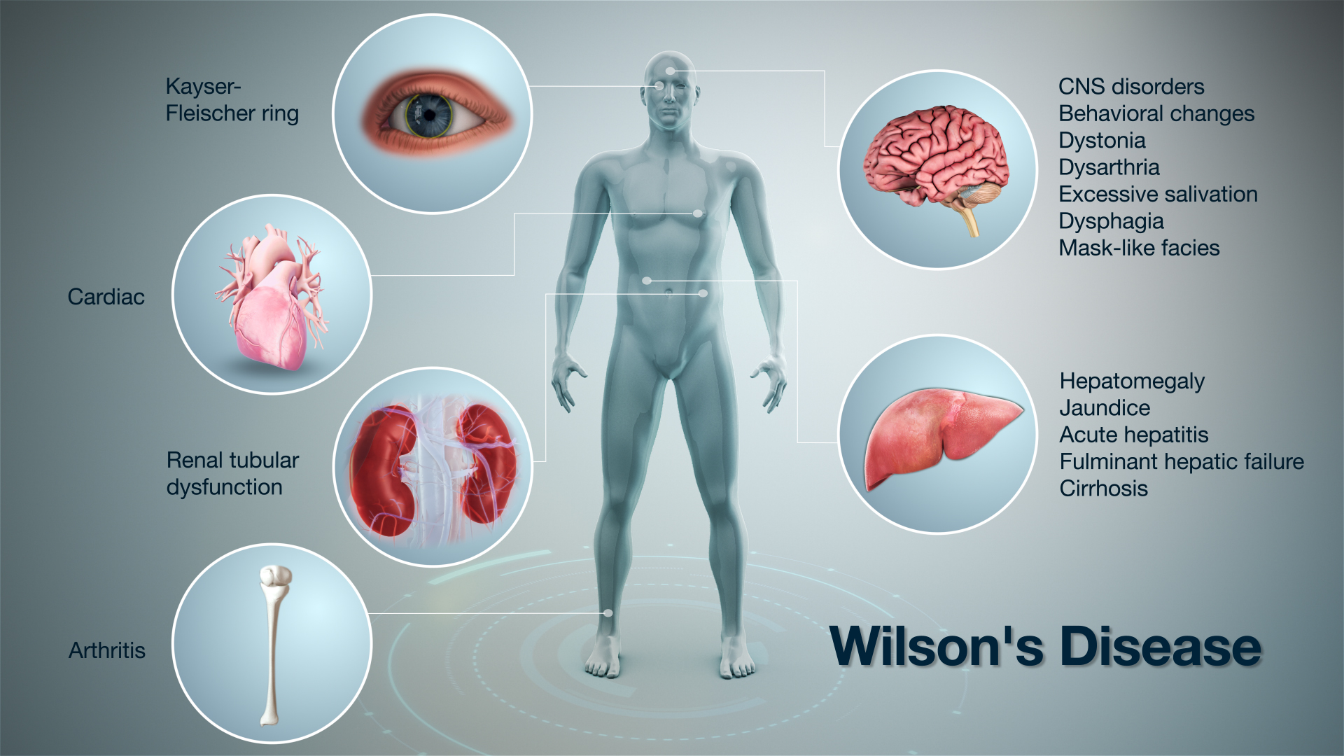
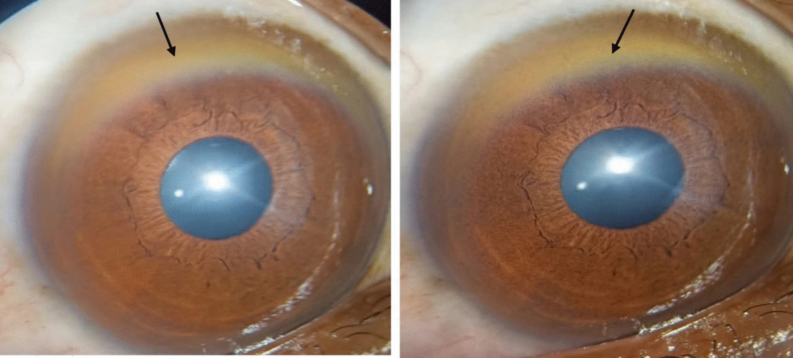
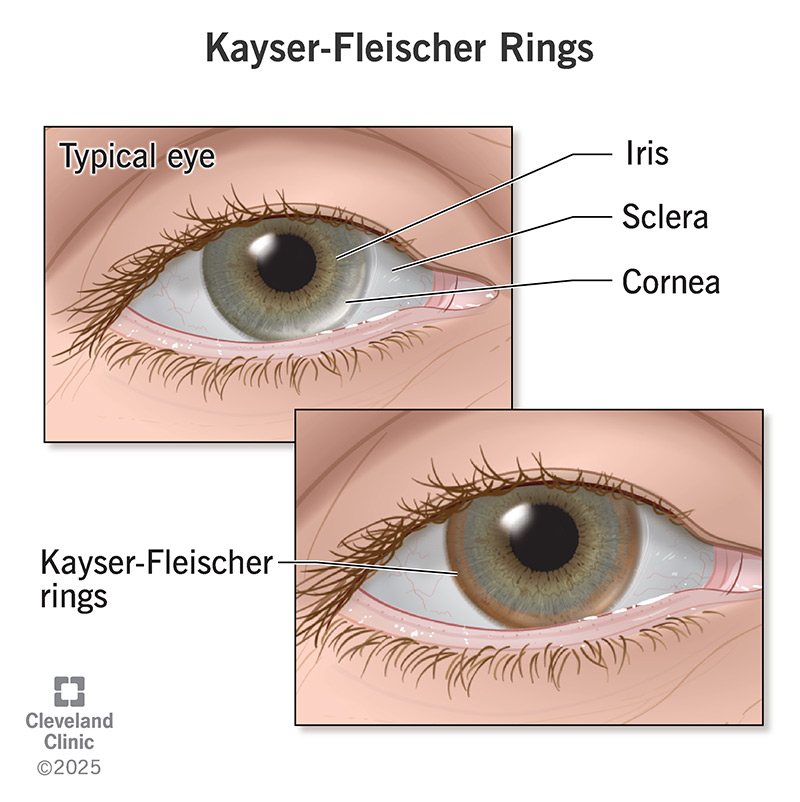
- Kayser-Fleischer rings on slit lamp examination (corneal copper deposition in Descemet membrane)
- Movement disorder signs on neuroimaging correlating with dystonia, tremor, and parkinsonism
Key Image Findings
- Hepatic copper accumulation occurs first, with progressive cirrhosis development and risk of hepatocellular carcinoma due to oxidative damage from free radical generation
- CNS manifestations result from free copper deposition primarily affecting basal ganglia (putamen and globus pallidus), visible as hyperintense T2/FLAIR signal representing edema and neuronal damage
- Kayser-Fleischer rings are pathognomonic when present, appearing as brownish-green discoloration in the Descemet membrane at the periphery of the cornea, characteristic of copper deposition
- Sunflower cataracts are rare but pathognomonic findings showing a characteristic petal-like pattern of lens opacification due to copper deposition in the lens
- Basal ganglia signal changes may be reversible in early stages with treatment but can become irreversible with chronic copper deposition and neuronal loss
- Associated musculoskeletal findings include premature osteoarthritis, reflecting copper-related bone and cartilage damage
- Portal hypertension develops secondary to hepatic cirrhosis, visible on imaging as splenomegaly, ascites, and varices
- Brain atrophy may develop in severe or untreated cases, representing chronic neurodegeneration from persistent oxidative damage
Differential Diagnosis
- Hepatic cirrhosis from alcohol or viral hepatitis—distinguished by history, serologies, and absence of basal ganglia involvement or Kayser-Fleischer rings
- Other genetic movement disorders (Huntington, Parkinson disease)—distinguished by normal ceruloplasmin, absence of liver disease and copper markers
- Hepatic encephalopathy—distinguished by preserved basal ganglia signal on MRI and reversibility with hepatic support
- Mitochondrial disorders (MELAS, Leigh syndrome)—distinguished by different locations of signal abnormalities, different inheritance pattern, and negative copper workup
- Toxic/metabolic encephalopathy—distinguished by normal liver function tests, negative copper studies, and different distribution of CNS abnormalities
- Demyelinating disease (MS)—distinguished by absence of liver disease, negative copper studies, and different distribution of white matter changes
Discussion
Wilson disease is an autosomal recessive disorder affecting approximately 1 in 30,000 individuals, making it rare but crucial to diagnose early as treatment can prevent progression and even reverse some neurologic manifestations
The ATP7B gene defect prevents both biliary copper excretion (95% of normal excretion) and coupling of copper to ceruloplasmin, resulting in toxic-free copper accumulation in liver and brain
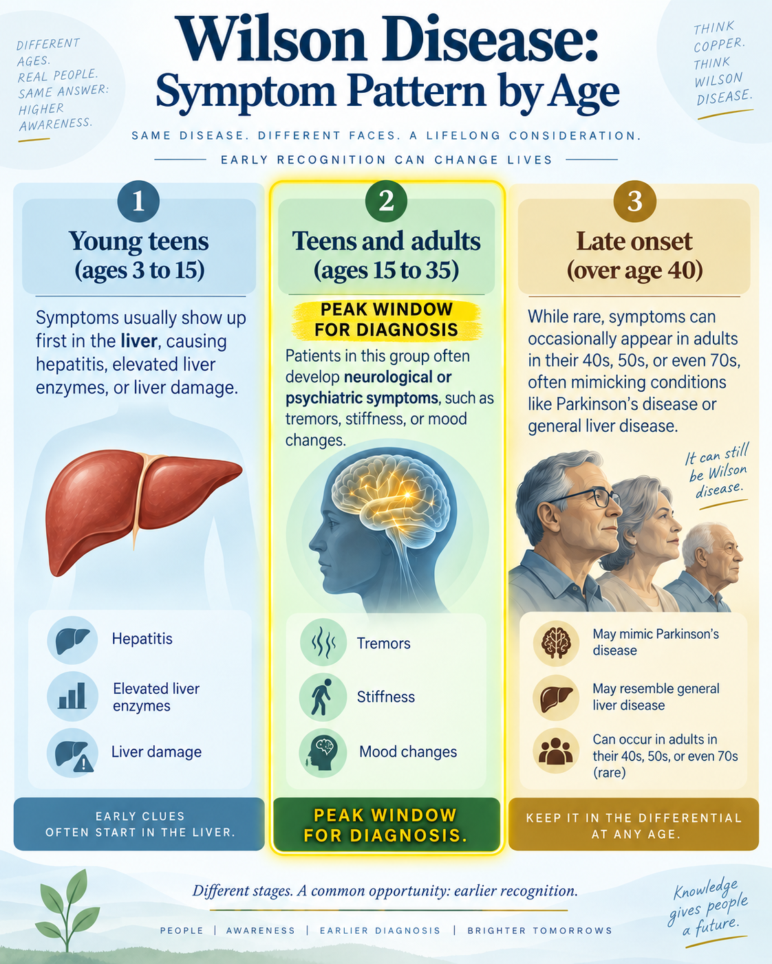
Hepatic manifestations precede neuropsychiatric symptoms, but neuropsychiatric disease may be the first recognized presentation in young adults, particularly affecting basal ganglia function
Free copper generates oxidative damage through free radical generation, affecting mitochondria, nuclei, and peroxisomes, explaining the multi-organ involvement and neurologic manifestations
Diagnosis combines clinical features with laboratory markers (low ceruloplasmin <20 mg/dL, elevated 24-hour urinary copper >100 mcg/dL) and imaging findings, with liver biopsy showing elevated copper >250 mcg/gm as gold standard
Treatment with zinc supplementation and chelating agents (trientine preferred over D-penicillamine) can halt disease progression, but chelation may temporarily worsen neuropsychiatric symptoms, requiring close clinical monitoring
Reporting Pearls
When reporting basal ganglia abnormalities in a young patient presenting with movement disorder, specifically document the signal intensity (T2 hyperintensity) and location (putamen/globus pallidus) while correlating with serum ceruloplasmin and urinary copper levels—describe as "signal abnormalities in the basal ganglia consistent with copper deposition" rather than assuming primary neurodegeneration, as early recognition enables copper-specific treatment before irreversible damage occurs.
Pitfalls
- Missing the diagnosis by attributing cirrhosis solely to alcohol or viral hepatitis without checking ceruloplasmin levels—Wilson disease must be excluded in any young patient with unexplained liver disease
- Confusing hepatic encephalopathy with Wilson disease neuropsychiatric manifestations; hepatic encephalopathy shows no specific basal ganglia involvement on MRI and is not caused by copper deposition
- Overlooking early-stage disease because Kayser-Fleischer rings are absent in some patients, particularly those presenting with pure hepatic disease before CNS involvement
- Assuming basal ganglia signal abnormalities are irreversible; early copper chelation therapy can potentially reverse imaging findings and clinical symptoms if initiated before advanced neurodegeneration