Parkinson disease
Key Images
Look For First
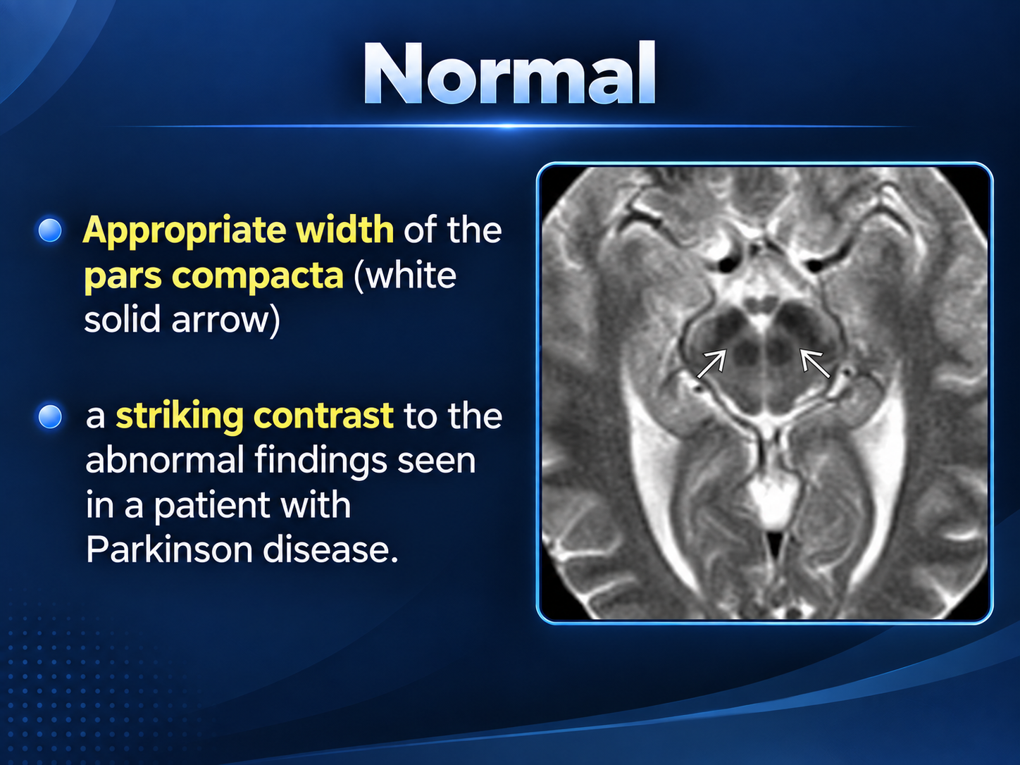
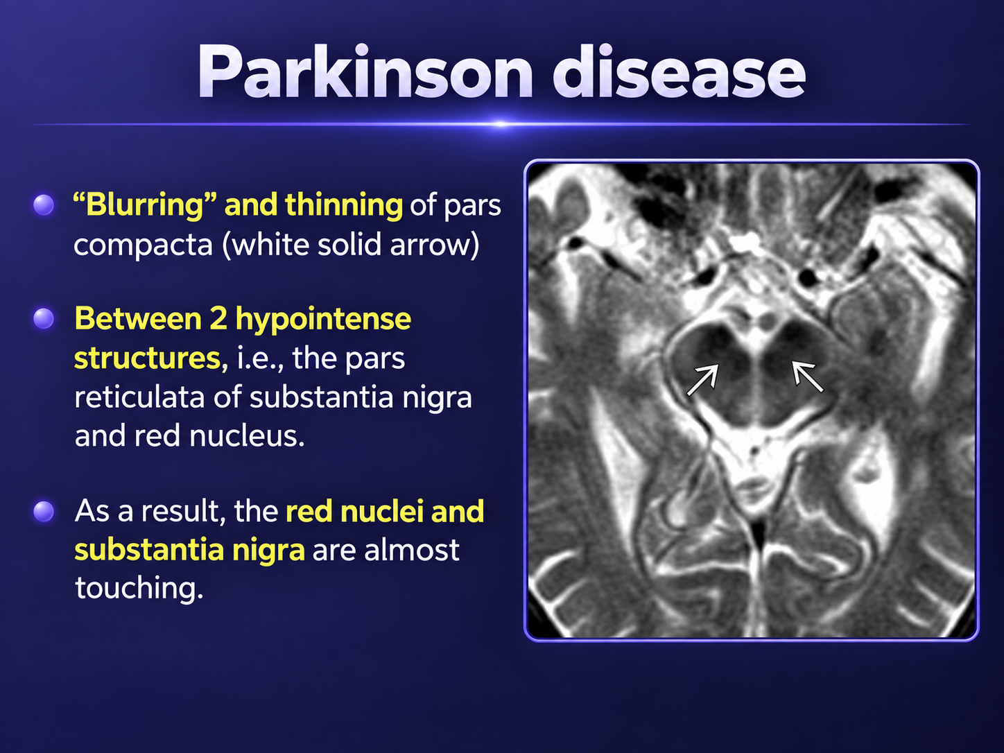
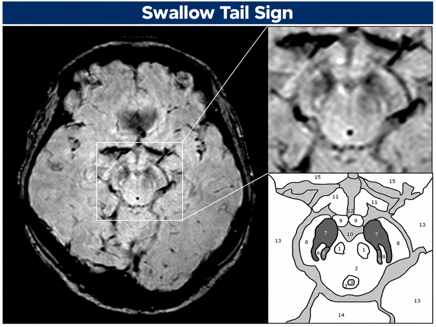
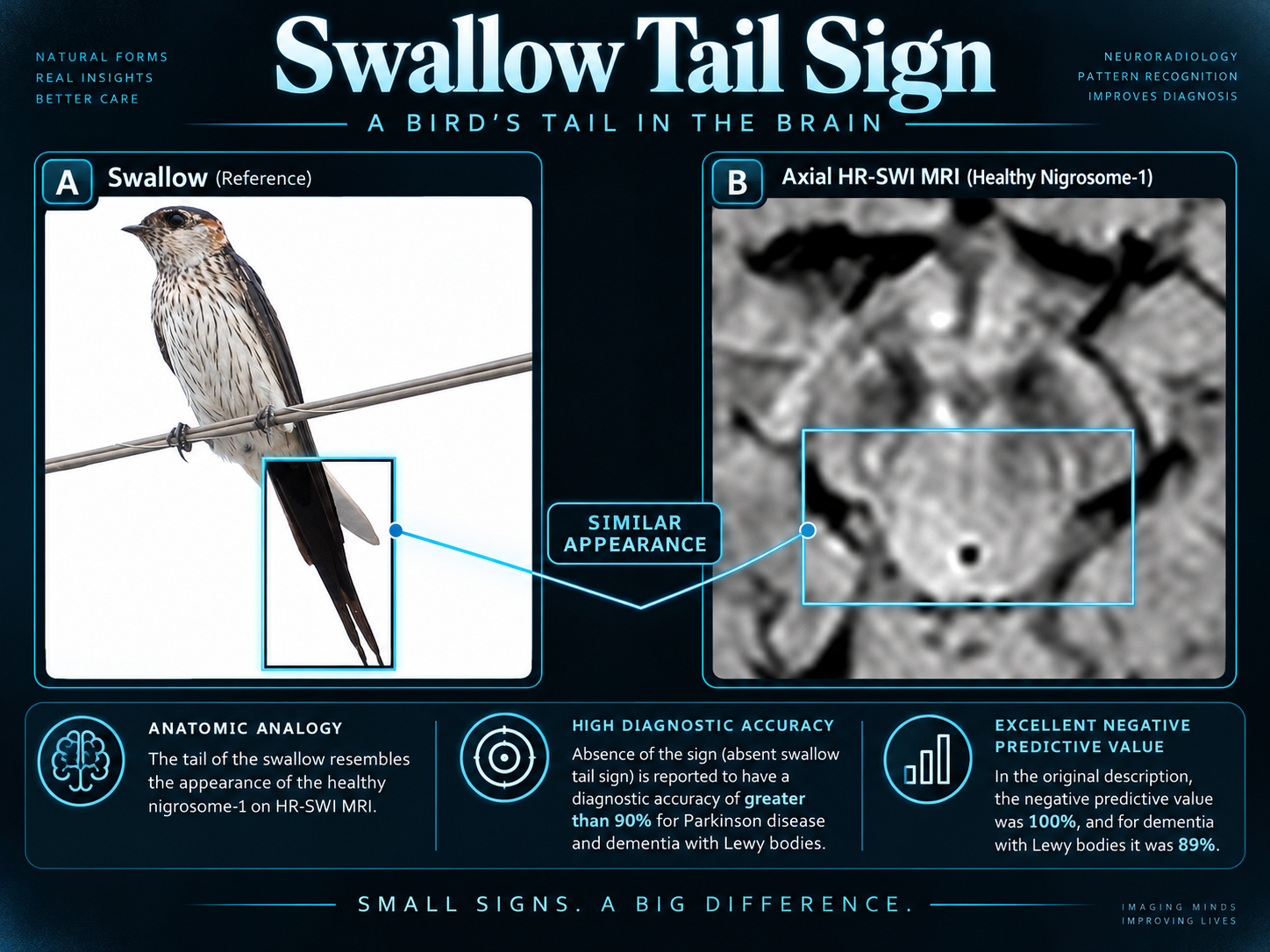
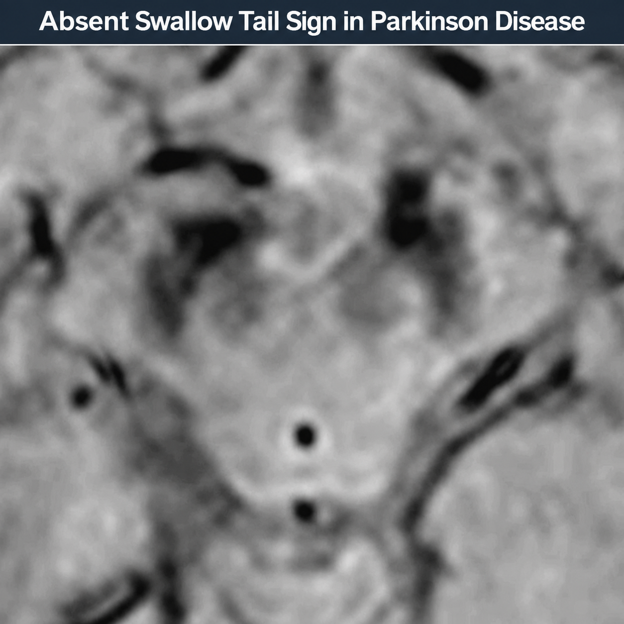
- Loss of the normal swallow tail sign on axial T2*/SWI MRI of the substantia nigra
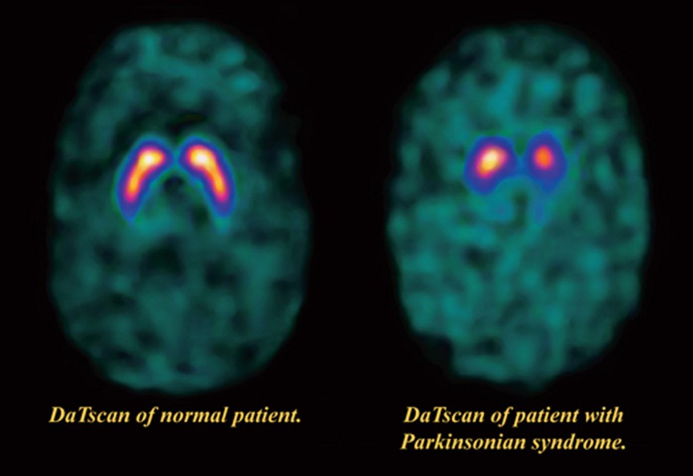
- Absence of normal comma-shaped or crescent-shaped tracer uptake in the putamen on DaTscan with period-shaped or oval-shaped uptake limited to the caudate nucleus
- Increased echogenicity size of the substantia nigra on transcranial B-mode ultrasound
- Mild hyperintensity of the substantia nigra and red nuclei on T1 MRI due to iron accumulation
Key Image Findings
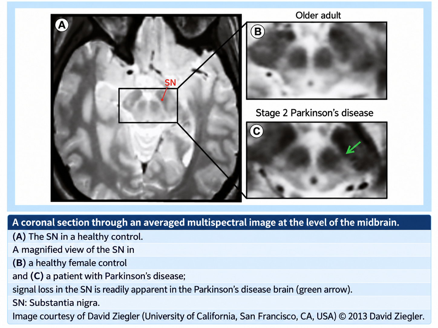
- On axial T2*/GRE/SWI MRI, loss of the normal swallow tail appearance of susceptibility signal in the substantia nigra is the most promising diagnostic sign, with reported diagnostic accuracy exceeding 90% including 100% sensitivity and negative predictive value.
- T1 MRI may show mild hyperintensity of the substantia nigra compact and reticular parts and red nuclei due to iron accumulation, and loss of normal slight hyperintensity in the substantia nigra due to loss of neuromelanin.
- T2*/GRE/SWI MRI demonstrates dot-like areas of hyperintensity in the compact part of the substantia nigra and may show confluence of normally hypointense regions due to iron accumulation.
- I-123 ioflupane SPECT (DaTscan) shows loss of normal comma-shaped or crescent-shaped tracer uptake in the striatum, replaced by period-shaped or oval-shaped uptake limited to the caudate nucleus head without putaminal uptake, progressing to eventual reduction in caudate uptake.
- Quantitative assessment of I-123 ioflupane SPECT reveals significantly reduced uptake in the putamen compared to normative values, reflecting loss of presynaptic dopaminergic terminals.
- B-mode transcranial sonography demonstrates increased echogenicity size of the substantia nigra, though sensitivity varies depending on the area cutoff values used and ultrasound equipment.
- Advanced disease may show non-specific generalized minor cerebral volume loss on conventional MRI.
- MRI also identifies secondary causes of parkinsonism such as extensive small vessel ischemic change, helping exclude primary versus secondary parkinsonism.
Differential Diagnosis
- Dementia with Lewy bodies: distinguished by cognitive impairment preceding or occurring within 12 months of parkinsonian symptom onset, whereas primary Parkinson disease typically shows bradyphrenia and dementia as late features.
- Multiple system atrophy (MSA): may show different striatal uptake patterns on DaTscan and distinct brainstem/cerebellar atrophy features not typical of primary Parkinson disease.
- Progressive supranuclear palsy (PSP): demonstrates vertical gaze palsy and distinctive midbrain atrophy on MRI, distinguishing it from Parkinson disease.
- Corticobasal degeneration: shows prominent cortical and white matter atrophy with asymmetric changes not typical of Parkinson disease.
- Vascular parkinsonism (cerebrovascular disease): identified by extensive small vessel ischemic changes and vascular lesions in the basal ganglia and brainstem on MRI.
- Metabolic diseases with parkinsonian signs (Wilson disease, manganism, chronic hepatitis): characterized by more pronounced basal ganglia signal abnormalities on conventional imaging.
Discussion
Parkinson disease accounts for approximately 80% of all parkinsonism cases and is encountered in 2-4% of individuals older than 65 years, making it the most common neurodegenerative cause of movement disorder.
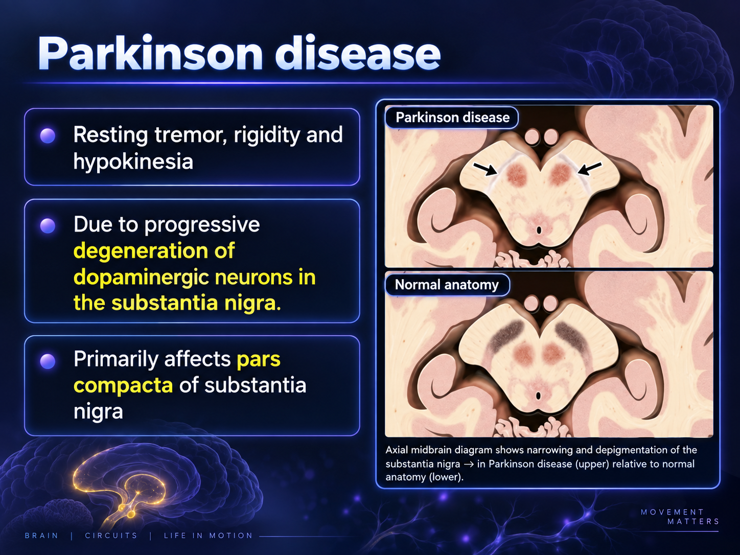
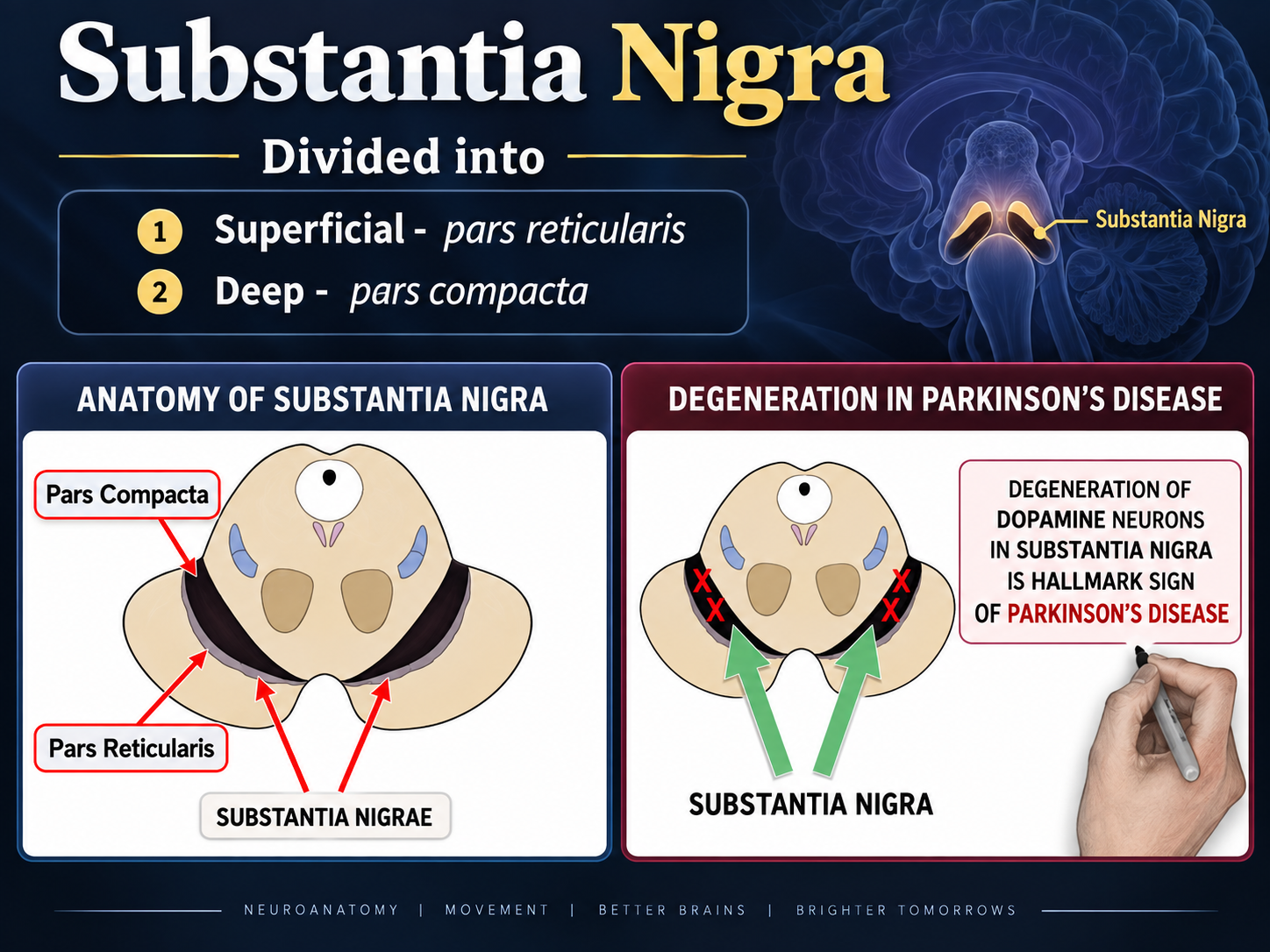
The pathology is characterized by selective nigrostriatal dopaminergic degeneration, most conspicuous in the ventrolateral tier neurons of the substantia nigra pars compacta, reflecting the loss of neuromelanin and iron accumulation.
While 85-90% of cases are sporadic, 10-15% have positive family history with rare familial forms caused by genetic mutations such as LRRK2, and juvenile-onset forms may occur without Lewy bodies when PARK2 mutations impair Parkin function.
Non-motor features such as constipation, anosmia, REM sleep behavior disorder, and anxiety often precede cardinal motor features, making them important early diagnostic clues.
The swallow tail sign on T2*/SWI MRI is the most promising imaging biomarker for diagnosis, with over 90% diagnostic accuracy, making it particularly useful for confirming suspected disease and ruling out secondary parkinsonism.
DaTscan provides high sensitivity for assessment of presynaptic dopaminergic deficits and aids differentiation between primary Parkinson disease and atypical parkinsonian syndromes based on characteristic striatal uptake patterns.
Reporting Pearls
When reporting suspected Parkinson disease on MRI, specifically comment on the presence or absence of the swallow tail sign on axial T2*/SWI sequences, noting that loss of this sign strongly supports the diagnosis; additionally describe the iron content appearance and signal intensity changes in the substantia nigra to avoid confusion with secondary causes of parkinsonism.
Pitfalls
- Failing to specifically evaluate for the swallow tail sign on T2*/SWI sequences, which is the most sensitive and specific MRI finding for Parkinson disease diagnosis.
- Over-interpreting generalized minor cerebral volume loss as diagnostic, since this finding is non-specific and can occur in other neurodegenerative conditions and normal aging.
- Misidentifying increased substantia nigra echogenicity on transcranial ultrasound as pathognomonic, since sensitivity varies considerably based on technical factors and cutoff values used.
- Confusing the stromal signal changes in the substantia nigra on T1 MRI (due to iron and neuromelanin loss) with pathologic findings indicating secondary parkinsonism, when specific imaging of secondary causes (small vessel disease, atrophy patterns) is needed to distinguish primary from secondary disease.