Opioid-associated amnestic syndrome
Key Images
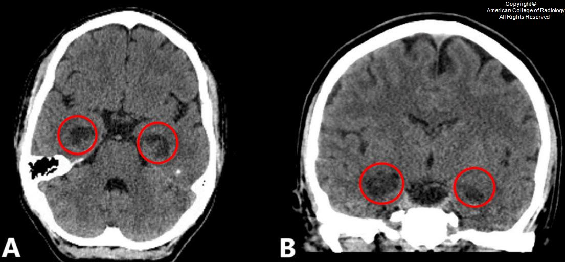
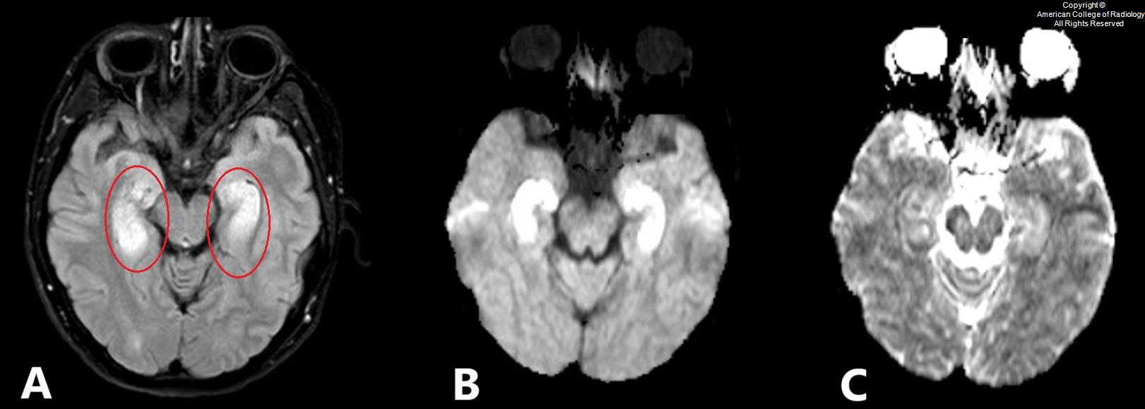
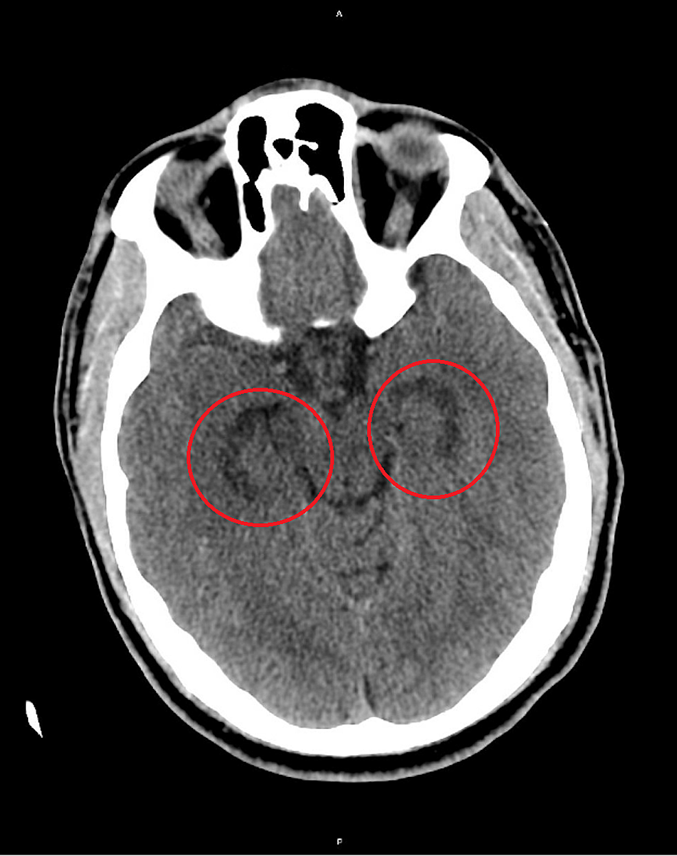
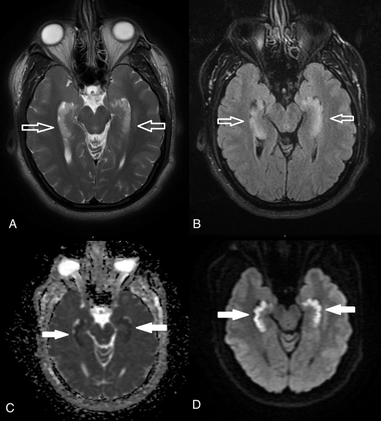
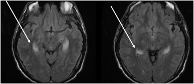
Look For First
- Bilateral hippocampal hypoattenuation on CT or diffusion restriction on MRI
- Anterograde amnesia with new-onset memory impairment lasting >24 hours
- Symmetric involvement without unilateral predominance (distinguishes from TGA and seizures)
- Positive opioid history with otherwise negative workup for other encephalopathies
Key Image Findings
- Bilateral hippocampal hypoattenuation is the hallmark finding on unenhanced CT, representing acute injury from opioid toxicity.
- MRI shows bilateral hippocampal diffusion restriction (high signal on DWI, low ADC) indicating cytotoxic edema with acute injury.
- T2/FLAIR hyperintensity in bilateral hippocampi and may extend to other temporal lobe structures, reflecting edema and inflammation.
- Vascular imaging (CTA/MRA) is typically unremarkable, helping exclude acute stroke or vasculitis as alternative diagnoses.
- Concomitant cortical and deep nuclear structures involvement can occur, including basal ganglia, thalami, and cerebral cortex in more severe cases.
- The pattern of injury is bilateral and symmetric, distinguishing OAS from unilateral processes.
- No enhancement pattern is described, helping differentiate from infectious encephalitis or autoimmune limbic encephalitis.
- Associated cerebral edema and swelling are typically milder than in CHANTER syndrome, without hydrocephalus.
Differential Diagnosis
- Hypoxic-ischemic injury (HII): presents with similar history of time down but involves gray matter structures including basal ganglia, thalami, and diffuse cortical involvement; distinguished by clinical context (cardiac arrest, drowning) and pattern of injury.
- CHANTER syndrome: severe end of OAS spectrum with bilateral cerebellar hypoattenuation and diffusion restriction, swelling, and obstructive hydrocephalus—the cerebellar predominance and hydrocephalus distinguish it from OAS.
- Transient global amnesia (TGA): presents with anterograde amnesia but shows small unilateral CA1 hippocampal diffusion restriction, whereas OAS is bilateral and typically more extensive.
- Status epilepticus: can mimic OAS clinically but typically presents with unilateral hippocampal involvement rather than bilateral symmetric changes.
- Autoimmune limbic encephalitis (paraneoplastic and non-paraneoplastic): has unilateral or bilateral hippocampal involvement but usually with enhancement and positive autoimmune workup, whereas OAS lacks enhancement.
- Toxic and metabolic encephalopathy: may have unilateral or bilateral hippocampal involvement but clinical context and laboratory abnormalities (electrolytes, liver function) distinguish from OAS.
Discussion
OAS amnesia is characteristically anterograde (inability to form new memories) with a milder retrograde component, often accompanied by confusion, inability to focus, and poor executive dysfunction.
Diagnosis is challenging because multiple entities mimic OAS clinically and radiographically, making comprehensive history, laboratory workup, and imaging essential for exclusion of alternative diagnoses.
Routine urine drug screening (UDS) has a critical limitation: it does not detect synthetic opioids such as fentanyl and its analogs, so negative UDS does not exclude opioid use as the causative agent.
OAS has been reported in patients with concurrent benzodiazepine use and negative UDS, indicating polysubstance abuse may be involved and routine screening is insufficient.
Currently, no formal diagnostic criteria exist; Barash et al. proposed provisional criteria: positive opioid toxicology, bilateral hippocampal injury on imaging, and new-onset amnesia >24 hours, categorized as confirmed, probable, or possible.
Recovery varies widely from complete resolution within weeks to months, although chronic cognitive deficits can persist; patients often require physical rehabilitation, cognitive therapy, and social support for addiction and long-term impairment.
Reporting Pearls
Describe bilateral hippocampal involvement specifically: "Bilateral symmetric hippocampal diffusion restriction without associated hydrocephalus or enhancement, consistent with opioid-associated amnestic syndrome in the appropriate clinical context. Vascular imaging is unremarkable, and there is no evidence of status epilepticus, encephalitis, or other alternative causes."
Pitfalls
- Mistaking unilateral hippocampal involvement for OAS: TGA and seizures typically present with unilateral changes, whereas OAS is bilateral and symmetric.
- Over-relying on negative UDS to exclude opioid toxicity: synthetic opioids and benzodiazepines are not detected on routine screens, so clinical history and imaging context are essential.
- Confusing OAS with CHANTER syndrome: CHANTER has preferential cerebellar involvement with obstructive hydrocephalus, whereas OAS primarily affects the hippocampus without hydrocephalus.
- Attributing acute hippocampal changes to ischemia without assessing vascular patency: OAS has unremarkable vascular imaging, whereas acute stroke shows arterial occlusion or perfusion deficit.