Multiple system atrophy
Key Images
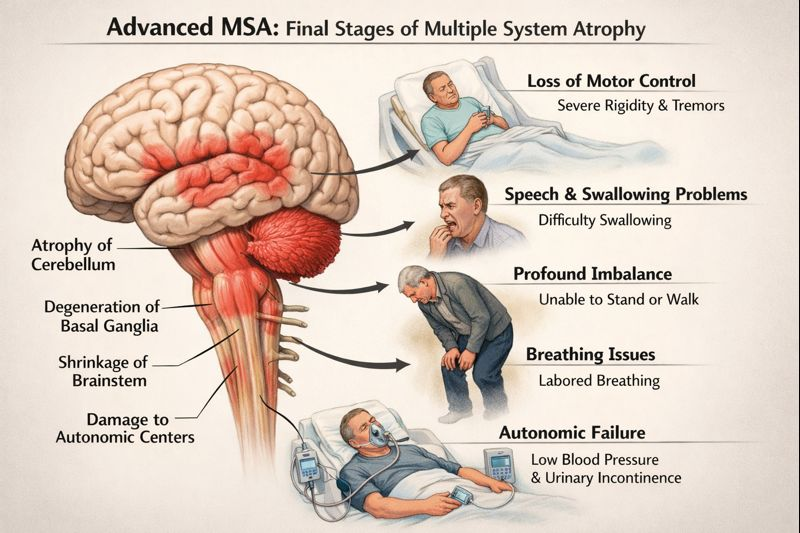
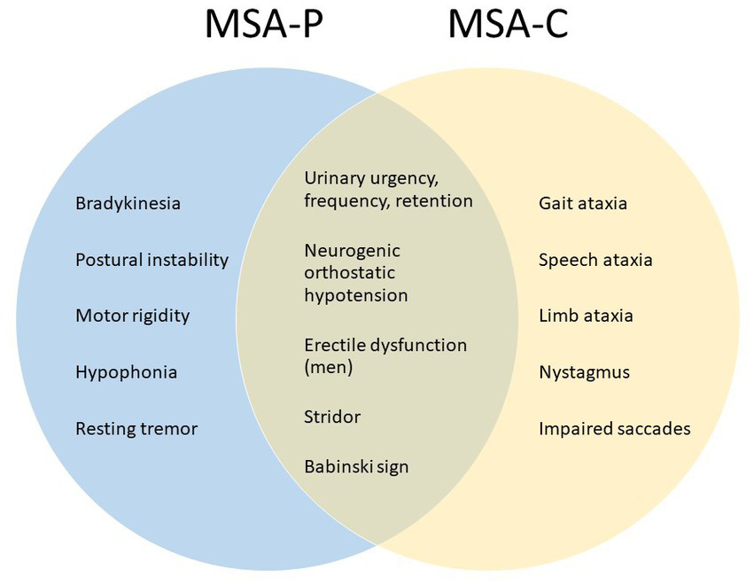
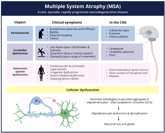
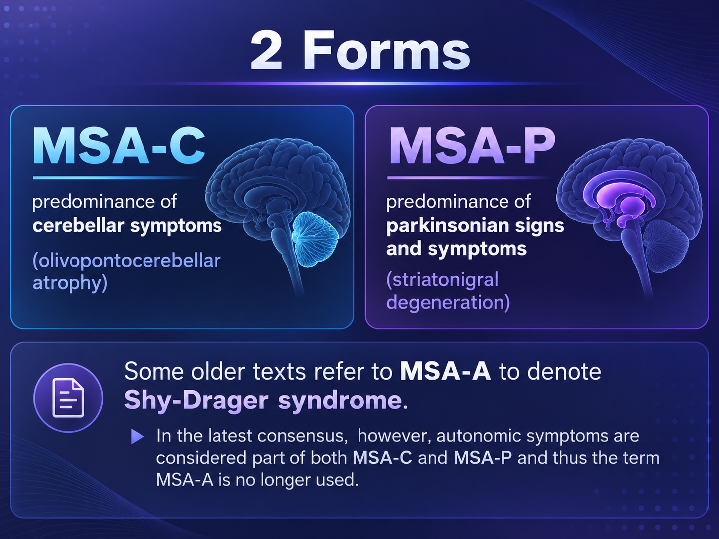
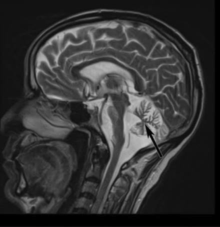
Look For First
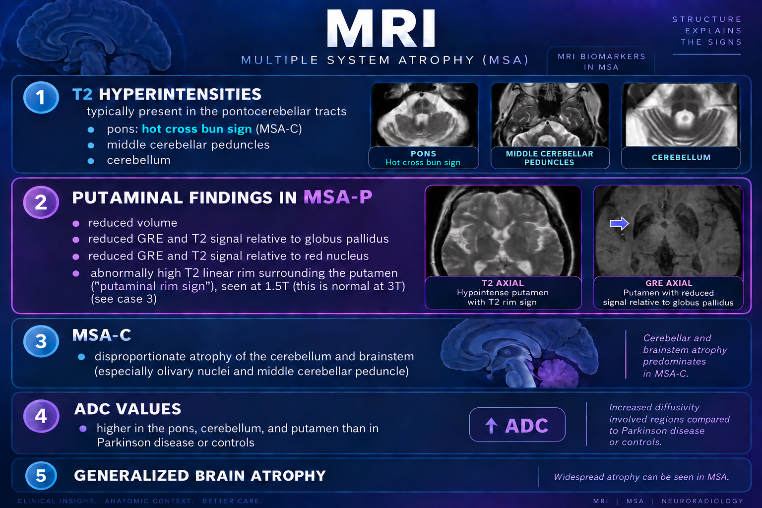
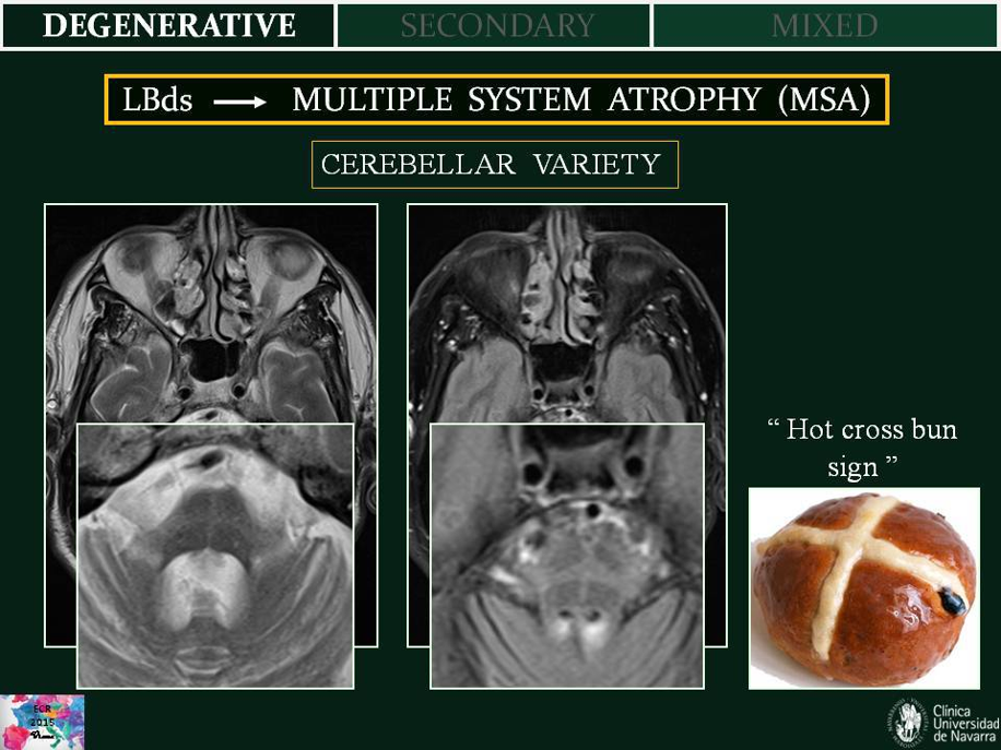
- Hot cross bun sign: T2 hyperintense pontine pyramids crossed by T2 hypointense corticospinal tracts forming a cross pattern
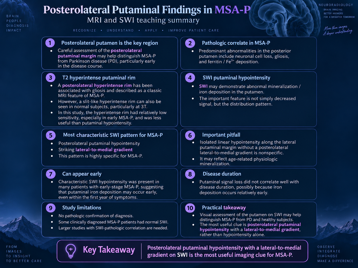
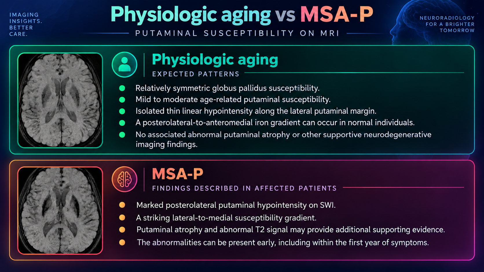
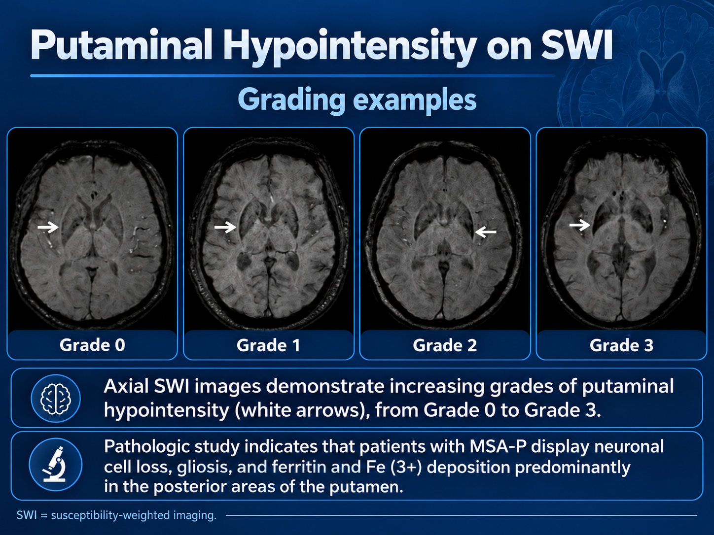
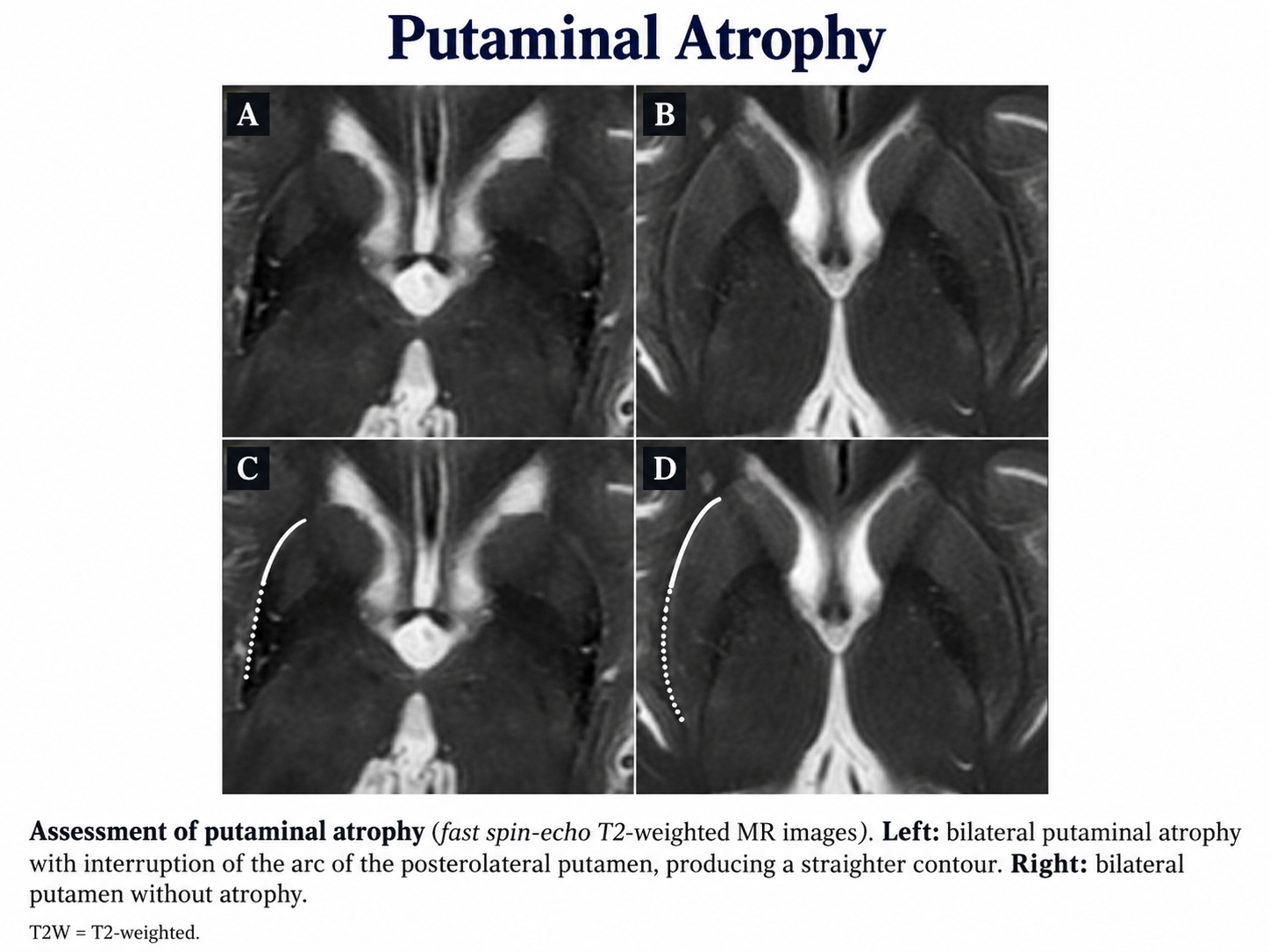
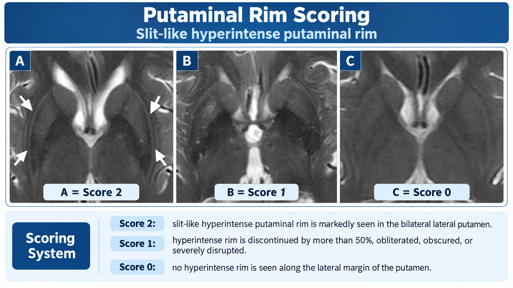
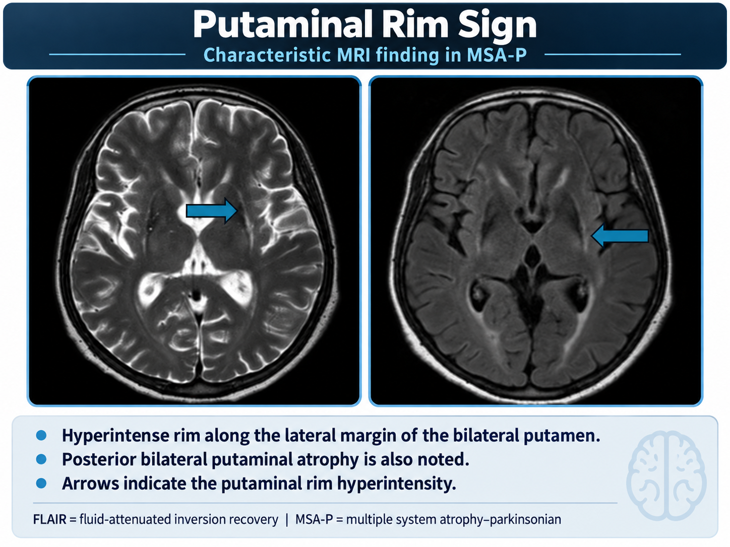
- Putaminal abnormalities: reduced signal on T2*, GRE, or SWI sequences (susceptibility effect) and/or putaminal rim sign with abnormally high T2 linear rim
- Middle cerebellar peduncle atrophy and T2 hyperintensity
- Disproportionate pontine atrophy relative to midbrain
Key Image Findings
- Hot cross bun sign on axial T2 MRI at the level of the pons: T2 hyperintensity in the pontine pyramids intersected by a T2 hypointense or normal vertical line representing the corticospinal tracts, or its precursor vertical line sign showing only the hypointense vertical line without full cross pattern
- Putaminal susceptibility effect: reduced signal intensity on T2*, gradient-recalled echo (GRE), or susceptibility-weighted imaging (SWI) sequences relative to the globus pallidus and red nucleus, reflecting iron deposition and neurodegeneration
- Putaminal rim sign on T2 MRI at 1.5 Tesla: abnormally high signal linear rim surrounding the putamen indicating preferential peripheral neurodegeneration; note this appears normal at 3 Tesla field strength
- Middle cerebellar peduncle (MCP) atrophy and T2 hyperintensity, often asymmetric, with characteristic atrophy rate of -0.5 mm/year having 85% sensitivity and 79% specificity for MSA-C
- Pontine atrophy, particularly in the anterior-posterior dimension, with characteristic atrophy rate of -0.4 mm/year showing 92% sensitivity and 87% specificity for MSA-C
- T2 hyperintensity in the pontocerebellar tracts including inferior cerebellar peduncles and cerebellum, reflecting gliosis and neurodegeneration of these pathways
- Elevated apparent diffusion coefficient (ADC) values in the pons, cerebellum, and putamen compared with Parkinson disease and normal controls, indicating increased water diffusion from neuronal loss
- Reduced fractional anisotropy (FA) on diffusion tensor imaging in the pons, cerebellum, and putamen reflecting disrupted white matter microstructure and decreased fiber organization
Differential Diagnosis
- Parkinson disease: distinguished by preserved putaminal signal on susceptibility-weighted sequences, absence of hot cross bun sign, normal or only mild pontine atrophy, and normal DTI metrics in affected regions
- Progressive supranuclear palsy (PSP): distinguished by midbrain atrophy (midbrain to pons ratio <0.5), hummingbird sign, absence of putaminal abnormalities, and different pattern of motor signs with early vertical gaze palsy
- Spinocerebellar ataxia (SCA): distinguished by genetic testing and inheritance pattern, often more prominent cerebellar vermis atrophy, variable putaminal changes depending on subtype, and lack of autonomic dysfunction
- Dementia with Lewy bodies: excluded by presence of dementia within 3 years of motor onset, early visual hallucinations, and fluctuating cognition; putaminal signal changes less prominent
- Corticobasal degeneration: distinguished by focal cortical atrophy (asymmetric), lack of hot cross bun sign, absence of putaminal rim sign, and prominent motor asymmetry
- Paraneoplastic cerebellar degeneration: distinguished by presence of malignancy and paraneoplastic antibodies, more diffuse cerebellar atrophy without pontine or putaminal changes
Discussion
Alpha-synuclein pathology in MSA involves not only neurons but also oligodendroglia (glial cytoplasmic inclusions), differentiating it from Parkinson disease and Lewy body dementia where inclusions are primarily neuronal
MSA-C and MSA-P cannot be reliably differentiated on MRI due to significant overlap of imaging findings, despite clinically distinguishable presentations; classification is based on predominant motor syndrome rather than imaging pattern
MRI has high specificity for differentiating MSA from Parkinson disease and progressive supranuclear palsy (important clinical mimics) but lower sensitivity especially in early disease, so normal MRI does not exclude MSA diagnosis
Atrophy rates of specific structures such as the pons and middle cerebellar peduncles may be more characteristic of MSA-C than static measurements, requiring serial imaging for optimal assessment
While autonomic dysfunction is a core diagnostic feature, approximately 40% of MSA patients lack autonomic symptoms initially, and 8% never develop autonomic dysfunction during long-term follow-up
MSA is relentlessly progressive with death typically occurring within 10 years of diagnosis, and no disease-modifying treatments are currently available, making early accurate diagnosis important for prognosis discussion
Reporting Pearls
When describing the key findings in MSA, clearly identify the specific pathologic MRI markers: state whether hot cross bun sign is present (and which pattern: full cross vs. vertical line sign), measure and comment on putaminal signal changes on susceptibility sequences, assess pontine and middle cerebellar peduncle atrophy relative to baseline or expected age-matched norms, and correlate the pattern of abnormalities (pontocerebellar-predominant vs. putaminal-predominant) with clinical phenotype to support the diagnostic impression of MSA.
Pitfalls
- Over-relying on absent MRI findings to exclude MSA: low sensitivity of imaging especially in early disease means normal or minimal MRI findings do not exclude clinically established or probable MSA diagnosis based on clinical criteria
- Misinterpreting putaminal rim sign at 3 Tesla as pathologic when this finding is a normal artifact at higher field strength; rim sign is a useful diagnostic marker only at 1.5 Tesla
- Failing to distinguish MSA from Parkinson disease on putaminal imaging: both conditions show putaminal changes, but MSA-specific findings include the susceptibility effect relative to globus pallidus/red nucleus and the characteristic putaminal rim sign
- Not recognizing that hot cross bun sign, while highly specific for MSA, can also be seen in spinocerebellar ataxias and other neurodegenerative conditions; clinical correlation with additional imaging features and laboratory workup is essential for accurate diagnosis