Mild encephalopathy with reversible splenial lesion (MERS)
Key Images
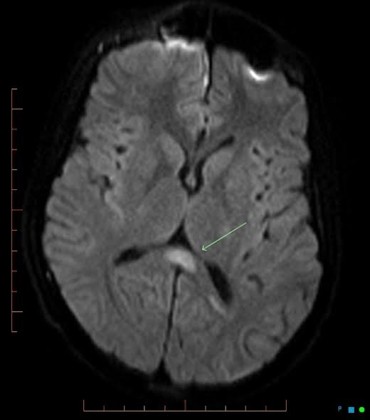
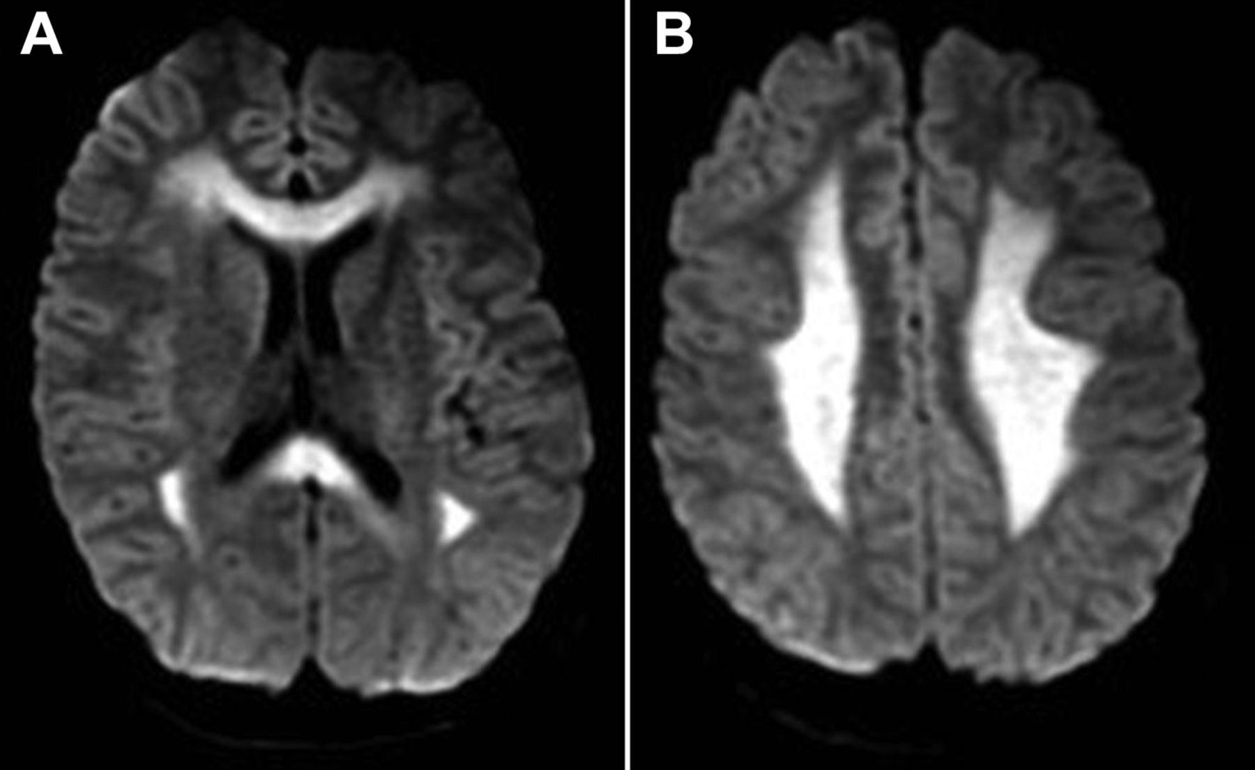
Look For First
- High signal intensity restricted to the splenium of the corpus callosum on T2/FLAIR and DWI
- Diffusion restriction (high DWI signal with low ADC values) in the splenial lesion, indicating cytotoxic edema
- No contrast enhancement following gadolinium administration
- Bilateral symmetrical white matter/centrum semiovale involvement in MERS type 2
Key Image Findings
- Splenium of corpus callosum (SCC) lesion showing high T2/FLAIR signal intensity with iso/low T1 signal, typically appearing as cytotoxic edema within the tightly packed fibers of the splenium.
- Diffusion restriction demonstrated by high signal on DWI with corresponding low ADC values, reflecting restricted water movement consistent with intra-myelinic and interstitial edema.
- No enhancement observed on T1 post-gadolinium sequences, distinguishing MERS from demyelinating diseases or other inflammatory conditions that may enhance.
- MERS type 1 presents as isolated splenial involvement, while MERS type 2 shows diffuse corpus callosum involvement with bilateral symmetrical white matter and centrum semiovale high signal lesions.
- Lesions are typically transient, resolving completely within days to weeks (often within 7-10 days) without residual cerebral atrophy or chronic changes on follow-up imaging.
- The differential timing and pattern—splenial predominance with diffusion restriction but no enhancement—helps distinguish MERS from ADEM (which typically enhances) and PRES (which shows vasogenic rather than cytotoxic edema).
Differential Diagnosis
- Acute disseminated encephalomyelitis (ADEM): distinguished by presence of gadolinium enhancement, more extensive white matter involvement, and typically more severe clinical presentation.
- Posterior reversible encephalopathy syndrome (PRES): differentiated by vasogenic rather than cytotoxic edema (high ADC values rather than restricted diffusion) and involvement of posterior temporal and parietal regions rather than corpus callosum.
- Toxic encephalopathy (e.g., methotrexate encephalopathy): may show bilateral centrum semiovale signal changes but typically has different clinical context (chemotherapy history) and lack of splenial predominance.
- Genetic disease such as Charcot-Marie-Tooth (CMT) disease: presents with chronic progressive symptoms rather than acute febrile onset and does not show diffusion restriction.
- Demyelinating disease: distinguished from MERS by the presence of enhancement on gadolinium-enhanced T1 imaging and lack of complete resolution without residual changes.
- Reversible splenial lesion syndrome (RESLES) broader category: MERS is a subtype of RESLES characterized specifically by infection-triggered presentation and complete reversibility within days to weeks.
Discussion
MERS is an uncommon syndrome most frequently affecting children and young adults, typically associated with viral infections including influenza, human herpesvirus-6, rotavirus, and mumps.
The pathophysiology involves transient intra-myelinic and interstitial edema in the tightly packed fibers of the corpus callosum, possibly precipitated by hypotonic hyponatremia or transient inflammatory infiltrates.
Diagnosis requires five criteria: neurological symptoms within one week of fever, complete resolution within 10 days, splenial/corpus callosum high signal intensity on T2, possible bilateral white matter involvement, and complete disappearance of lesions without atrophy.
The reversible nature of imaging findings is crucial for diagnosis—complete radiological recovery without residual changes distinguishes MERS from permanent demyelinating or neurodegenerative processes.
Treatment is supportive, and the disease carries an excellent prognosis with near-complete clinical and radiological recovery in the large majority of patients.
MERS falls under the broader category of Infection Triggered Encephalopathy Syndromes (ITES) and is part of the reversible splenial lesion syndrome (RESLES) spectrum.
Reporting Pearls
Report the characteristic finding as: "Reversible splenial lesion with restricted diffusion—high T2/FLAIR and DWI signal intensity with low ADC values in the splenium of the corpus callosum, without enhancement, consistent with cytotoxic edema in the clinical context of acute febrile illness. Note the absence of contrast enhancement and commit to follow-up imaging at 1-2 weeks to document complete resolution, which is diagnostic for MERS."
Pitfalls
- Mistaking MERS for ADEM by overlooking the lack of enhancement on gadolinium sequences—MERS shows no enhancement while ADEM typically does, which is a key distinguishing feature.
- Confusing cytotoxic edema (restricted diffusion with low ADC) with vasogenic edema seen in PRES—MERS shows DWI restriction while PRES shows elevated ADC values in posterior regions.
- Failing to perform or recommend follow-up MRI to document complete resolution—documentation of lesion disappearance within 7-10 days without residual changes is essential for confirming the diagnosis and excluding chronic demyelinating disease.
- Overlooking the splenial predominance and considering diffuse white matter disease as a primary concern—the characteristic splenial involvement in MERS type 1 is the key diagnostic clue that should trigger recognition of the syndrome.