Hypoglycemic encephalopathy
Key Images
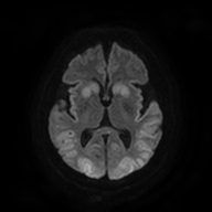
Look For First
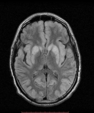
- Bilateral symmetric T2/FLAIR hyperintensity in posterior limb of internal capsule
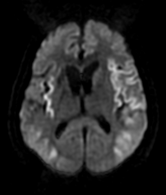
- Involvement of parieto-occipital cortex and insula
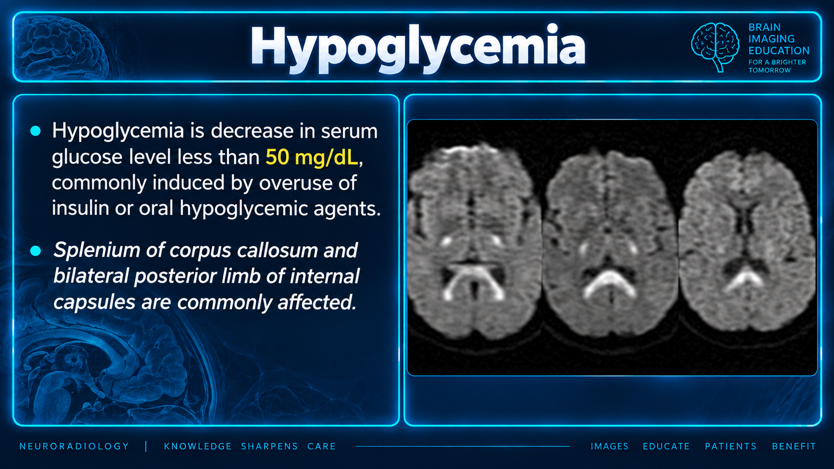
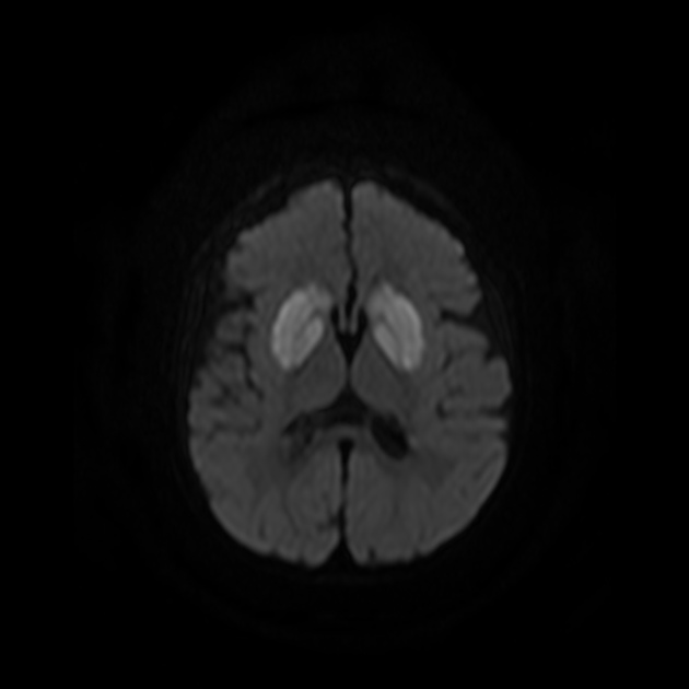
- Restricted diffusion on DWI as an early and sensitive marker
Key Image Findings
- T1-weighted imaging shows low signal (hypointense) in affected regions including posterior limb of internal capsule, cerebral cortex, hippocampus, and basal ganglia.
- T2-weighted and FLAIR images demonstrate high signal (hyperintense) in characteristic bilateral distribution affecting posterior limb of internal capsule, parieto-occipital and insular cortex, hippocampus, and basal ganglia.
- DWI/ADC sequences show reversible diffusion restriction, which is often an earlier and more sensitive finding than conventional T2/FLAIR changes.
- Lesions are typically bilateral and symmetric, distinguishing them from focal ischemic infarcts.
- The splenium of the corpus callosum may be affected, producing the characteristic 'boomerang sign' appearance.
- The cerebellum, brainstem, and thalami are typically spared in adults but may show involvement in neonates.
- Restricted diffusion is often reversible, reflecting the potentially reversible nature of hypoglycemic injury.
- Signal changes reflect cellular energy failure and tissue swelling secondary to neuronal death and altered cellular physiology.
Differential Diagnosis
- Hypoxic-ischemic brain injury: typically shows bilateral symmetric thalamic lesions, whereas hypoglycemia preferentially affects basal ganglia and cortex while sparing thalami in adults.
- Creutzfeldt-Jakob disease (CJD): distinguished by different clinical presentation; CJD shows progressive dementia rather than acute neurological crisis from metabolic derangement.
- Ischemic infarct: usually focal and unilateral distribution, whereas hypoglycemic changes are bilateral and symmetric.
- Seizure-related changes: may show cortical FLAIR hyperintensity but lack the characteristic bilateral symmetric pattern and involvement of deep gray matter seen in hypoglycemia.
- Status epilepticus-related injury: can mimic hypoglycemic injury but typically presents with different clinical context and history.
Discussion
Hypoglycemia causes cellular energy failure since the brain is an obligate glucose metabolizer, leading to sodium/potassium pump failure, cellular swelling, and tissue alkalosis.
Restricted diffusion on DWI is an earlier and more sensitive imaging marker than T2/FLAIR hyperintensity, making it particularly valuable in acute presentations.
Many hypoglycemic injuries show reversible diffusion restriction, suggesting the potential for recovery if the underlying metabolic derangement is corrected promptly.
MRI is typically not performed unless there is complicated clinical recovery, as most cases of hypoglycemia are recognized and managed promptly.
The distribution of signal abnormalities preferentially affecting basal ganglia, cortex, hippocampus, and posterior internal capsule while sparing thalami is a key distinguishing feature in adults.
Prognosis directly correlates with severity and duration of the hypoglycemic insult, making early recognition and treatment critical for outcomes.
Pathophysiology and vulnerable regions
Because the brain depends heavily on glucose oxidation to generate ATP, severe hypoglycemia causes cerebral energy failure. This can result in neuronal degeneration and necrosis with glial-cell infiltration.
The most metabolically active—and therefore most vulnerable—regions include:
- Cerebral cortex
- Hippocampi
- Cerebellum
- Caudate nuclei
- Globus pallidi
The thalami, hypothalamus, brainstem, and cranial nerve nuclei are generally less vulnerable, although they may also be affected in severe cases
Reporting Pearls
Describe the finding as 'bilateral symmetric T2/FLAIR hyperintensity in the posterior limbs of the internal capsules, basal ganglia, hippocampi, and parieto-occipital cortex consistent with hypoglycemic encephalopathy, with DWI showing reversible-appearing diffusion restriction' and note the sparing of thalami in adults as a key distinguishing feature from hypoxic-ischemic injury.
Pitfalls
- Mistaking hypoglycemic injury for hypoxic-ischemic injury when thalami are spared; remember that hypoxic-ischemic injury preferentially affects thalami in adults, whereas hypoglycemia typically spares them.
- Overlooking early restricted diffusion on DWI as a sensitive sign; DWI changes may precede conventional T2/FLAIR findings, making them critical to evaluate in acute presentation.
- Failing to recognize the bilateral symmetric distribution; unilateral or focal changes should prompt consideration of ischemic stroke or other focal pathology rather than hypoglycemia.
- Assuming findings are irreversible; reversible diffusion restriction is characteristic of hypoglycemic injury, and improvement or resolution on follow-up imaging is expected with appropriate treatment.