Huntington disease
Key Images
Look For First
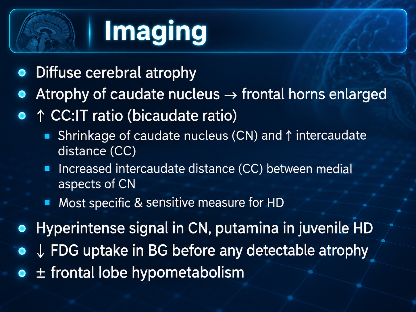
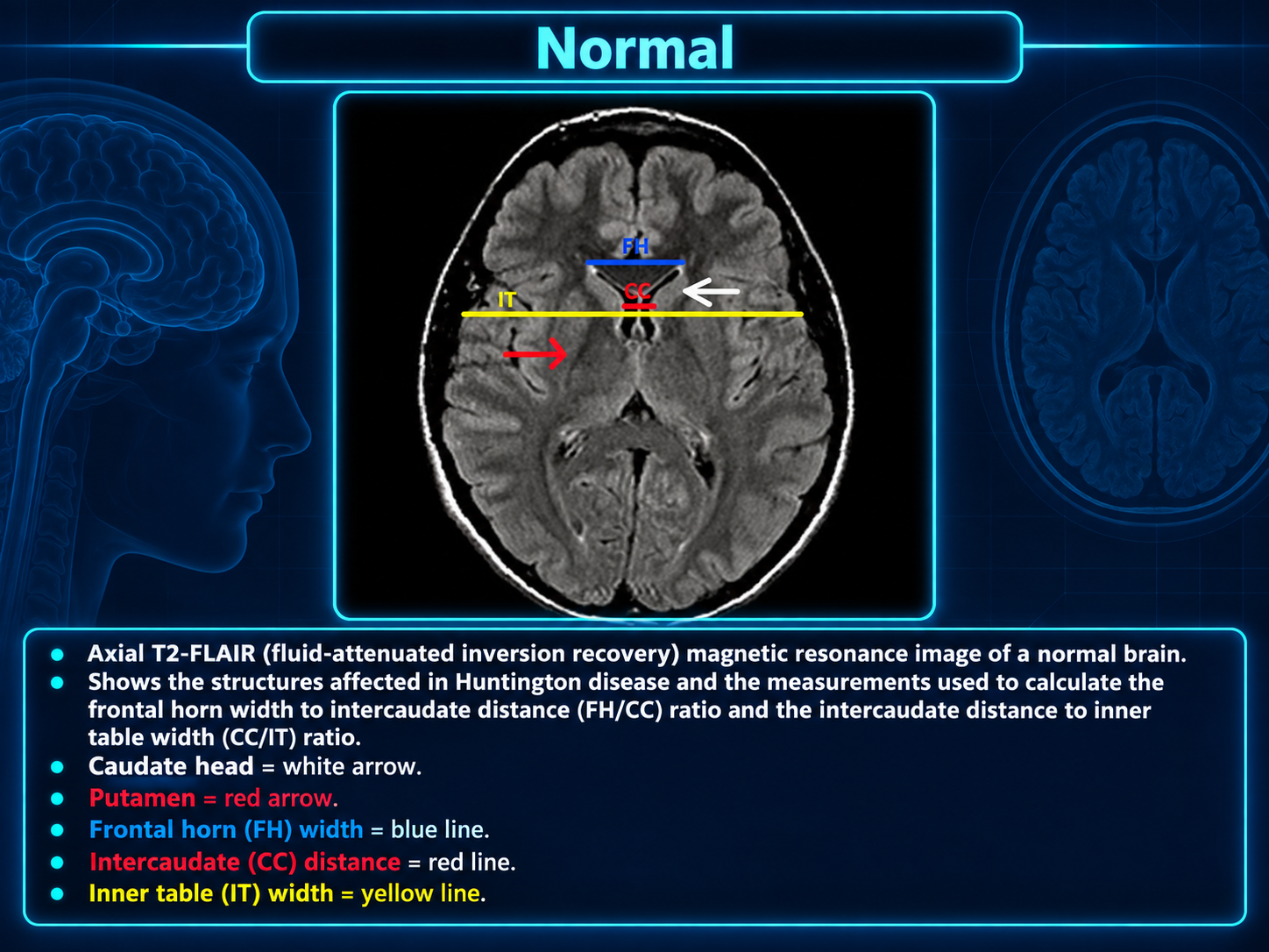
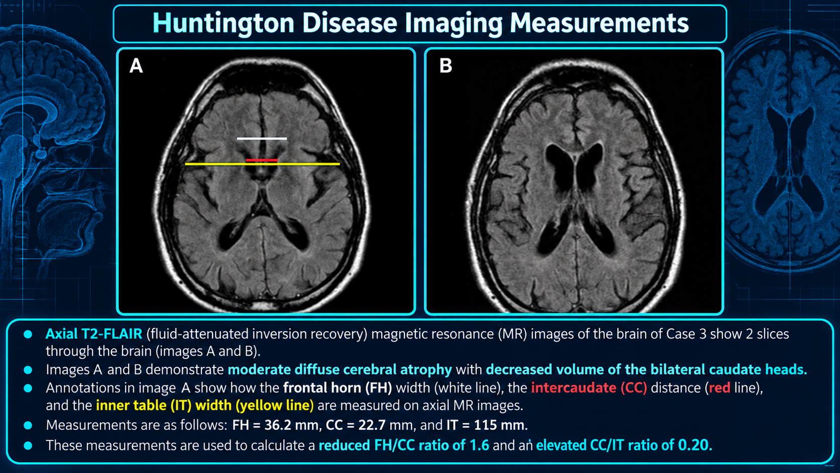
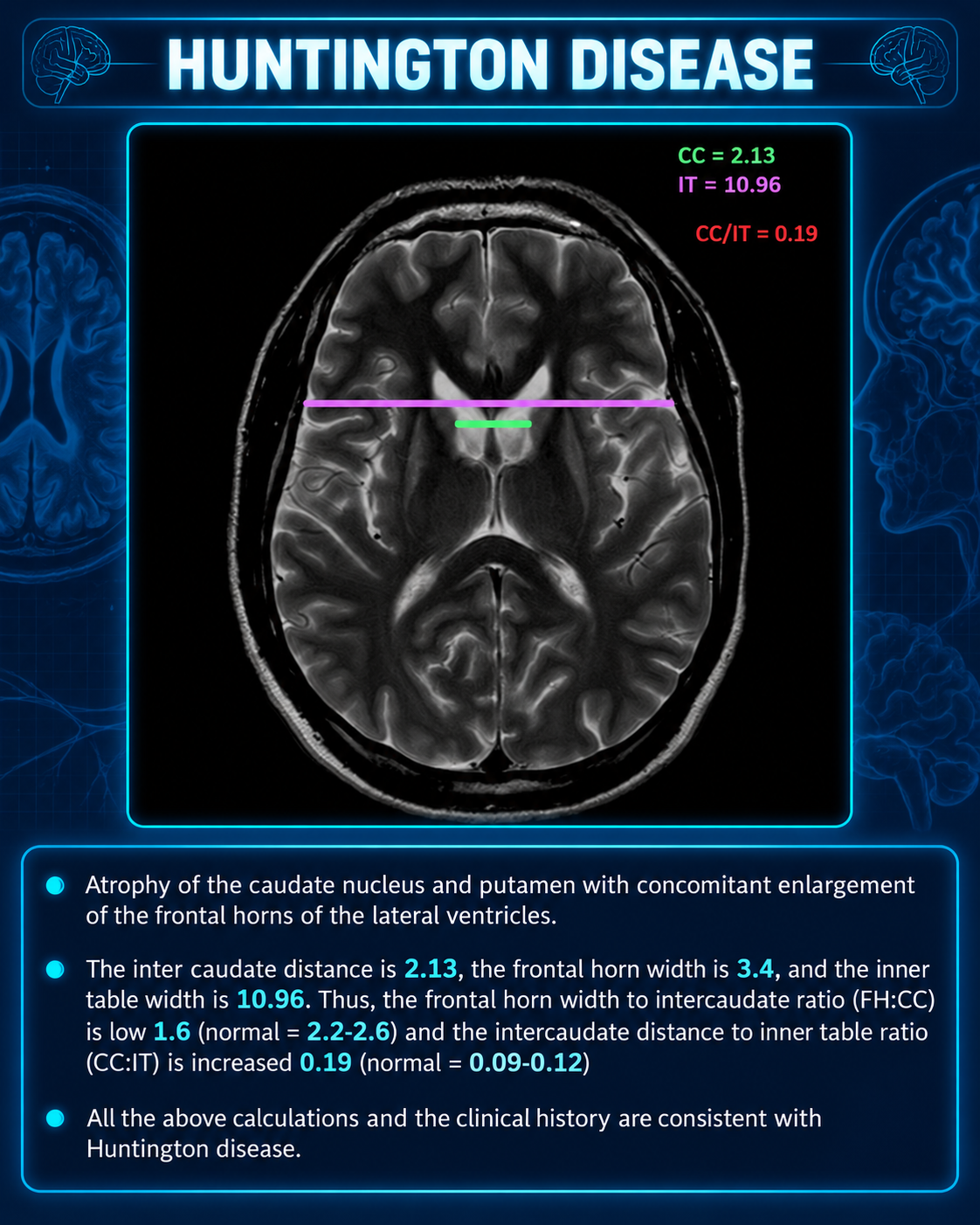
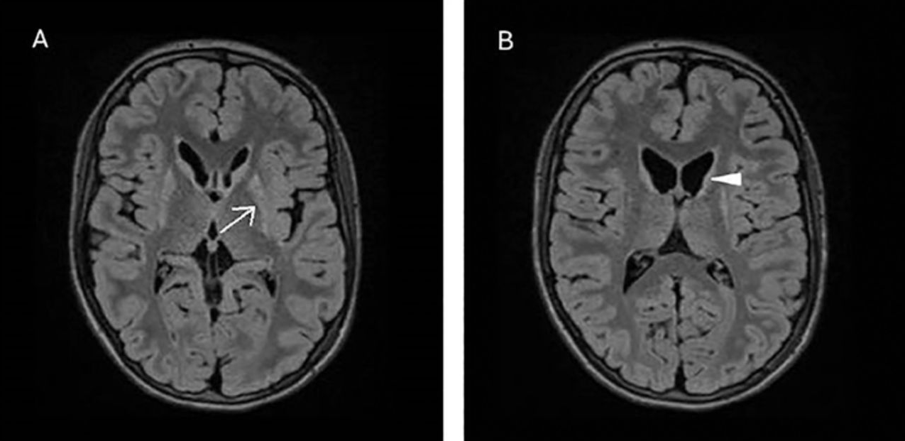
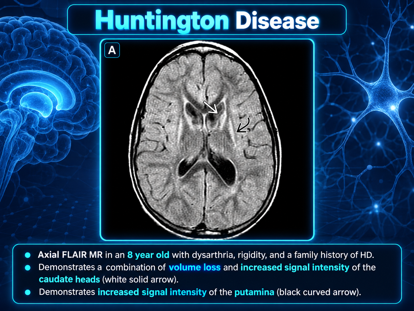
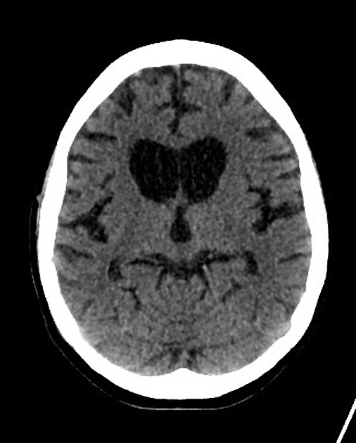
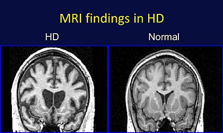
- Caudate head atrophy with enlargement of frontal horns creating a characteristic 'boxcar' configuration on axial images
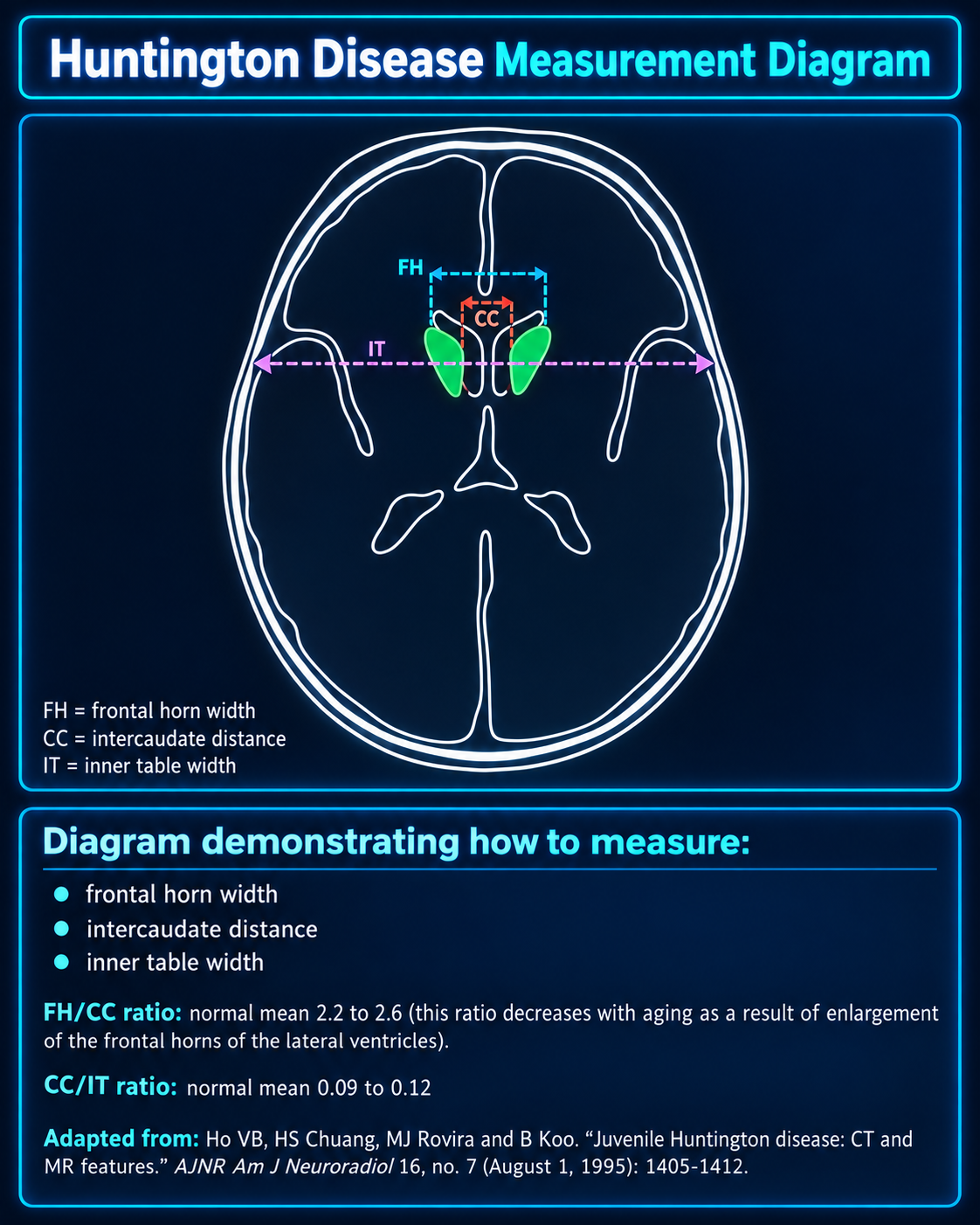
- Increased intercaudate distance relative to inner table width (CC/IT ratio elevated)
- Prominent putaminal volume loss visible on morphometry or careful visual inspection
- Age-inappropriate cortical thinning and generalized brain volume loss
Key Image Findings
- MRI demonstrates striking caudate nucleus head atrophy as the most recognizable feature, with bilateral involvement.
- Putaminal volume loss is present but often less visually obvious than caudate atrophy; this is particularly prominent in younger patients and the juvenile (Westphal) variant.
- The combination of caudate and putamen atrophy results in enlargement of the frontal horns with a characteristic 'box' or 'boxcar-like' configuration on axial T1-weighted imaging.
- Frontal horn width to intercaudate distance ratio (FH/CC) is reduced, while intercaudate distance to inner table width ratio (CC/IT) is increased, providing quantifiable metrics of striatal atrophy.
- Basal ganglia may demonstrate decreased T2 signal and SWI blooming artifact consistent with iron deposition in some cases.
- MR spectroscopy may show elevation of lactate (variably present, most often in basal ganglia) and decreased NAA/creatine ratio particularly in the basal ganglia, reflecting neuronal loss.
- Generalized age-inappropriate cortical thinning and volume loss are recognized, indicating broader neocortical involvement beyond the striatum.
- Changes may be detected on FDG-PET as striatal and cortical hypometabolism, which can appear even in the presymptomatic phase before clinical symptom onset.
Differential Diagnosis
- Neuroacanthocytosis syndromes (chorea-acanthocytosis, McLeod syndrome): differentiate by presence of circulating acanthocytes on blood smear and genetic testing; these conditions show similar striatal atrophy but have distinct hematologic findings.
- Frontotemporal lobar degeneration (FUS variant, FTLD-FUS): can show frontal lobe atrophy and behavioral changes, but typically shows more prominent anterior temporal and frontal lobe volume loss rather than selective striatal atrophy.
- Wilson disease: predominantly involves white matter, thalamus, cerebellum, and brainstem rather than selective caudate/putamen atrophy; also presents with Kayser-Fleischer rings on slit-lamp examination.
- Leigh disease: metabolic disorder affecting white matter, thalamus, cerebellum, and brainstem; lacks the selective striatal atrophy pattern of Huntington disease.
- Pantothenate kinase-associated neurodegeneration (PKAN): shows the characteristic 'eye-of-the-tiger' sign with iron deposition in globus pallidus; striatal involvement is not the primary pattern.
- Acute toxic or metabolic encephalopathy (hypoxia, carbon monoxide poisoning, hypoglycemia): can produce basal ganglia changes but lack the chronic progressive selective striatal atrophy and are associated with acute clinical presentation rather than insidious midlife onset.
Discussion
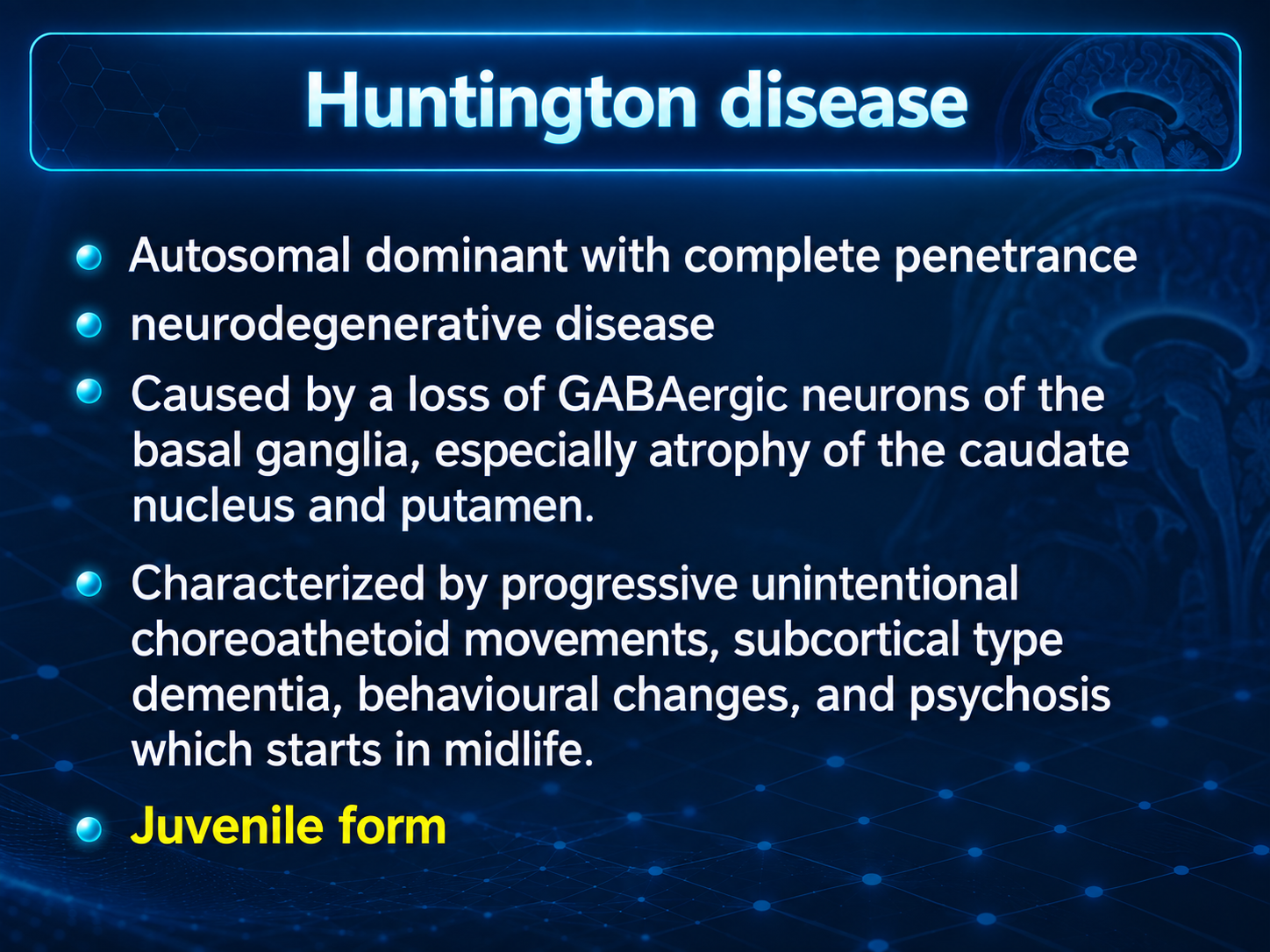
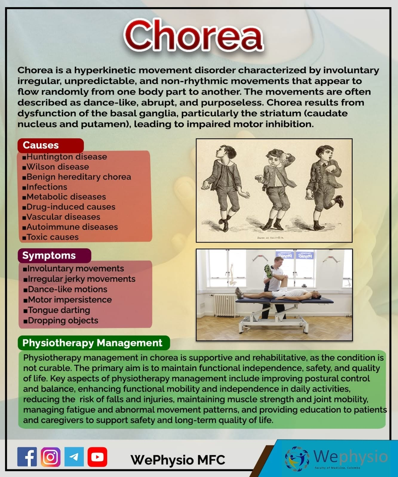
Huntington disease results from progressive loss of GABAergic neurons in the basal ganglia, particularly affecting the dorsal striatum (caudate and putamen); the absence of inhibitory striatal neurons leads to increased motor output causing the characteristic chorea.
The disease is caused by pathologic CAG trinucleotide repeat expansion on chromosome 4p16.3; normal individuals have <27 repeats while affected patients have >36 repeats, with higher repeat numbers correlating with earlier symptom onset.
Genetic anticipation is a hallmark feature, particularly with paternal inheritance, resulting in earlier disease presentation and greater severity in successive generations due to progressive CAG repeat amplification.
The juvenile (Westphal) variant occurs in 1-6% of cases with onset before age 20, typically from paternal inheritance with >60 CAG repeats, and presents with early parkinsonism, cognitive impairment, ataxia, and myoclonus rather than pure chorea.
MRI changes correlate with clinical severity: putaminal atrophy is particularly prominent in younger patients and the juvenile variant, whereas the classic 'boxcar' ventricular enlargement may be more striking in older-onset disease.
No disease-modifying treatment currently exists; adult-onset disease is fatal in 14-15 years while juvenile-onset is more rapidly progressive with death in 7-8 years, highlighting the importance of early imaging diagnosis and genetic counseling.
Reporting Pearls
When reporting Huntington disease, clearly describe the "bilateral caudate head atrophy with concomitant frontal horn enlargement resulting in a characteristic 'boxcar' configuration of the lateral ventricles." Provide quantitative measurements using the FH/CC and CC/IT ratios to objectively document striatal volume loss and support the diagnosis, as visual impression alone may underestimate putaminal involvement, particularly in younger patients.
Pitfalls
- Putaminal atrophy can be underrecognized on visual inspection alone, particularly in younger patients and the juvenile variant; careful measurement and morphometry are needed to appreciate the full extent of striatal involvement beyond the more obvious caudate atrophy.
- Confusing Huntington disease with other causes of basal ganglia iron deposition (such as PKAN with its pathognomonic 'eye-of-the-tiger' sign) or Wilson disease with thalamic involvement; careful attention to the pattern of atrophy and distribution of abnormality is essential.
- Misinterpreting age-appropriate enlargement of frontal horns as pathologic in older patients, or conversely missing mild caudate atrophy in young presymptomatic carriers; clinical context and genetic testing status should guide interpretation.
- Overlooking broader cortical atrophy and white matter changes that accompany striatal pathology; Huntington disease is a multifocal neurodegenerative disorder, not purely a basal ganglia disease.