Cytotoxic lesions of the corpus callosum (CLOCCs)
Key Images
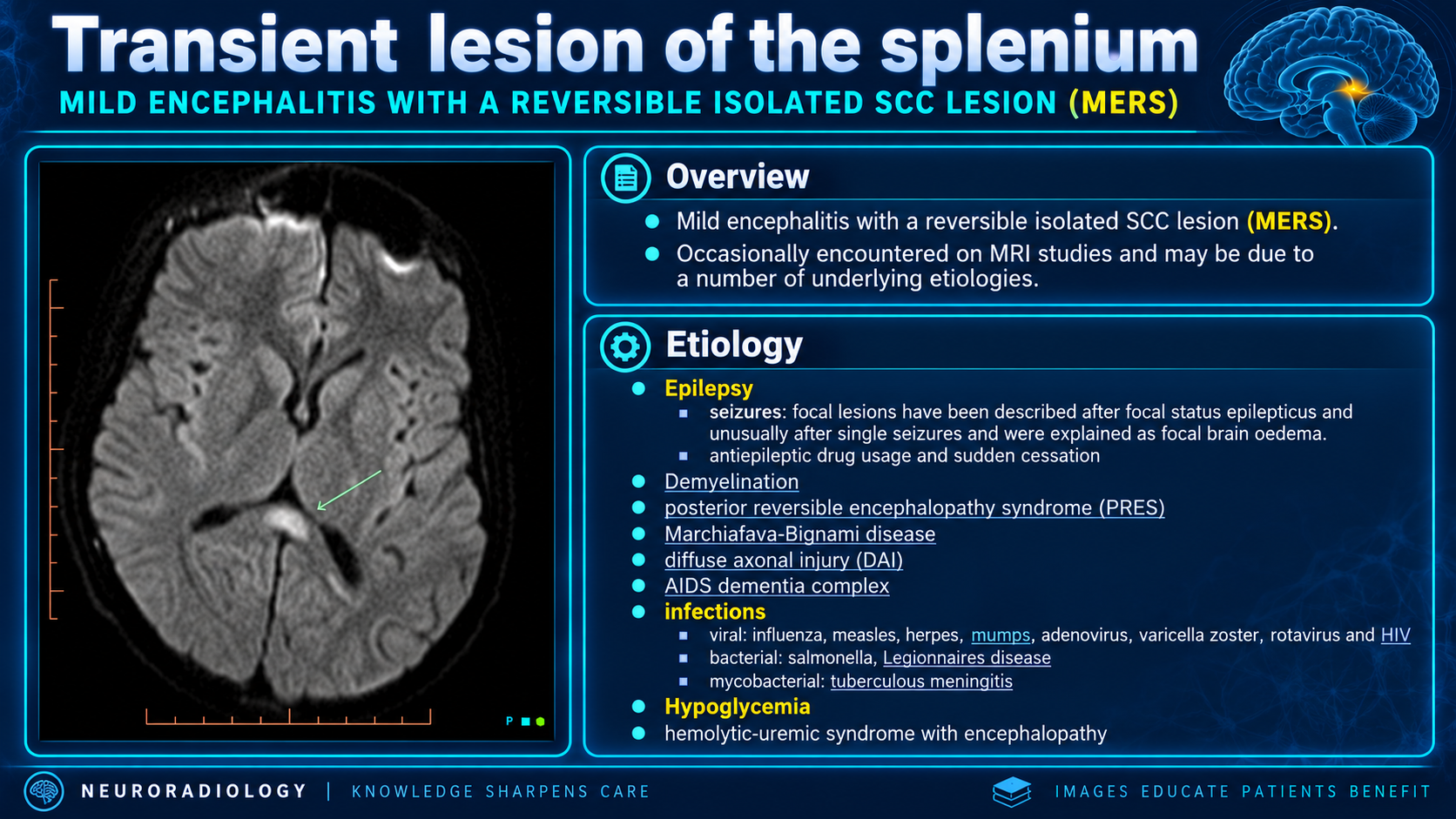
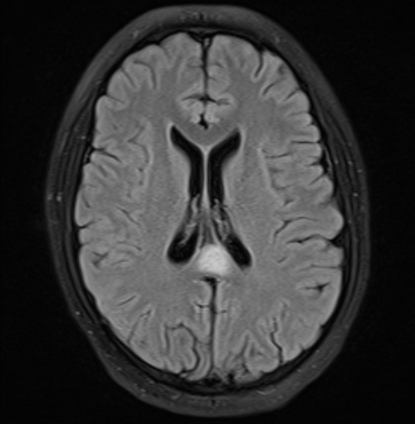
Look For First
- Small, well-circumscribed oval midline lesion within the splenium on T2 hyperintense imaging
- Restricted diffusion (reduced ADC 300-500 x 10-6 mm2/s) on DWI in the splenium
- T1 hypointense, T2 hyperintense lesion without post-gadolinium enhancement
- Boomerang sign: larger irregular lesion extending from splenium into adjacent hemispheres in non-seizure etiologies
Key Image Findings
- T1-weighted imaging shows hypointense signal in the affected corpus callosum, most commonly the splenium.
- T2-weighted imaging demonstrates hyperintense signal change within the splenium with sharply circumscribed margins in typical seizure-related cases.
- DWI imaging reveals restricted diffusion with ADC values typically ranging 300-500 x 10-6 mm2/s, confirming cytotoxic edema rather than vasogenic edema.
- No enhancement is observed on T1 post-gadolinium imaging, distinguishing CLOCCs from inflammatory or neoplastic lesions.
- Well-circumscribed small oval lesions in the midline splenium are most characteristic of seizure-related or antiseizure medication-related CLOCCs.
- Boomerang sign refers to larger, more irregular lesions extending from the splenium into the body and adjacent hemispheres, typically seen with metabolic, infectious, or toxic etiologies.
- Complete radiographic resolution typically occurs within 1 week to 1 month following clinical neurological recovery.
- The splenium is preferentially affected due to high density of oligodendrocytes expressing abundant glutamate receptors, making this region vulnerable to excitotoxic injury.
Differential Diagnosis
- Multiple sclerosis: demyelinating plaques are typically periventricular and show varying enhancement patterns, while CLOCCs lack enhancement and are usually transient.
- Acute disseminated encephalomyelitis (ADEM): multifocal white matter involvement with various enhancement patterns, whereas CLOCCs are typically confined to corpus callosum with no enhancement.
- Acute infarction (pericallosal artery occlusion): shows vascular distribution and arterial territory pattern, while CLOCCs are centrally located in splenium without vascular territory specificity.
- Posterior reversible encephalopathy syndrome (PRES): predominantly involves parietal and occipital white matter with vasogenic edema, whereas CLOCCs are focal to corpus callosum with cytotoxic edema pattern.
- Diffuse axonal injury from trauma: typically affects corpus callosum body in addition to splenium with gradient echo blooming, while CLOCCs are often limited to splenium without hemorrhagic components.
- Tumors: mass effect, enhancement, and mass-like morphology distinguish neoplasms from the transient, non-enhancing nature of CLOCCs.
Discussion
CLOCCs result from a stereotyped pathophysiologic cascade involving inflammatory cytokine release (IL-1, IL-6, TNF-α), blood-brain barrier breakdown, astrocyte activation, and massive extracellular glutamate accumulation leading to cytotoxic edema.
The splenium's vulnerability reflects the high concentration of oligodendrocytes with abundant glutamate receptors, explaining the stereotyped preference for this location despite diverse underlying etiologies.
Unlike other corpus callosum lesions, CLOCCs do not produce hemispheric disconnection syndromes such as alien hand syndrome, apraxia, or visual field defects.
Seizure-related CLOCCs (from seizures themselves or antiseizure medication cessation) typically present as small, well-circumscribed lesions, whereas metabolic or infectious etiologies tend to produce larger, more irregular boomerang-shaped lesions.
Complete radiographic resolution within 1 week to 1 month post-recovery favors CLOCCs, distinguishing them from chronic demyelination or infarction.
Prognosis is favorable in seizure-related and antiseizure medication-related cases but depends on underlying etiology in metabolic, infectious, or toxic contexts.
Reporting Pearls
Describe CLOCCs as transient cytotoxic lesions with restricted diffusion in the splenium (and potentially extending into the body or hemispheres in boomerang pattern), emphasizing the lack of enhancement and the specific ADC reduction to support the cytotoxic rather than vasogenic edema pattern, while commenting on the expected complete resolution within weeks in the appropriate clinical context.
Pitfalls
- Misinterpreting hyperintense T2 signal in the splenium as demyelination without confirming restricted diffusion; cytotoxic edema (CLOCCs) shows reduced ADC while vasogenic edema in demyelination shows elevated ADC.
- Overlooking seizures, antiseizure medication cessation, and metabolic derangements as etiologies and pursuing aggressive investigation for malignancy or demyelination unnecessarily.
- Failing to correlate imaging findings with clinical context and etiologic workup; the same splenial lesion appearance can reflect seizures, infection, metabolic disturbance, drug toxicity, or other insults.
- Assuming all splenial lesions are reversible; while CLOCCs are typically transient, Marchiafava-Bignami disease and post-traumatic corpus callosum damage can produce permanent lesions in similar distributions.