Corticobasal Degeneration
Key Images
Look For First
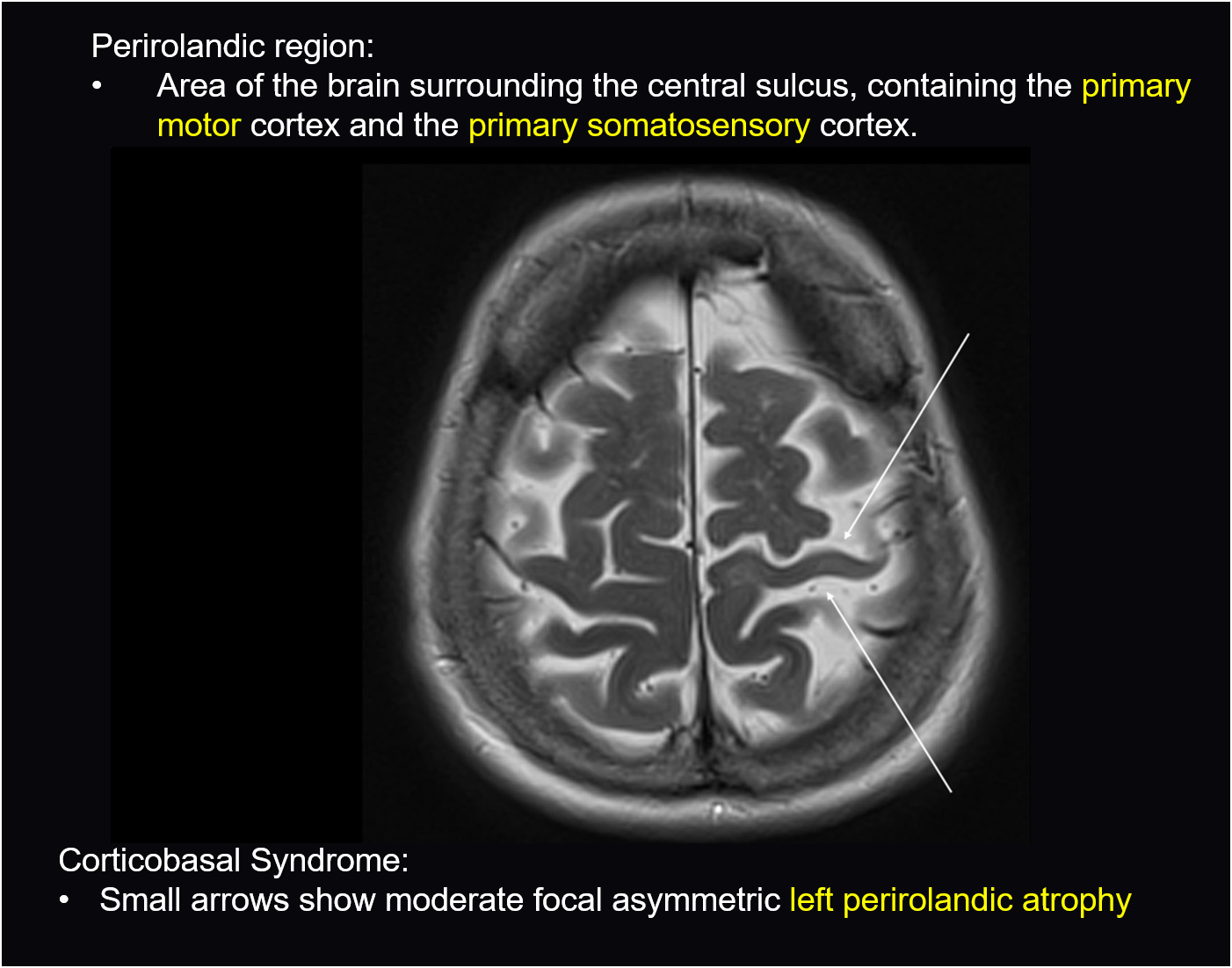
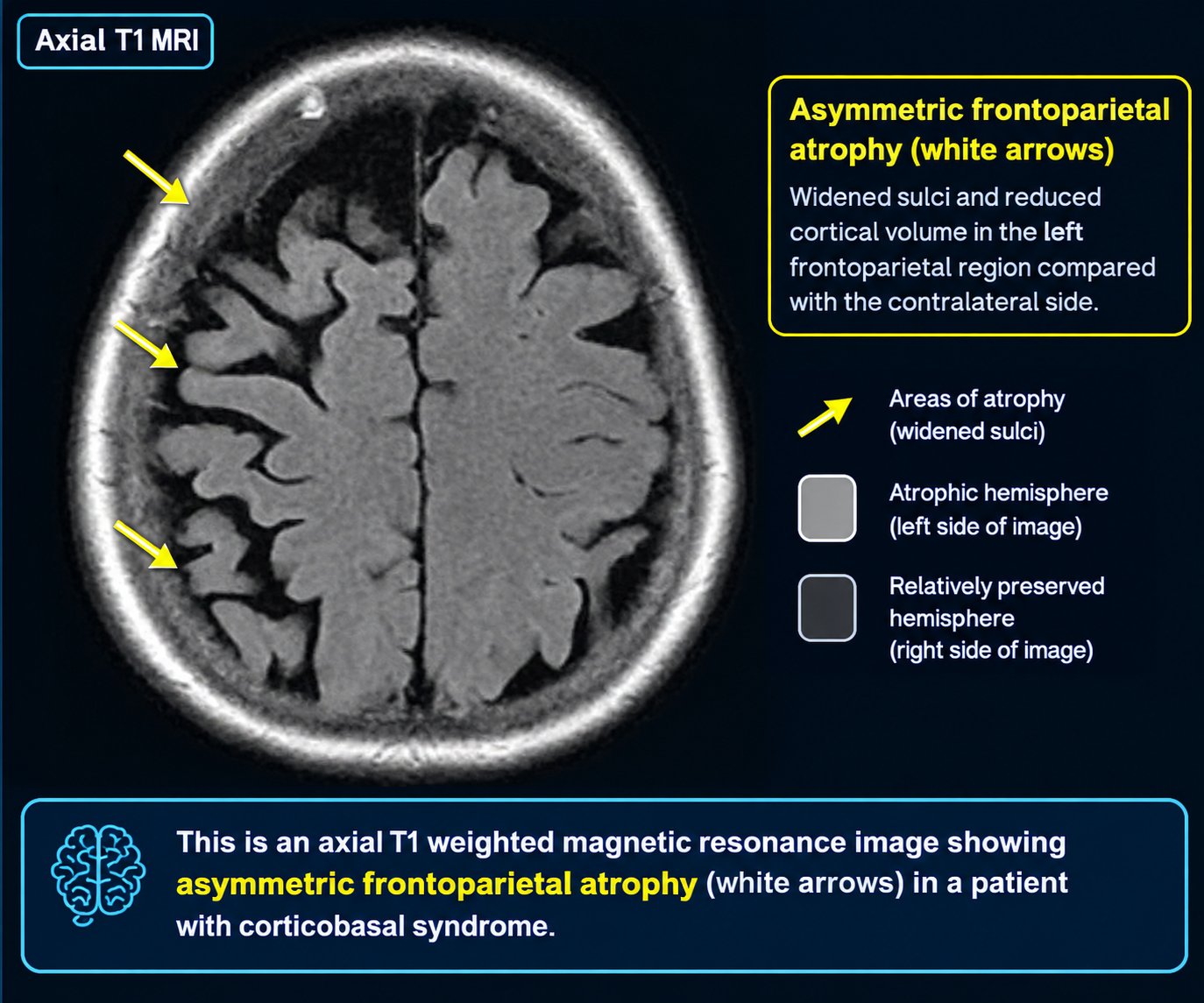
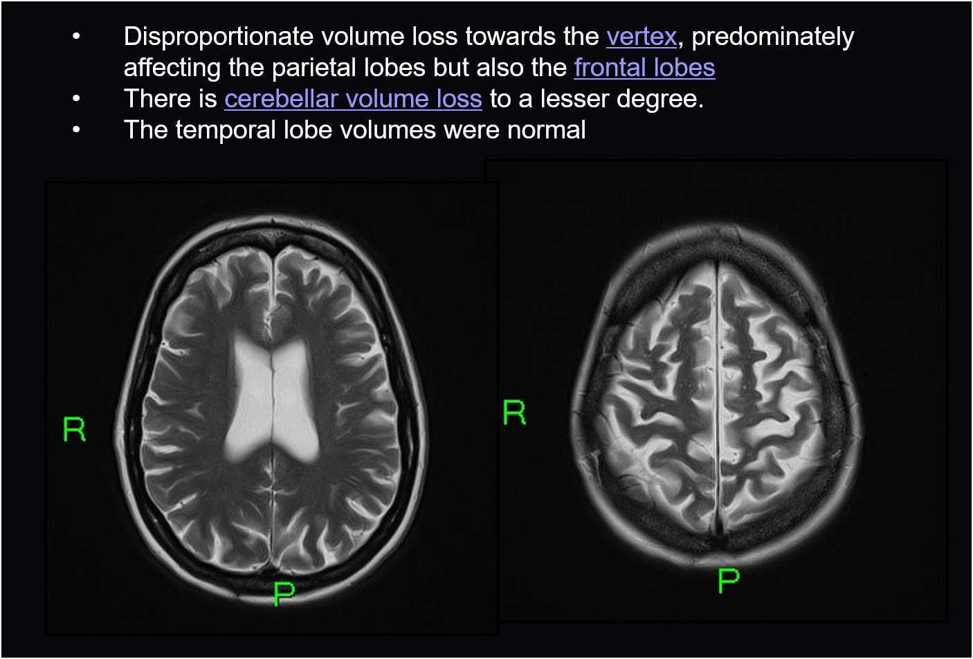
- Asymmetric cortical atrophy, particularly in superior parietal lobule (most constant feature)
- Perirolandic and precentral gyral atrophy
- Preserved brainstem anatomy distinguishing it from PSP
Key Image Findings
- Asymmetric cortical atrophy is the hallmark MRI finding, most prominently affecting the superior parietal lobule which represents the most constant feature of CBD.
- Perirolandic gyri, precentral gyri, postcentral gyri, and superior frontal gyri demonstrate characteristic atrophy that may be asymmetric in distribution.
- T2 hyperintensity in subcortical white matter of affected gyri reflects underlying pathology and gliosis in regions of cortical loss.
- Bilateral basal ganglia atrophy is present, with T2 hyperintensity particularly evident in the posterolateral putamen.
- Corpus callosum atrophy may be observed on MRI.
- The atrophy pattern in CBD is distinguishable from PSP by relatively preserved brainstem anatomy; CBD tends to show posterolateral and medial frontal cortical atrophy whereas PSP shows prominent midbrain atrophy.
- Nuclear medicine imaging with SPECT and PET demonstrates hypometabolism in superior parietal and superior frontal regions, with less frequent involvement of basal ganglia and thalamus.
- Early metabolic changes on functional imaging tend to be asymmetrical, mirroring the asymmetric clinical presentation.
Differential Diagnosis
- Progressive supranuclear palsy (PSP-CBS): PSP shows prominent brainstem atrophy (midbrain and superior pontine atrophy) whereas CBD has relatively preserved brainstem with frontoparietal cortical predominance.
- Pick disease: May present similarly with frontal atrophy but typically shows more symmetric anterior temporal involvement and ballooning of pick bodies on histology.
- Multiple system atrophy (MSA): Presents with different basal ganglia involvement patterns and cerebellar atrophy; lacks the characteristic asymmetric cortical atrophy seen in CBD.
- Parkinson disease: Does not show the asymmetric cortical atrophy pattern of CBD; Parkinsonism in CBD is levodopa-unresponsive unlike idiopathic Parkinson disease.
- Posterior cortical atrophy (PCA): Affects posterior parietal and occipital regions but typically shows more symmetric involvement; CBS has earlier motor and limb apraxia features.
- Creutzfeldt-Jakob disease: Can present with CBS clinically but shows different MRI patterns including cortical ribboning and basal ganglia signal changes.
Discussion
Corticobasal syndrome (CBS) is the clinical diagnosis while corticobasal degeneration (CBD) is the pathologically confirmed tauopathy; CBS has multiple etiologies including PSP, frontotemporal dementia, and Creutzfeldt-Jakob disease.
The asymmetric akinetic-rigid syndrome in CBD is resistant to levodopa, which helps differentiate it from idiopathic Parkinson disease and guides treatment decisions.
The characteristic histopathology includes neuronal loss and ballooned achromatic neurons primarily affecting the frontoparietal cortex, subcortical structures, striatum, and substantia nigra.
The preserved brainstem on MRI is a key feature distinguishing CBD from PSP, where midbrain atrophy is prominent; this distinction is critical for differential diagnosis.
Prognosis is poor with gradual neurological decline and death typically occurring 5-10 years after diagnosis; no disease-modifying therapies exist, with treatment focused on symptomatic relief and supportive services.
Emerging imaging techniques such as 3D volumetric MRI and diffusion tensor imaging may provide additional diagnostic utility in differentiating CBD from mimics.
Reporting Pearls
In CBD, clearly describe the pattern of atrophy as "asymmetric cortical atrophy predominantly involving the superior parietal lobule and perirolandic regions with relative sparing of the brainstem," as this pattern both confirms the diagnosis and helps exclude PSP which shows prominent midbrain atrophy.
Pitfalls
- Confusing corticobasal syndrome (clinical diagnosis) with corticobasal degeneration (pathologic diagnosis); multiple other conditions including PSP cause CBS, so MRI pattern is essential for narrowing the differential.
- Failing to identify the asymmetric nature of cortical atrophy, which is a defining characteristic; subtle asymmetry may be missed on casual inspection and requires careful comparison of bilateral structures.
- Overinterpreting basal ganglia atrophy in isolation; CBD has bilateral basal ganglia involvement, but the distinctive feature is the cortical atrophy pattern rather than basal ganglia changes alone.
- Missing the brainstem preservation as a distinguishing feature from PSP; PSP shows characteristic midbrain atrophy and the Mickey Mouse sign, whereas CBD has relatively preserved brainstem anatomy.