Basal ganglia T1 hyperintensity
Key Images
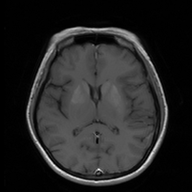
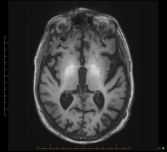
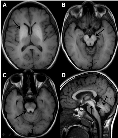
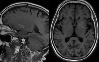
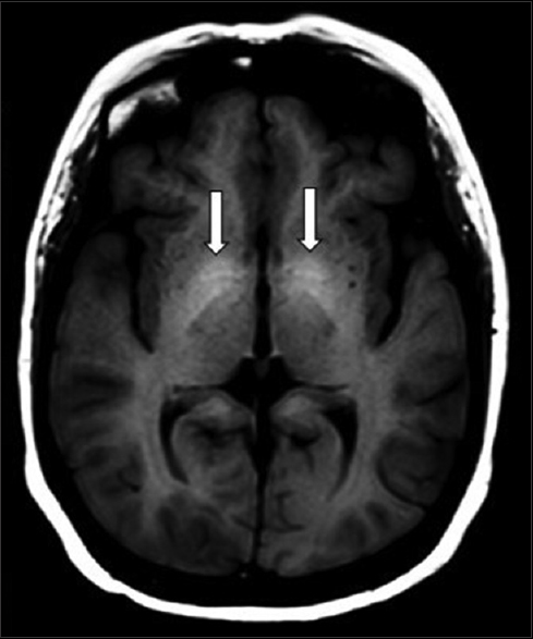
Look For First
- Bilateral vs. unilateral T1 hyperintensity — this distinction is the primary decision point
- Anatomic location: globus pallidus (pallidal pattern suggests manganese/calcification) vs. striatum/caudate/putamen (striatal pattern suggests hemichorea, hemorrhage, or hypoxia)
- T2/SWI phase and non-contrast CT correlation — essential to distinguish calcium (hyperdense on CT, diamagnetic/negative on QSM) from hemorrhage/iron (paramagnetic/positive on QSM)
Key Image Findings
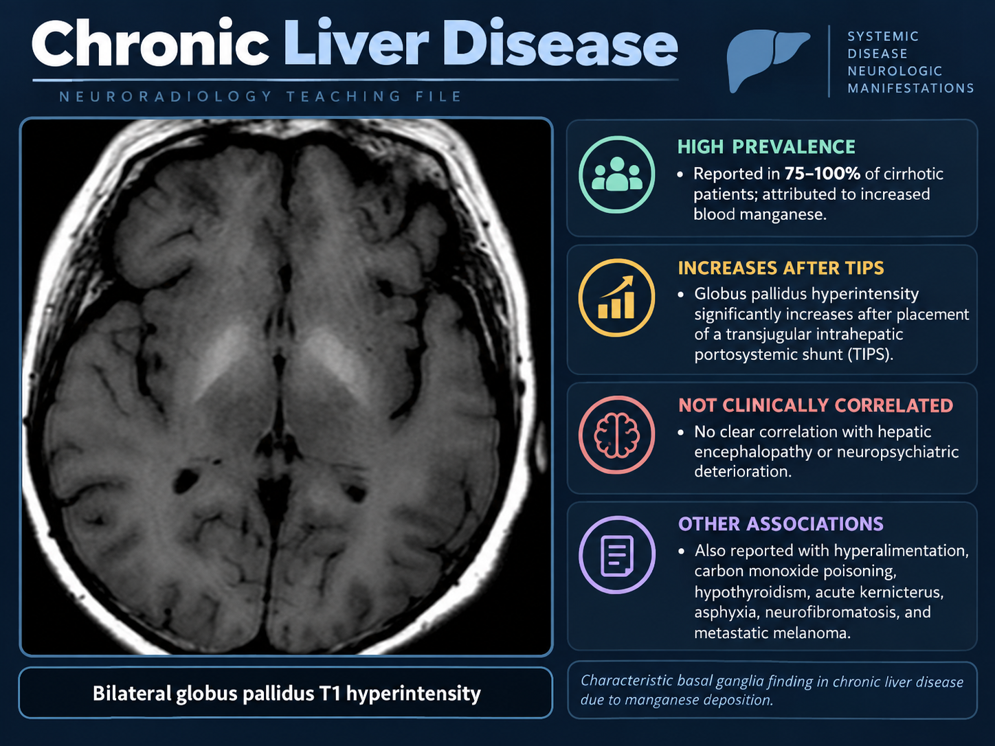
- Chronic hepatic encephalopathy shows bilateral symmetric globus pallidus T1 hyperintensity without CT correlate or T2 abnormality, reflecting manganese accumulation that can extend to substantia nigra, subthalamic nucleus, dentate nucleus, and tectal plate, typically reversing after liver transplantation.
- Manganese deposition from other sources (total parenteral nutrition, occupational welding/mining, ephedrone/methcathinone abuse, Crohn disease, or chronic PPI use) produces the same bilateral symmetric pallidal T1 pattern; the pallidal index quantifies severity and signal fades within 1–2 years of exposure cessation.
- Wilson disease presents with mixed T1/T2 signal abnormality in putamen, globus pallidus, caudate, and thalamus, with SWI hypointensity from copper deposition and characteristic findings of concurrent tectal-plate sign and 'face of the giant panda' sign.
- Primary familial brain calcification (Fahr disease) and other calcification produce T1 hyperintensity confirmed by CT hyperdensity and SWI/phase imaging; QSM with diamagnetic (negative susceptibility) signature reliably distinguishes calcium from paramagnetic hemorrhage/iron.
- Nonketotic hyperglycemic hemichorea–hemiballism (diabetic striatopathy) shows unilateral striatal T1 hyperintensity contralateral to symptoms, characteristically sparing the internal capsule and lacking mass effect, distinguishing it from hemorrhage; MRI sensitivity is 95% vs. 79% on CT with slower resolution.
- Subacute basal ganglia hemorrhage produces T1 hyperintensity from methemoglobin; hypertensive hemorrhage is the main cause and presents with unilateral lesions showing mass effect and edema.
- Carbon monoxide poisoning classically produces bilateral globus pallidus lesions that are hypodense on CT with T2/DWI/FLAIR brightness; T1 hyperintensity appears mainly when associated hemorrhage is present.
- Methanol poisoning causes bilateral putaminal necrosis that is often hemorrhagic (T1-bright) with FLAIR and CT showing putaminal involvement.
Differential Diagnosis
- Manganese deposition (hepatic encephalopathy, TPN, occupational exposure) — bilateral symmetric pallidal T1 hyperintensity without CT correlate, improving with source removal or liver transplantation.
- Primary familial brain calcification (Fahr) vs. other calcification — T1 hyperintensity confirmed by CT hyperdensity and diamagnetic QSM signature (negative susceptibility).
- Nonketotic hyperglycemic hemichorea–hemiballism — unilateral striatal T1 hyperintensity sparing internal capsule without mass effect, in diabetic patients with acute chorea.
- Subacute hemorrhage — unilateral T1 hyperintensity from methemoglobin showing mass effect and edema, primarily in hypertensive basal ganglia bleed.
- Wilson disease — mixed T1/T2 signal with SWI hypointensity (copper), concurrent panda sign and tectal findings in young patients with ceruloplasmin deficiency.
- Calcium vs. hemorrhage/iron — QSM quantitative susceptibility mapping is superior (κ 0.91) to GRE phase (κ 0.55), with cutoffs: <68 HU on CT, <15 mg/mL on dual-energy CT, and >38 ppb on QSM identifying calcium as diamagnetic.
Discussion
Bilateral symmetric T1 hyperintensity in the globus pallidus is the prototypical pattern for manganese accumulation and should prompt evaluation for liver disease, TPN history, occupational exposure, or chronic PPI use.
Unilateral striatal T1 hyperintensity in a diabetic patient with acute hemichorea/hemiballism has high MRI sensitivity (95%) and is distinguished from hemorrhage by the absence of mass effect and sparing of the internal capsule.
T1 signal alone is rarely specific for the underlying substance; correlation with non-contrast CT (calcification), T2/SWI phase imaging (iron/hemorrhage), and clinical history is essential for accurate diagnosis.
Wilson disease must be considered in young patients presenting with mixed basal ganglia T1/T2 signal, and slit-lamp examination for Kayser-Fleischer rings plus ceruloplasmin and 24-hour urinary copper testing confirms the diagnosis.
QSM (quantitative susceptibility mapping) with diamagnetic signature reliably distinguishes calcium from paramagnetic blood products and iron, superior to conventional GRE phase imaging for characterization of basal ganglia lesions.
Toxic/acute causes (carbon monoxide, methanol, acute hyperammonemia) require urgent recognition: CO produces bilateral globus pallidus hypodensity on CT with DWI brightness; methanol shows bilateral putaminal necrosis; hyperammonemia shows insular/cingulate changes with MRS glutamine elevation.
Reporting Pearls
Report the basal ganglia T1 hyperintensity by specifying: (1) laterality (unilateral vs. bilateral/symmetric), (2) location (pallidal vs. striatal), (3) T2/SWI appearance (hypointense, isointense, or hyperintense), (4) CT density (hyperdense suggesting calcium, hypodense suggesting edema/toxin), and (5) associated features (mass effect, edema, extension to other nuclei) — then anchor differential to the clinical context (liver disease, diabetes, exposure history, age).
Pitfalls
- Assuming T1 hyperintensity alone indicates a specific substance — always correlate with CT (for calcium), T2/SWI phase imaging (for iron/hemorrhage), and QSM (for quantitative susceptibility characterization).
- Confusing calcium and subacute hemorrhage on conventional imaging — QSM with diamagnetic (negative) susceptibility signature confirms calcium, while paramagnetic (positive) susceptibility indicates blood/iron.
- Missing unilateral striatal T1 hyperintensity in a diabetic with acute chorea as hemichorea–hemiballism and mistaking it for hemorrhage based on T1 brightness alone without assessing mass effect and internal capsule involvement.
- Overlooking toxic/acute encephalopathies (CO poisoning, methanol, acute hyperammonemia) that present with basal ganglia changes but require urgent recognition — always check CT density, DWI/FLAIR pattern, and clinical history for toxic exposure or metabolic derangement.