Basal ganglia calcification
Key Images
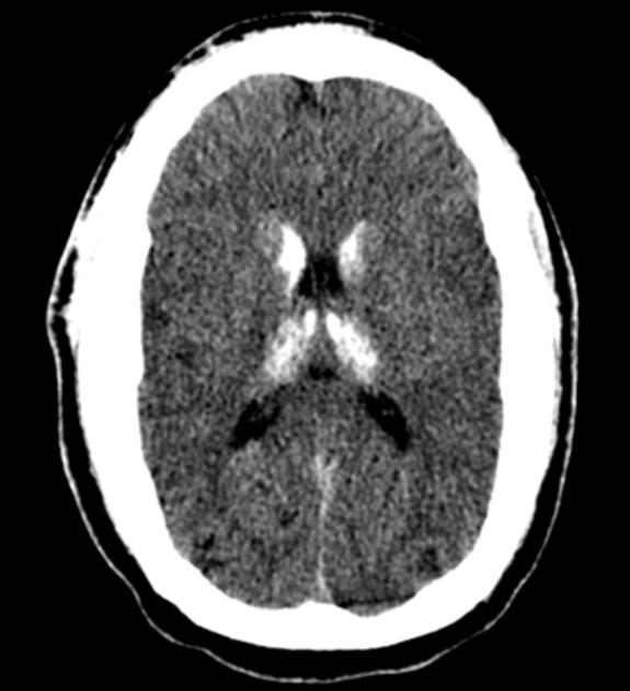
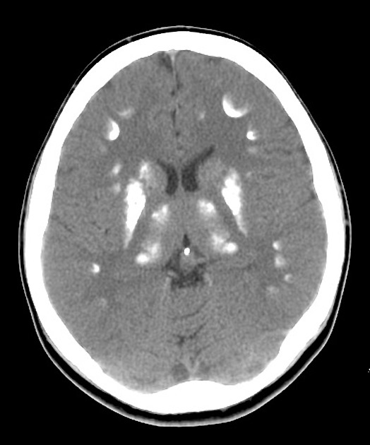
Look For First
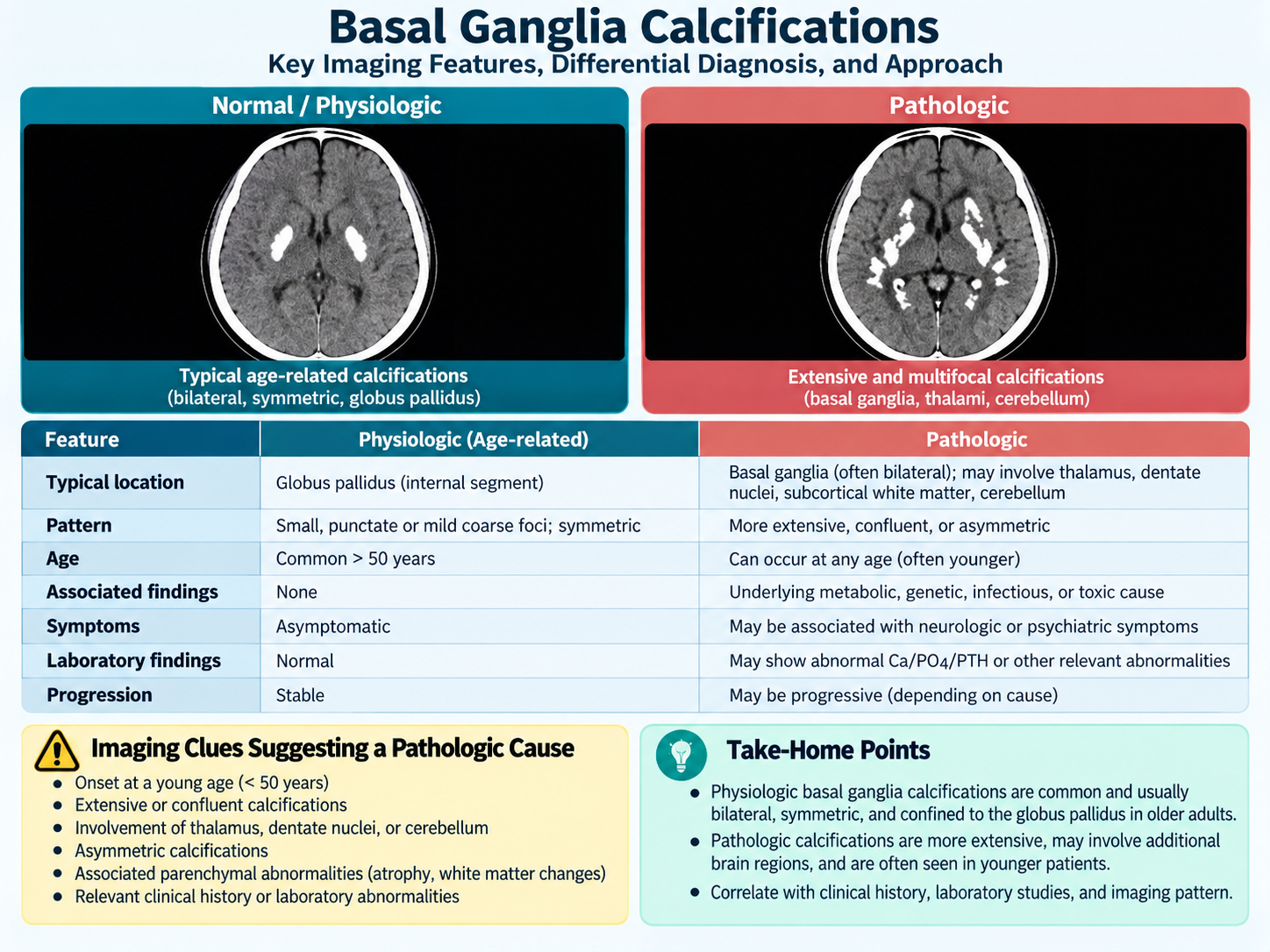
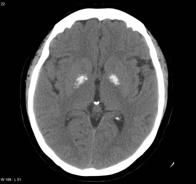
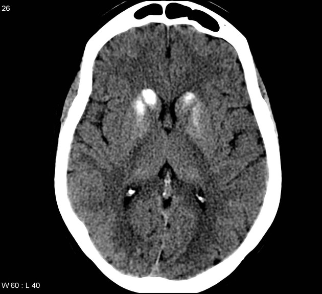
- Symmetric hyperdense deposits in the globus pallidus (internal segment) on CT brain
- Patient age: incidental finding if >60 years, pathologic finding unless proven otherwise if <40 years
- Bilaterality of calcification: bilateral suggests primary familial brain calcification; unilateral suggests alternative diagnoses
Key Image Findings
- CT is the gold standard for detection of basal ganglia calcification, showing hyperdense deposits in the basal ganglia nuclei with density indicating calcium-phosphate deposition.
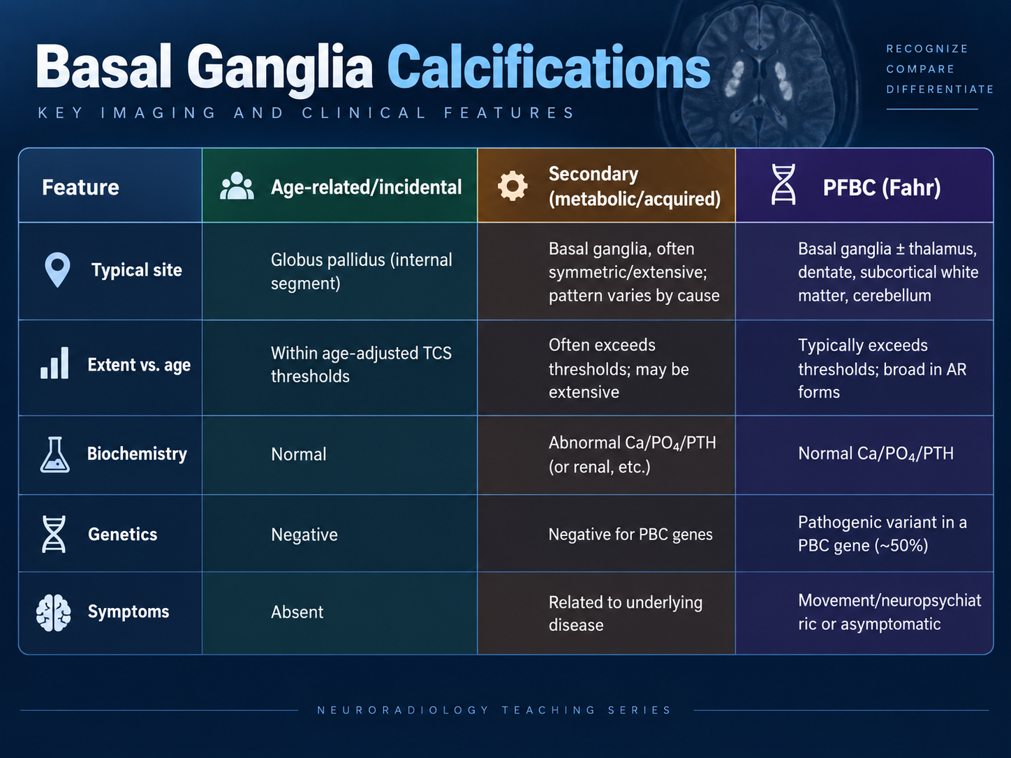
- Age-related/idiopathic calcification is predominantly located in the globus pallidus (internal segment), appears symmetric, is vascular in nature, and is found in 15-20% of elderly patients (up to 20-30% in patients >60 years) without clinical correlates.
- Primary familial brain calcification (PFBC/Fahr disease) presents as bilateral calcifications predominantly affecting the microvascular system, diagnosed after exclusion of secondary causes, with onset typically in the fourth to sixth decades.
- Secondary acquired calcification can occur in >50 systemic conditions including hypoparathyroidism, pseudohypoparathyroidism, hyperparathyroidism, chronic kidney disease, post-radiotherapy, post-chemotherapy, carbon monoxide poisoning, TORCH infections, HIV, and mitochondrial disorders.
- Phosphocalcic metabolism disorders (low PTH with hypocalcemia and hyperphosphatemia) promote calcium-phosphate deposition in up to 74% of idiopathic hypoparathyroidism patients, producing calcifications indistinguishable from PFBC on imaging.
- Total Calcification Score is used for age-adjusted quantification to distinguish incidental age-related calcification from pathologic causes.
- Associated imaging findings may include changes from toxic exposures (carbon monoxide, lead poisoning), infectious etiologies (tuberculosis, toxoplasmosis, neurocysticercosis), or radiation/chemotherapy effects on the brain.
- Unilateral calcification may suggest developmental venous anomalies or other alternative diagnoses rather than primary familial brain calcification.
Differential Diagnosis
- Age-related incidental calcification (globus pallidus predominant, symmetric, >60 years, no clinical symptoms) vs. pathologic PFBC/Fahr disease (bilateral, <40 years, family history, progressive neuropsychiatric symptoms)
- Hypoparathyroidism/pseudohypoparathyroidism (low PTH with hypocalcemia, hyperphosphatemia, biochemical abnormalities) vs. PFBC (normal calcium-phosphate metabolism, genetic disorder)
- Primary familial brain calcification (bilateral, monogenic disorder, family history or asymptomatic) vs. secondary metabolic causes (unilateral or atypical distribution, systemic metabolic dysfunction)
- Post-infectious calcification (history of TORCH, tuberculosis, toxoplasmosis, or neurocysticercosis) vs. idiopathic age-related changes
- Toxic exposure calcification (carbon monoxide poisoning, chronic lead encephalopathy, mineralizing microangiopathy from radiation/chemotherapy) vs. inherited metabolic disorders (MELAS, mitochondrial diseases, pantothenate kinase-associated neurodegeneration)
- Unilateral or atypical calcification patterns (developmental venous anomaly, Down syndrome, tuberous sclerosis, Cockayne syndrome) vs. symmetric bilateral basal ganglia distribution
Discussion
Basal ganglia calcification occurs in approximately 1% of all CT brain scans and up to 15-20% (20-30% in patients >60 years), making it a common incidental finding that must be age-contextually interpreted.
Age is the critical discriminator: calcification in patients <40 years should be considered pathologic until proven otherwise, while the same finding in patients >60 years is typically benign and age-related.
The diagnostic approach requires integration of imaging (CT calcification pattern and distribution), biochemistry (calcium, phosphate, PTH levels), clinical presentation, and genetic testing when indicated.
Primary familial brain calcification is a monogenic disorder with incomplete penetrance (asymptomatic rates 9-56%), so family history and symptoms may be absent, and diagnosis requires bilateral basal ganglia calcification plus exclusion of secondary causes.
More than 50 systemic conditions can cause secondary basal ganglia calcification, spanning metabolic disorders, infections, toxic exposures, autoimmune conditions, and acquired insults, making comprehensive clinical history essential.
Hypoparathyroidism and pseudohypoparathyroidism account for a significant proportion of secondary calcification (up to 74% of idiopathic hypoparathyroidism patients develop CT calcifications), and biochemical screening is mandatory.
Reporting Pearls
Describe the location (globus pallidus vs. other nuclei), distribution (symmetric vs. asymmetric, bilateral vs. unilateral), and extent (using Total Calcification Score if quantifying) of basal ganglia calcification, always noting patient age and recommending biochemical (calcium, phosphate, PTH) and genetic evaluation if calcification is present in a patient <40 years or has atypical features.
Pitfalls
- Overcalling incidental age-related calcification in elderly patients as pathologic, leading to unnecessary workup and patient anxiety when the finding is benign physiologic mineralization.
- Underevaluating basal ganglia calcification in younger patients (<40 years) as merely incidental without pursuing secondary causes such as hypoparathyroidism, metabolic disorders, or genetic PFBC.
- Relying on imaging alone without biochemical evaluation (calcium, phosphate, PTH) to distinguish idiopathic age-related calcification from metabolic disorders such as hypoparathyroidism.
- Failing to recognize atypical calcification patterns (unilateral, extranuclear locations, asymmetric distribution) that may suggest alternative diagnoses including developmental venous anomalies, infectious etiologies, or inherited neurodegenerative conditions.