Transient physiological myometrial contraction
Key Images
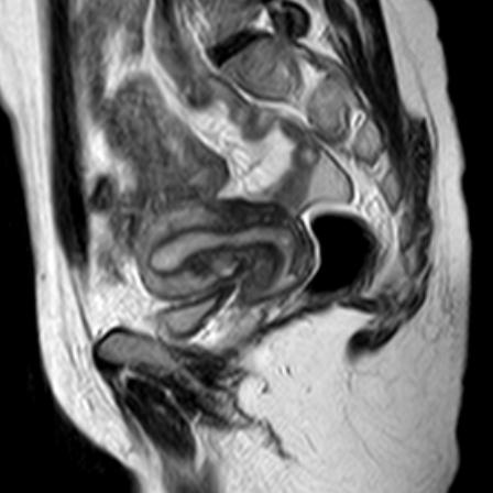
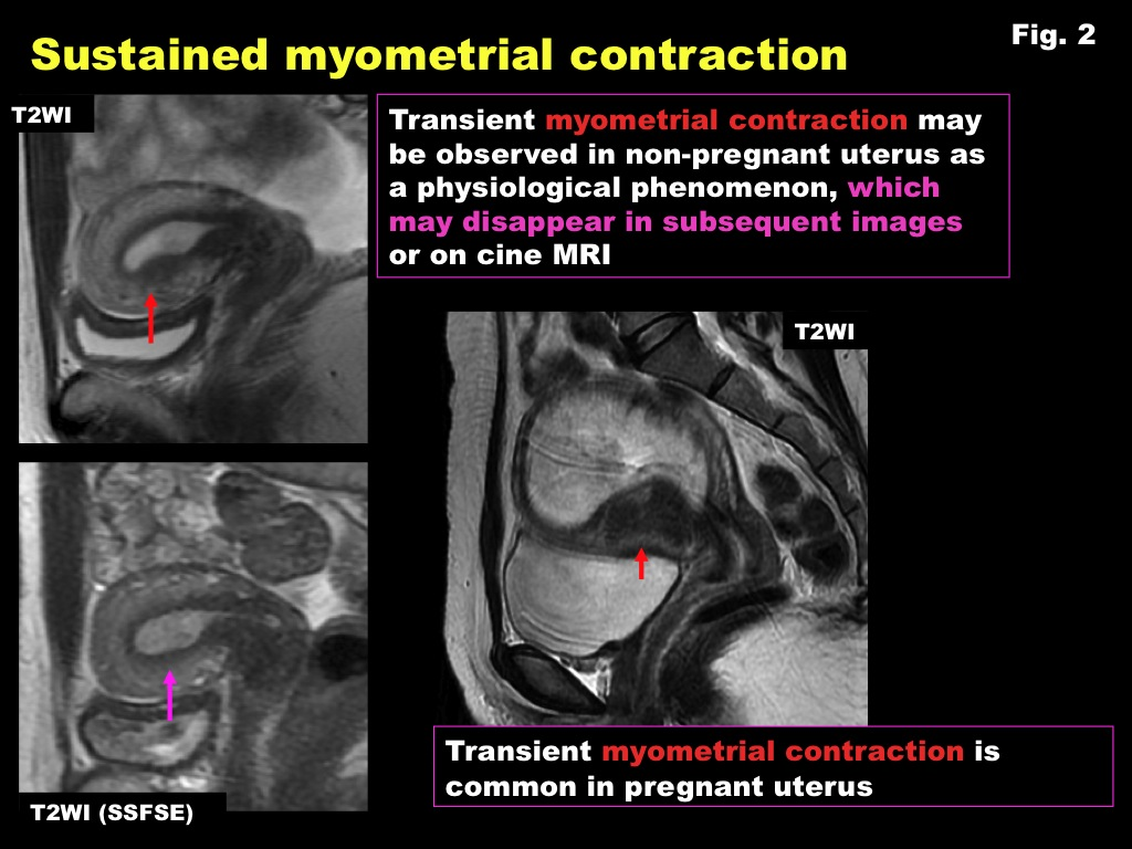
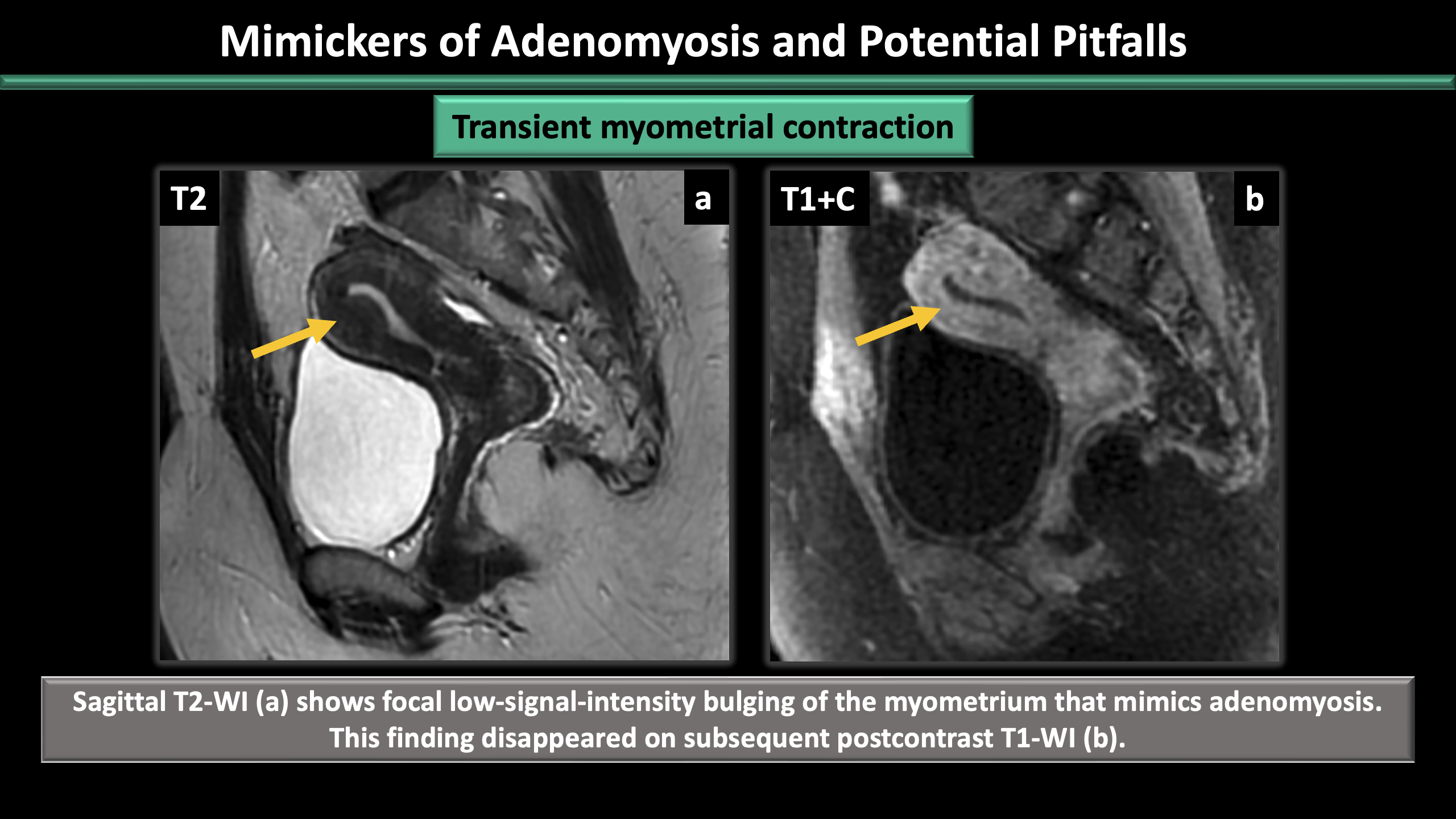
Look For First
- Focal low-T2 myometrial band that changes appearance, resolves, or shifts position on sequences acquired minutes apart
- Absence of internal T2-hyperintense foci and T1-hyperintense foci that would indicate adenomyosis
- Homogeneous low-T2 signal without tissue abnormality — a temporal signature rather than pathology
Key Image Findings
- Focal low-T2 band that thickens the myometrium and can bulge into or efface the junctional zone or indent the endometrial cavity.
- Signal is homogeneous and low on T2 with no internal bright foci, reflecting muscle contraction rather than structural disease.
- Key distinguishing feature: the finding appears on one sequence (e.g., sagittal T2) and is absent, shifted, or altered in size and shape on another sequence acquired minutes later (e.g., subsequent axial T2).
- Smoothly tapering margins that preserve the underlying junctional zone once the contraction relaxes.
- Resolution or change in morphology on repeat imaging confirms the diagnosis and excludes adenomyosis.
- May focally thicken the myometrium but lacks the fixed, reproducible appearance and internal foci characteristic of adenomyosis.
- Multiplanar imaging is essential; a single isolated sequence showing low-T2 myometrial thickening cannot reliably exclude adenomyosis without temporal confirmation.
- The contraction is a masquerade of transient muscle activity, not a tissue abnormality, and should resolve with patient relaxation or on delayed imaging.
Differential Diagnosis
- Adenomyosis: distinguished by fixed, reproducible low-T2 signal across all sequences and planes, junctional zone thickening ≥12 mm, ill-defined margins, and presence of T2-hyperintense foci (ectopic endometrial glands) and T1-hyperintense foci (microhemorrhage).
- Subacute leiomyoma: would show internal signal heterogeneity and T1 hyperintensity from hemorrhage; remains fixed on repeated imaging.
- Focal myometrial edema: typically shows diffuse or multifocal distribution and T2 hyperintensity rather than low-T2 signal.
- Endometrial carcinoma: demonstrates invasion across the junctional zone with abnormal enhancement and does not change with repeated imaging.
Discussion
Transient contractions are a normal physiologic phenomenon during pelvic MRI and occur due to spontaneous uterine muscle activity, most commonly in reproductive-age women.
The temporal behavior on multiplanar or sequential imaging is the gold standard for distinguishing contraction from adenomyosis; absence of internal T2 and T1 bright foci further supports transient contraction.
Adenomyosis shows a junctional zone thickening ≥12 mm (diagnostic) or 8–12 mm (indeterminate), with ancillary criteria including globular uterine enlargement, striations radiating from the endometrium, and pseudowidening of the endometrium.
A sustained contraction imaged on a limited protocol represents a genuine pitfall; multiplanar T2 and, when equivocal, delayed repeat sequencing is the reliable arbiter.
Junctional zone-to-total myometrial thickness ratio >40% supports a diagnosis of adenomyosis and is a useful ancillary measurement when fixed JZ thickening is present.
Resolution of the focal finding on repeat imaging confirms the transient nature and should prompt confidence in excluding adenomyosis.
Reporting Pearls
When reporting a focal low-T2 myometrial band, document its appearance on at least two different planes or sequences and note whether it persists unchanged (adenomyosis) or shows temporal change/resolution (transient contraction); if uncertain, explicitly recommend repeat imaging or note the limitation of a single-timepoint acquisition for definitive exclusion of adenomyosis.
Pitfalls
- Single-sequence diagnosis: relying on a single isolated sequence showing focal low-T2 myometrial thickening without multiplanar correlation can lead to false-positive adenomyosis diagnoses; always check orthogonal planes and prior timepoints.
- Sustained contraction on limited protocol: a contraction imaged only once on a limited acquisition cannot be distinguished from adenomyosis and represents the main reason multiplanar T2 imaging is essential.
- Absence of internal foci does not exclude adenomyosis: while T2-hyperintense and T1-hyperintense foci are fairly specific for adenomyosis, their absence does not rule it out; JZ thickening criteria and temporal behavior must be integrated.
- Overcalling adenomyosis based on mild focal JZ thickening: JZ thickening 8–12 mm is indeterminate and requires integration with other imaging features and clinical context; transient contractions may mimic mild JZ abnormality.