Cystic adenomyosis
Key Images
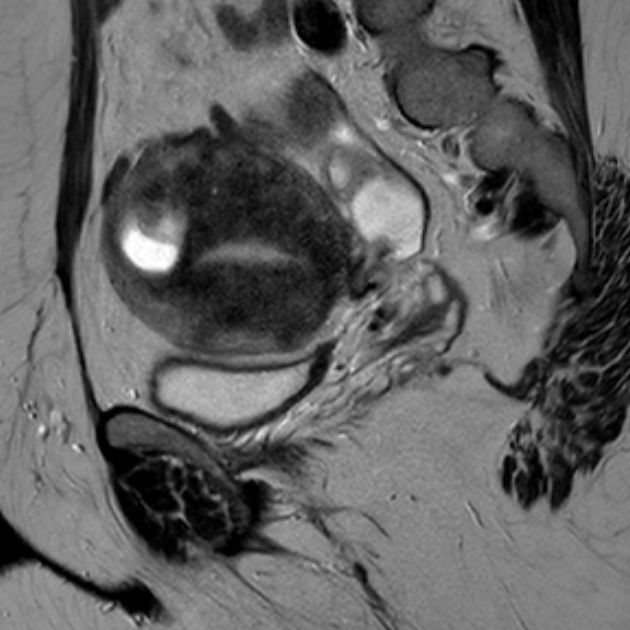
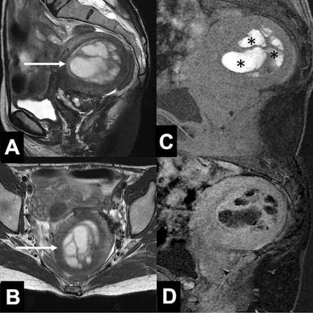
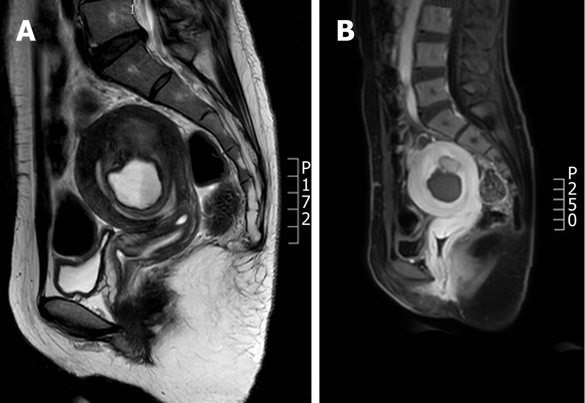
Look For First
- Grossly visible intramyometrial cyst >1 cm with T1-hyperintense (hemorrhagic) central content that does not suppress on fat saturation
- Thick low-T2 signal hypertrophied myometrial rim surrounding the cavity
- Blood products on T2* or SWI with blooming artifact, indicating hemosiderin and repeated bleeding
- Absence of enhancing mural nodule or soft-tissue component
Key Image Findings
- Central cystic cavity typically >1 cm in maximum diameter, filled with hemorrhagic fluid at varying stages of degradation from repeated cyclic bleeding of ectopic endometrium.
- T1-weighted imaging shows high signal intensity centrally (hallmark finding), representing methemoglobin and other paramagnetic blood products that do not suppress on fat-saturation sequences.
- T2-weighted imaging demonstrates variable signal (often high signal) with potential fluid–fluid levels or low-signal hemosiderin/iron deposition products from chronic hemorrhage.
- Surrounding rim of hypertrophied myometrium with persistently low T2 signal, reflecting smooth muscle hyperplasia and chronic inflammatory response.
- T2* gradient echo and susceptibility-weighted imaging (SWI) show blooming artifact from blood products (hemosiderin), which supports the diagnosis and confirms hemorrhagic etiology.
- No enhancing soft-tissue nodule or mural enhancement on post-contrast imaging, which helps exclude cystic neoplasm or hemorrhagic malignancy.
- Juvenile cystic adenomyosis occurs in women <30 years (often <20) and may clinically mimic an obstructed müllerian anomaly but is distinguished by a normal endometrial cavity and normal overall uterine anatomy.
- Adult-onset cystic adenomyosis typically arises within the setting of more diffuse adenomyotic change and tends to involve older patients.
Differential Diagnosis
- Non-communicating rudimentary or cavitary uterine horn (accessory cavitated uterine mass, ACUM): principal mimic in young patients; distinguish by confirming a normal ipsilateral endometrial cavity and normal müllerian anatomy elsewhere
- Cystic degeneration of a leiomyoma: typically lacks intrinsic T1-hyperintense hemorrhagic content within the cyst; fibroids show less characteristic high T1 signal
- Endometrial or hemorrhagic cystic neoplasm: absence of enhancing mural nodule or solid component on post-contrast imaging argues strongly against malignancy
- Hematometra from müllerian obstruction: normal endometrial cavity is preserved in cystic adenomyosis, whereas hematometra distends the endometrial cavity
Discussion
Cystic adenomyosis is defined by a blood-filled cavity within the myometrium that arises from repeated hemorrhage into an ectopic endometrial focus—the cyclic nature of this bleeding produces the characteristic imaging appearance.
Juvenile cystic adenomyosis (age <30, often <20) presents with severe dysmenorrhea that is often refractory and disproportionate to the size of the lesion, frequently mimicking an obstructed müllerian anomaly.
The absence of enhancing nodular tissue is a critical feature that helps exclude cystic neoplasms and argues for a benign hemorrhagic process rather than malignancy.
T1-hyperintense content that does not suppress on fat-saturation is the diagnostic linchpin, reflecting the presence of blood products (methemoglobin and paramagnetic iron) rather than fat.
Management decisions depend on age and fertility goals, ranging from hormonal suppression and observation to local cystectomy or hysterectomy in refractory cases.
The combination of imaging findings—hemorrhagic T1-bright cyst, low-T2 myometrial rim, blood products on T2*, and lack of enhancement—in the appropriate clinical context (young patient with severe dysmenorrhea) makes the diagnosis highly specific.
Reporting Pearls
Describe the finding as: "An intramyometrial cyst measuring [size] with T1-hyperintense hemorrhagic content and a surrounding rim of low-signal myometrial hypertrophy, without enhancing soft-tissue component, consistent with cystic adenomyosis." Include note of the clinical context (age, symptoms) and confirm normal endometrial cavity and normal müllerian anatomy to exclude mimics. The diagnostic linchpin is a hemorrhagic (T1-bright) intramyometrial cyst with a hypertrophic low-T2 rim and no enhancing nodule, set in the appropriate clinical context.
Pitfalls
- Confusing cystic adenomyosis with a non-communicating rudimentary uterine horn (ACUM) in young patients presenting with dysmenorrhea; carefully assess the ipsilateral endometrial cavity and overall uterine anatomy to differentiate
- Misinterpreting a hemorrhagic cyst as a simple cyst if T1 weighting and hemosiderin blooming are not carefully reviewed; always check T1, T2*, and fat-sat sequences
- Overestimating the significance of a small T2-bright glandular focus and calling it cystic adenomyosis; true cystic adenomyosis requires a grossly visible cavity >1 cm
- Assuming an enhancing mural nodule or solid component indicates malignancy and missing the distinction from a cystic degenerated fibroid or neoplasm; the lack of enhancement favors benign adenomyosis