Pelvis / Uterus / MRI
Adenomyosis
Adenomyosis (or uterine adenomyosis) is a common uterine condition of ectopic endometrial tissue in the myometrium, sometimes considered a spectrum of endometriosis. Although most commonly asymptomatic, it may present with menorrhagia and dysmenorrhea.
Pelvic imaging (i.e. ultrasound, MRI) may show characteristic findings, commonly including focal or diffuse myometrial bulkiness, which may be asymmetrical, and heterogeneous myometrium.
Transient myometrial contraction is a physiological phenomenon which may mimic focal adenomyosis.
Key Images
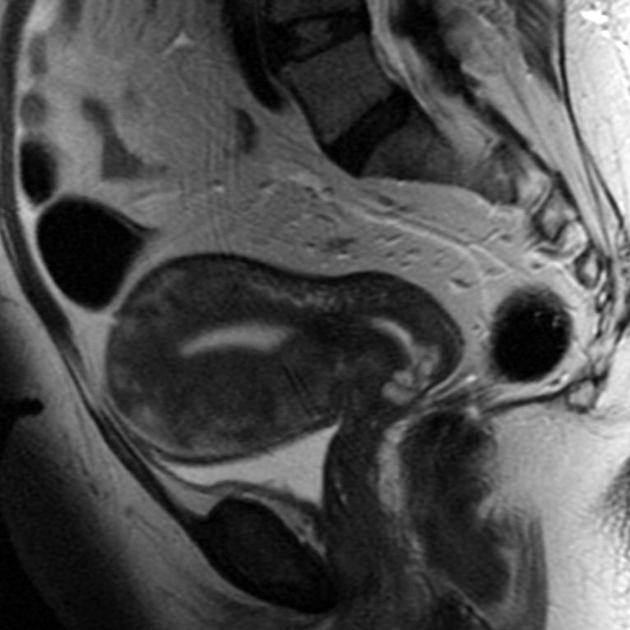
Adenomyosis 01
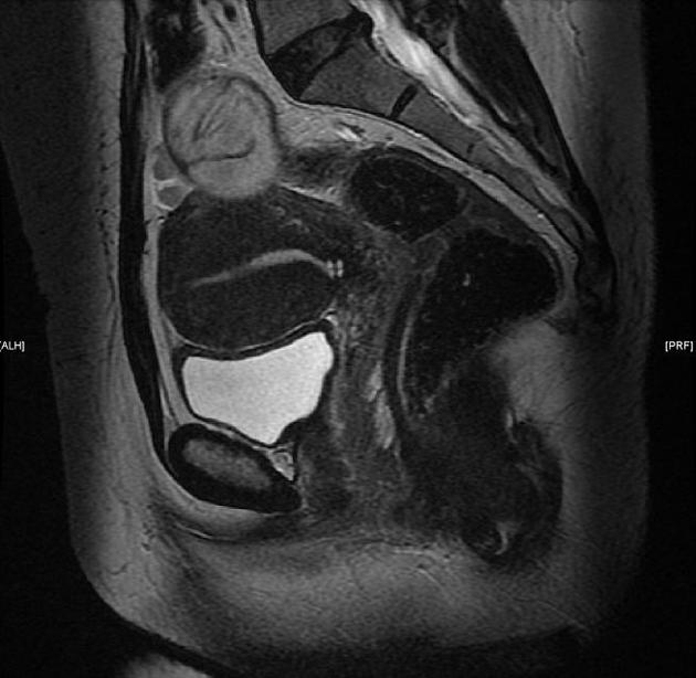
Adenomyosis 03
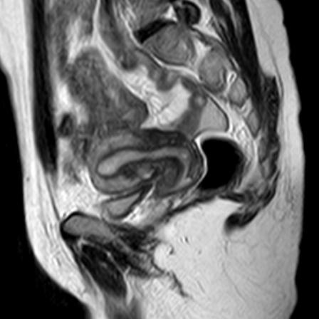
Transient physiological myometrial contraction
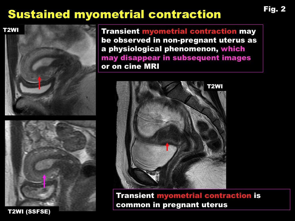
Transient physiological myometrial contraction
Look For First
- Junctional zone thickening, often greater than 12 mm. This is a test
- Ill-defined low T2 signal in the myometrium rather than a sharply marginated mass.
- Tiny internal T2 bright or T1 bright foci suggesting ectopic glands or hemorrhagic cysts.
Key Image Findings
- Test 3Diffuse or focal thickening of the junctional zone, often greater than 12 mm.
- Ill-defined low T2 signal within the myometrium rather than a sharply marginated mass.
- Punctate or small linear high T2 foci within the myometrium representing ectopic endometrial glands/cysts.
- T1 hyperintense foci may be present when hemorrhagic content is present.
- Globular enlargement of the uterus may be seen in diffuse disease.
- Subendometrial striations extending from endometrium into myometrium can support the diagnosis.
Differential Diagnosis
- Leiomyoma, especially when focal adenomyosis forms a pseudomass.
- Transient uterine contractions causing focal junctional zone thickening.
- Endometrial carcinoma with myometrial invasion in the appropriate clinical setting.
- Postsurgical or postprocedural myometrial change.
Discussion
Adenomyosis may be diffuse or focal and often coexists with leiomyomas and/or endometriosis.
MRI is particularly helpful when ultrasound is indeterminate or when mapping the extent of disease matters clinically.
A poorly defined low T2 myometrial abnormality with tiny internal bright foci generally favors adenomyosis over leiomyoma.
Always assess whether apparent junctional zone thickening could reflect transient uterine contraction; repeat or compare sequences when needed.
Reporting Pearls
When present on screening MRI, describe whether findings are diffuse or focal, whether there are internal cystic/hemorrhagic foci, and whether coexisting fibroids or endometriosis are also present.
Pitfalls
- Do not overcall adenomyosis on a single sequence if the abnormality may represent transient myometrial contraction.
- A sharply marginated T2 dark mass with typical whorled appearance favors leiomyoma over adenomyosis.
- Junctional zone assessment is less reliable after menopause, with hormonal therapy, or when image quality is limited.