Anastomosing hemangioma
Key Images
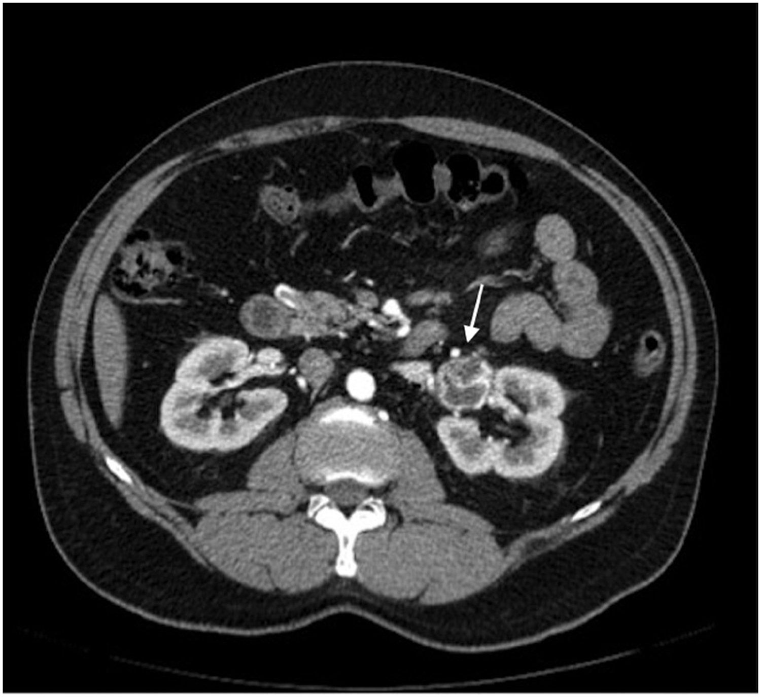
Look For First
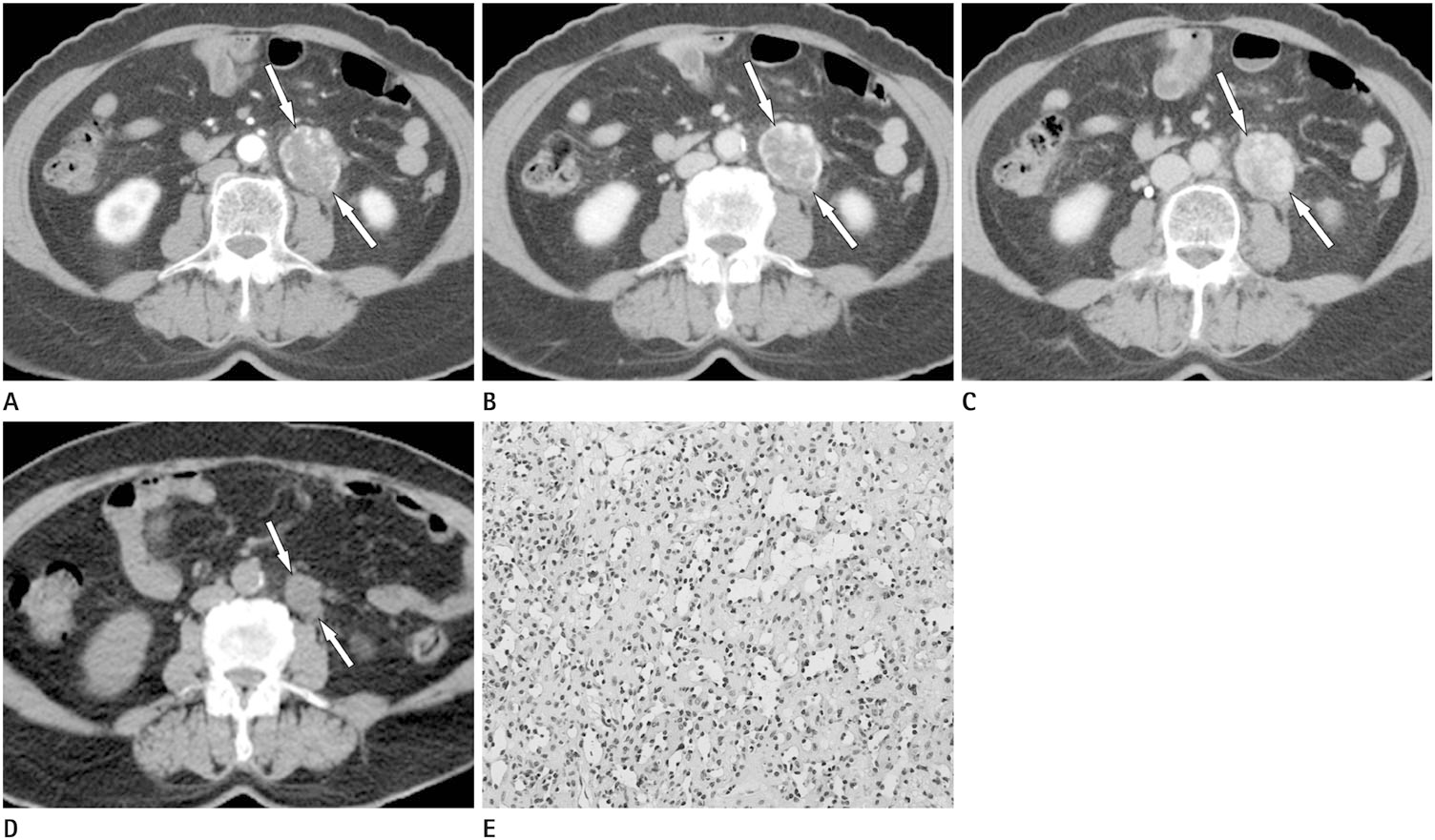
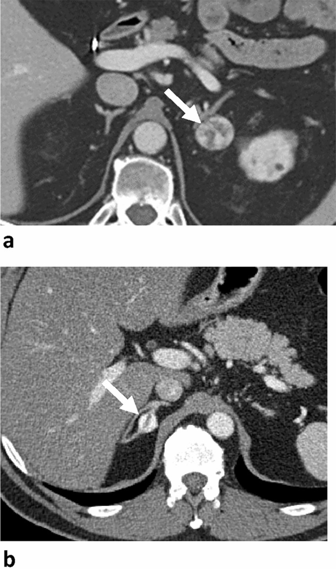
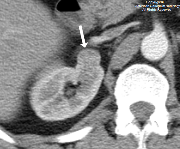
- Well-circumscribed hypervascular mass in kidney, retroperitoneum, or para-aortic region
- Peripheral nodular enhancement with progressive centripetal fill-in on multiphasic imaging—resembling a hepatic hemangioma pattern
- Slowly growing or stable lesion on follow-up imaging
Key Image Findings
- On CT: circumscribed hypervascular lesion with heterogeneous attenuation related to vascular spaces, soft-tissue elements, or intralesional fat; smaller lesions may be occult or difficult to characterize.
- Characteristic enhancement pattern is avid and shows early peripheral nodular enhancement with progressive centripetal fill-in on later phases, potentially becoming more homogeneous on delayed imaging.
- Larger renal lesions may appear as well-demarcated heterogeneous solid masses that can mimic renal cell carcinoma or other malignancies.
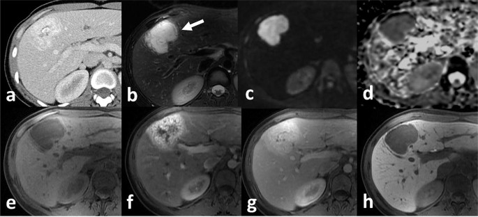
- On MRI: high T2 signal from vascular components and occasional high T1 signal related to intralesional fat.
- Lesions are typically unilateral and range from approximately 0.6 to 5 cm in size, with an indolent clinical course showing slow growth or stability during imaging surveillance.
Differential Diagnosis
- Renal cell carcinoma—distinguished by more aggressive imaging features, necrosis, and rapid growth, whereas AH shows slow growth and benign enhancement pattern
- Paraganglioma—typically larger, more likely to demonstrate necrosis or calcification, and located along sympathetic chain
- Solitary fibrous tumor—usually larger with heterogeneous signal and may show calcification; lacks the characteristic centripetal enhancement of AH
- Undifferentiated pleomorphic sarcoma—often larger with necrosis and more aggressive imaging features
- Hyaline vascular Castleman disease—shows more homogeneous enhancement rather than the peripheral nodular pattern of AH
- Angiosarcoma—demonstrates aggressive features, rapid growth, and local invasion, unlike the indolent course of AH
Discussion
Anastomosing hemangioma is histologically characterized by anastomosing, sinusoidal-like vascular channels lined by endothelial cells, distinguishing it from conventional cavernous hemangiomas which contain larger dilated vascular spaces.
The progressive centripetal fill-in enhancement pattern observed on multiphasic imaging resembles that of a typical hepatic hemangioma and is a key distinguishing feature that should raise suspicion for AH.
AH has an indolent clinical course and favorable prognosis, with reported lesions showing slow growth or stability during imaging surveillance over several years, with no documented local recurrences or metastases.
Imaging alone may not reliably distinguish AH from aggressive neoplasms, particularly when the lesion is large, heterogeneous, or in an unusual location, making tissue diagnosis important for definitive diagnosis.
When imaging features are sufficiently reassuring and the diagnosis is supported by clinical context or pathology, observation may be appropriate; surgical resection is often performed when malignancy cannot be excluded or when diagnostic uncertainty warrants tissue diagnosis.
AH is a recently recognized entity in the 2020 WHO classification and most commonly arises in the genitourinary tract, with additional reported cases in liver, gastrointestinal tract, retroperitoneum, and soft tissues.
Reporting Pearls
Describe a slowly growing, well-circumscribed hypervascular renal or retroperitoneal mass with peripheral nodular enhancement and progressive centripetal fill-in on multiphasic imaging as consistent with anastomosing hemangioma, emphasizing the benign enhancement pattern that resembles hepatic hemangioma and distinguishes it from aggressive malignancies.
Pitfalls
- Mistaking a larger, heterogeneous AH for renal cell carcinoma or other malignancy due to overlapping size and appearance; review multiphasic imaging for the characteristic centripetal fill-in pattern
- Failing to recognize the slow growth pattern on follow-up imaging as a benign feature that suggests AH rather than an aggressive neoplasm
- Overlooking intralesional fat on imaging, which is a reported feature that can support the diagnosis and help distinguish AH from other hypervascular masses
- Not considering AH in the differential diagnosis of hypervascular retroperitoneal or para-aortic masses; this location and imaging pattern should prompt consideration of benign vascular lesions before assuming malignancy