Incidental Mediastinal Masses
Reference Images
Localize / Describe
First step
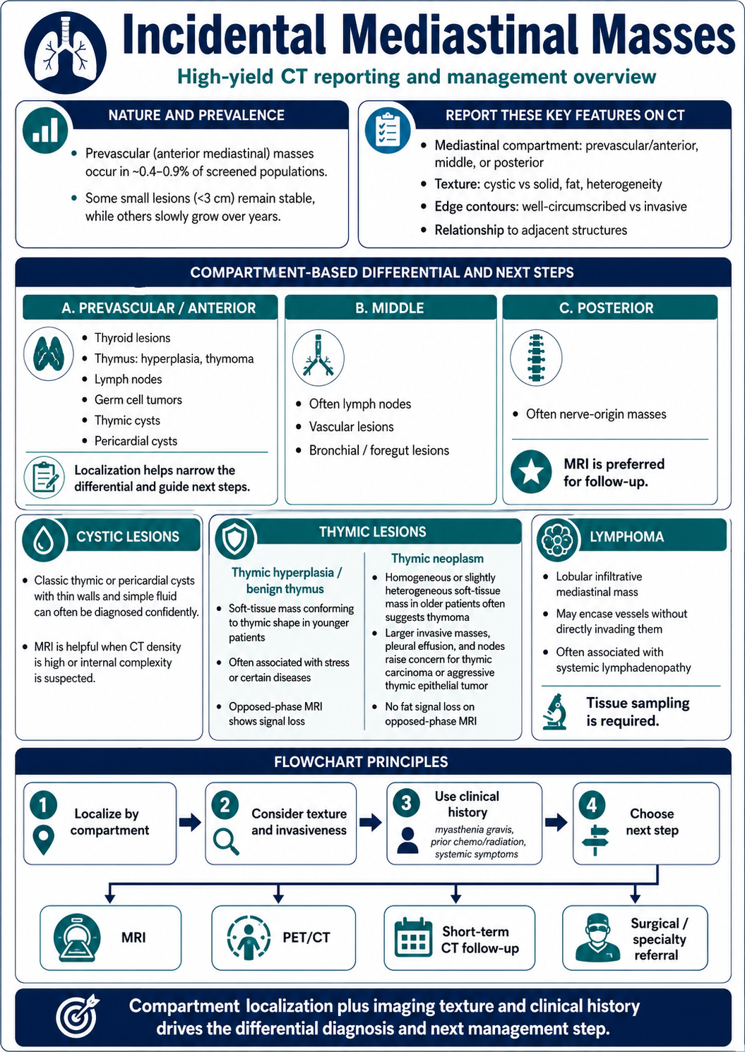
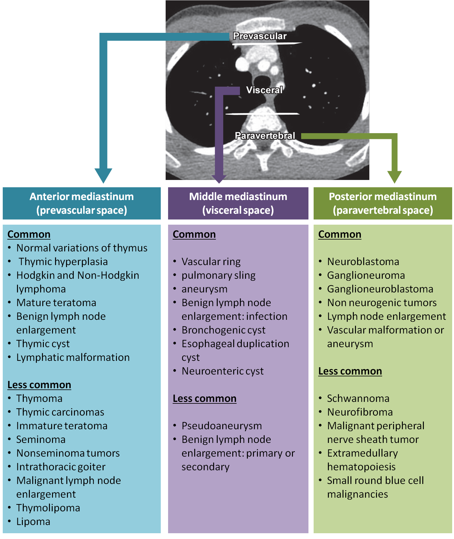
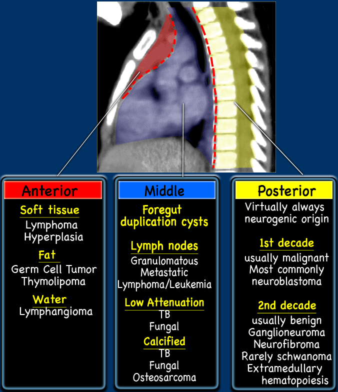
Localize by ITMIG CT compartment: prevascular/anterior, visceral/middle, or paravertebral/posterior. Localization drives the differential and the next test.
Report these CT features
- Mediastinal compartment: prevascular/anterior, visceral/middle, or paravertebral/posterior.
- Texture: cystic versus solid, fat, calcification, heterogeneity, or fluid attenuation.
- Edge contour: well-circumscribed versus infiltrative or invasive.
- Relationship to adjacent structures: airway, vessels, pericardium, pleura, chest wall, or spine.
- Clinical context: myasthenia gravis, prior chemo/radiation, systemic symptoms, malignancy history.
Compartment differential
- Prevascular/anterior: thymic cyst, thymic epithelial tumor, thymic hyperplasia, germ cell tumor, lymphoma, ectopic thyroid/parathyroid.
- Visceral/middle: lymphadenopathy, bronchogenic cyst, foregut duplication cyst, esophageal lesion, vascular lesion.
- Paravertebral/posterior: neurogenic tumor, meningocele, extramedullary hematopoiesis, paraspinal abscess, spine-related lesion.
Cystic / Benign
Classic cystic lesion
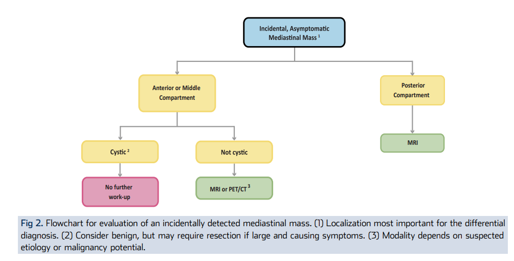
A well-circumscribed cystic anterior or middle mediastinal lesion without solid enhancement or invasive features often needs no immediate work-up once confidently characterized.
Benign cyst features
- Well-circumscribed margins.
- Simple fluid attenuation or homogeneous fluid signal on MRI.
- No enhancing solid component.
- No invasive features or suspicious lymphadenopathy.
- Typical thymic, pericardial, bronchogenic, or duplication cyst appearance.
When MRI helps
- CT density is higher than simple fluid and cyst versus solid lesion is uncertain.
- Internal complexity, proteinaceous/hemorrhagic content, or thymic cyst is suspected.
- Posterior compartment mass is present; MRI is generally preferred for follow-up/characterization.
- Anterior lesion is indeterminate and thymic cyst versus thymic epithelial lesion is the key question.
Escalation
Worrisome features
- Infiltrative or irregular margins.
- Enhancing solid component in a cystic lesion.
- Rapid growth or large invasive mass.
- Invasion of vessels, pericardium, pleura, chest wall, airway, or spine.
- Associated bulky lymphadenopathy, pleural nodules, or pleural effusion.
- Fat plus calcification in an anterior mass, suggesting germ cell tumor/teratoma.
- Symptoms or myasthenia gravis, suggesting clinically significant thymic disease.
Next steps
- Anterior or middle compartment cystic lesion: no further work-up if confidently benign; MRI if CT is indeterminate.
- Anterior or middle compartment noncystic lesion: MRI or PET/CT depending on suspected etiology and malignancy potential.
- Posterior compartment lesion: MRI for characterization and follow-up.
- Suspicious mediastinal mass: urgent thoracic surgery/oncology evaluation and tissue diagnosis as clinically appropriate.
Sources
- ACR incidental thoracic CT guidance for mediastinal findings.
- ITMIG CT-based mediastinal compartment system.
- Anterior mediastinal lesion literature supports MRI characterization and patient-tailored follow-up for small nonaggressive lesions.