Incidental Mediastinal Lymph Nodes
Reference Images
Assess / Report
Nature and prevalence
Incidental enlarged mediastinal nodes are seen in roughly 1-6% of screening CT populations. Etiologies range from benign reactive nodes to metastases, lymphoma, or germ cell tumors.
Reporting elements
- Measure and report short-axis diameter.
- Describe texture and density: homogeneous versus heterogeneous, necrosis, calcification, or fatty hilum.
- Note distribution: single station, multistation mediastinal/hilar, symmetric, or bulky.
- Report FDG uptake when PET/CT is available.
- Consider the clinical context: known cancer, infection, ILD, sarcoidosis, heart failure, or systemic symptoms.
Benign-favoring features
- Smooth borders, homogeneous attenuation, and central fatty hilum.
- Calcified nodes without a known calcifying malignancy usually suggest prior granulomatous disease.
15 mm Threshold
Key rule
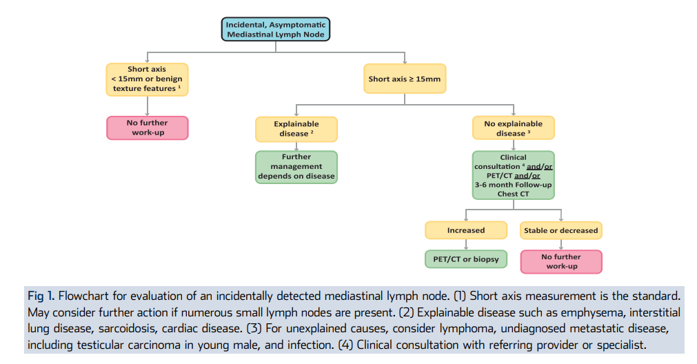
Short-axis 15 mm is the practical decision threshold for incidental, asymptomatic mediastinal lymph nodes.
Short axis <15 mm
- No further evaluation is usually needed if the chest is otherwise normal and morphology is benign.
- Use caution if there are numerous small nodes, suspicious morphology, or a known malignancy.
Short axis >=15 mm
- If explainable by ILD, infection, sarcoidosis, emphysema, or cardiac disease, management depends on that underlying condition.
- If unexplained, consider clinical consultation, PET/CT, and/or 3-6 month follow-up chest CT.
- If increased on follow-up, proceed to PET/CT or tissue sampling.
- If stable or decreased on follow-up, no further nodal work-up is usually needed.
Escalation / Staging
Escalate when
- Unexplained node is >=15 mm short axis.
- Node is enlarging, bulky, heterogeneous, necrotic, or highly FDG avid.
- Known malignancy or thoracic primary is present.
- Distribution or symptoms suggest lymphoma.
- Young male with suspicious bulky mediastinal nodes raises concern for seminoma or non-seminomatous germ cell tumor.
Next steps
- Clinical consultation or specialty referral.
- Short-interval chest CT, commonly 3-6 months.
- FDG PET/CT when metabolic characterization or staging is needed.
- Tissue sampling, usually EBUS-TBNA, when malignancy is suspected.
Sources
- ACR incidental thoracic findings guidance for incidental mediastinal lymph nodes.
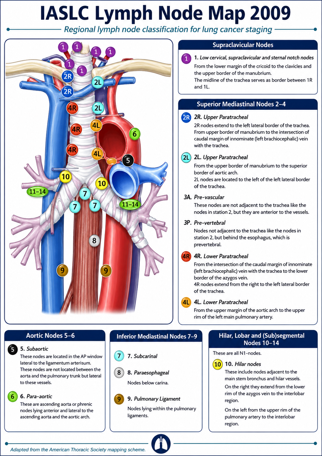
- IASLC lymph node map for anatomic station localization when lung cancer staging is relevant.
- Use oncologic staging pathways rather than the incidental algorithm when known malignancy is present.