Cervical Lymph Nodes on CT/MRI
Reference Images
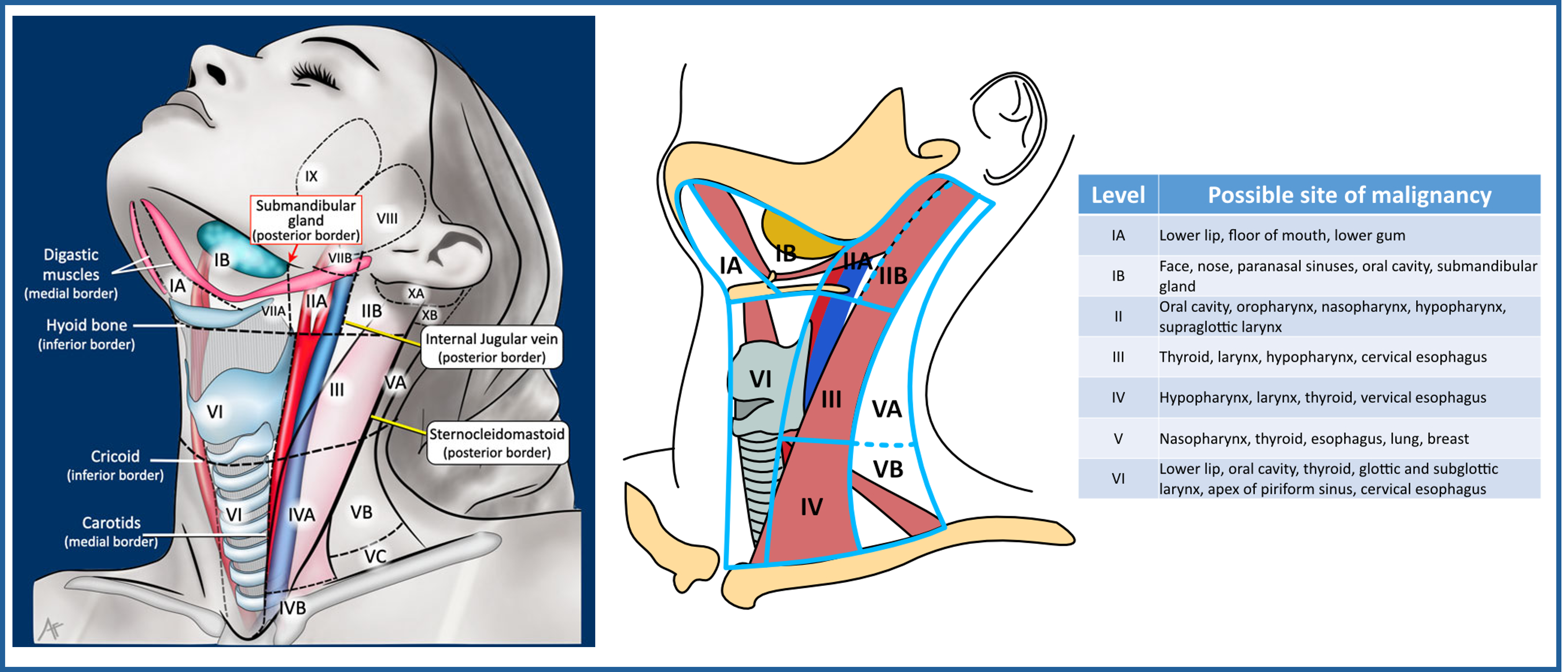
Measure / Thresholds
Key point
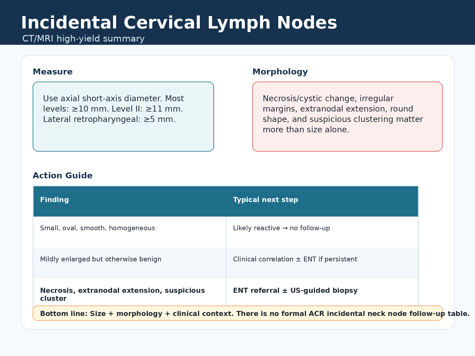
There is no dedicated ACR incidental cervical lymph node table. Use short-axis size, morphology, location, and clinical context.
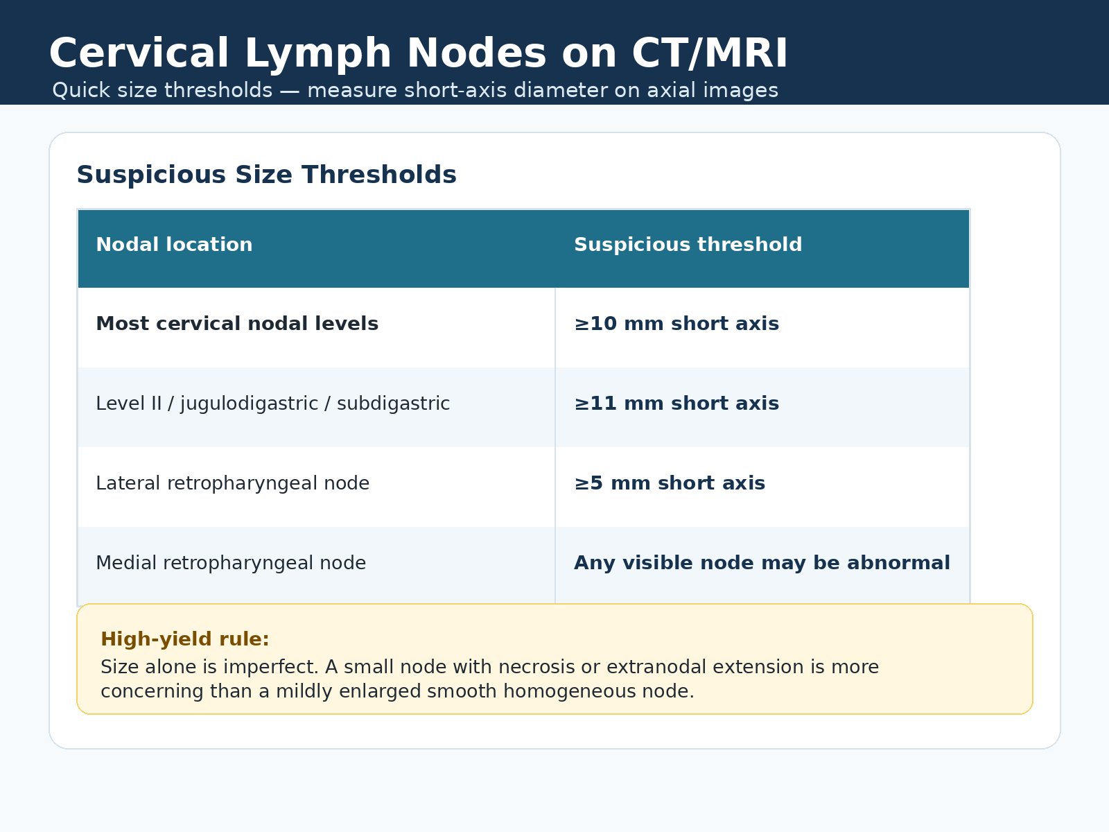
Size thresholds
- Measure cervical nodes by short-axis diameter on axial CT/MRI.
- Most cervical nodal levels: suspicious at 10 mm or greater short axis.
- Level II / jugulodigastric / subdigastric: suspicious at 11 mm or greater short axis.
- Lateral retropharyngeal node: suspicious at 5 mm or greater short axis.
- Medial retropharyngeal node: any visible node may be abnormal.
Clustered borderline nodes
- Three or more borderline nodes can be suspicious, especially in an expected drainage basin.
- Common cluster criterion: each node 8 mm or greater short axis.
- Level II / subdigastric cluster: each node greater than 9 mm short axis.
Morphology / Action
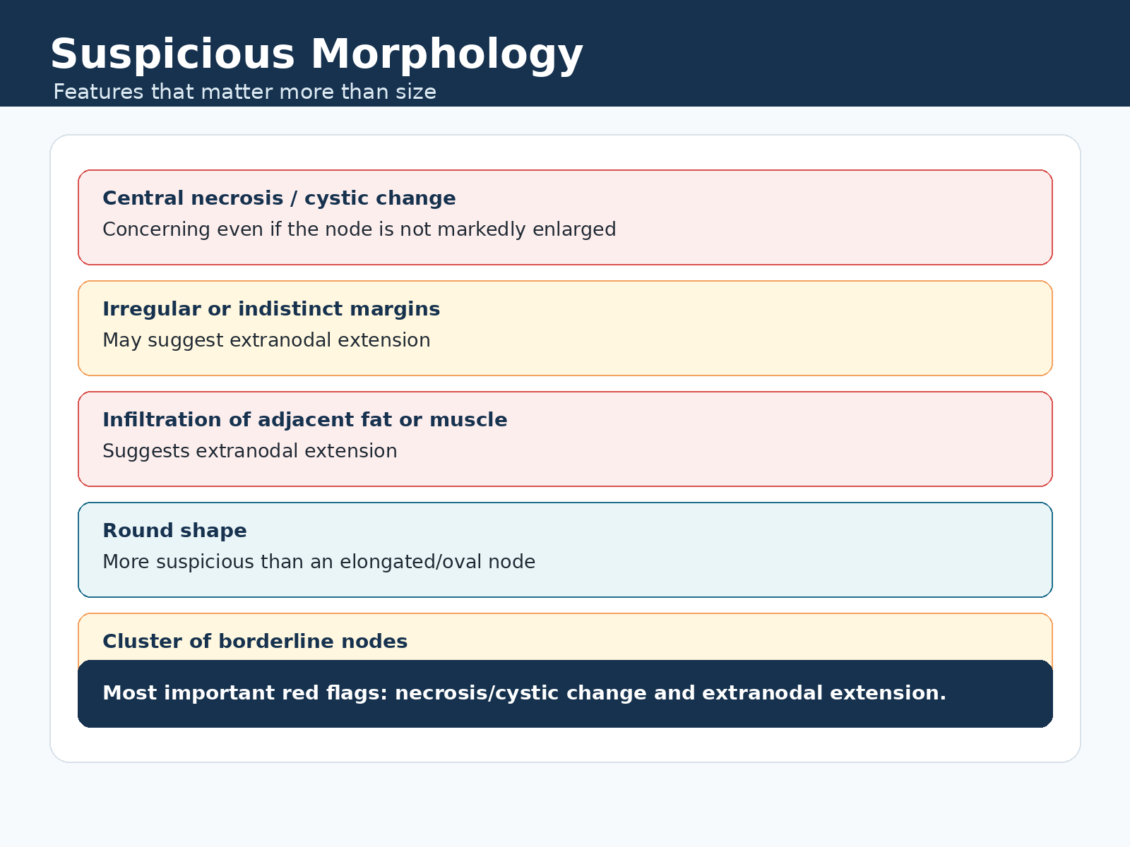
Suspicious morphology
- Central necrosis, cystic change, hypoenhancing center, or rim enhancement.
- Irregular or indistinct margins, infiltration of adjacent fat or muscle, or extranodal extension.
- Round shape is more suspicious than elongated/oval morphology.
- Suspicious clustering can matter even if no single node crosses the usual size threshold.
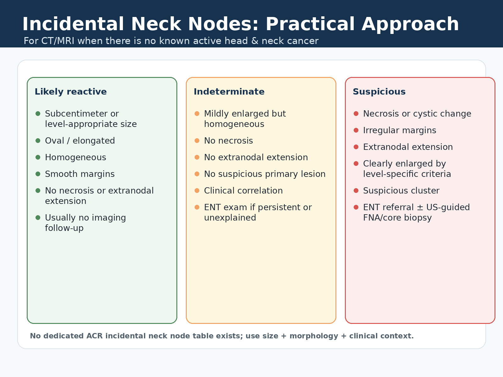
Practical categories
- Likely reactive: subcentimeter or level-appropriate, oval, homogeneous, smooth, and no necrosis or extranodal extension.
- Indeterminate: mildly enlarged but homogeneous, no necrosis, no extranodal extension, and no suspicious primary lesion.
- Suspicious: necrosis/cystic change, irregular margins, extranodal extension, clearly abnormal size, suspicious cluster, or associated primary lesion.
Report Language
Reusable phrases
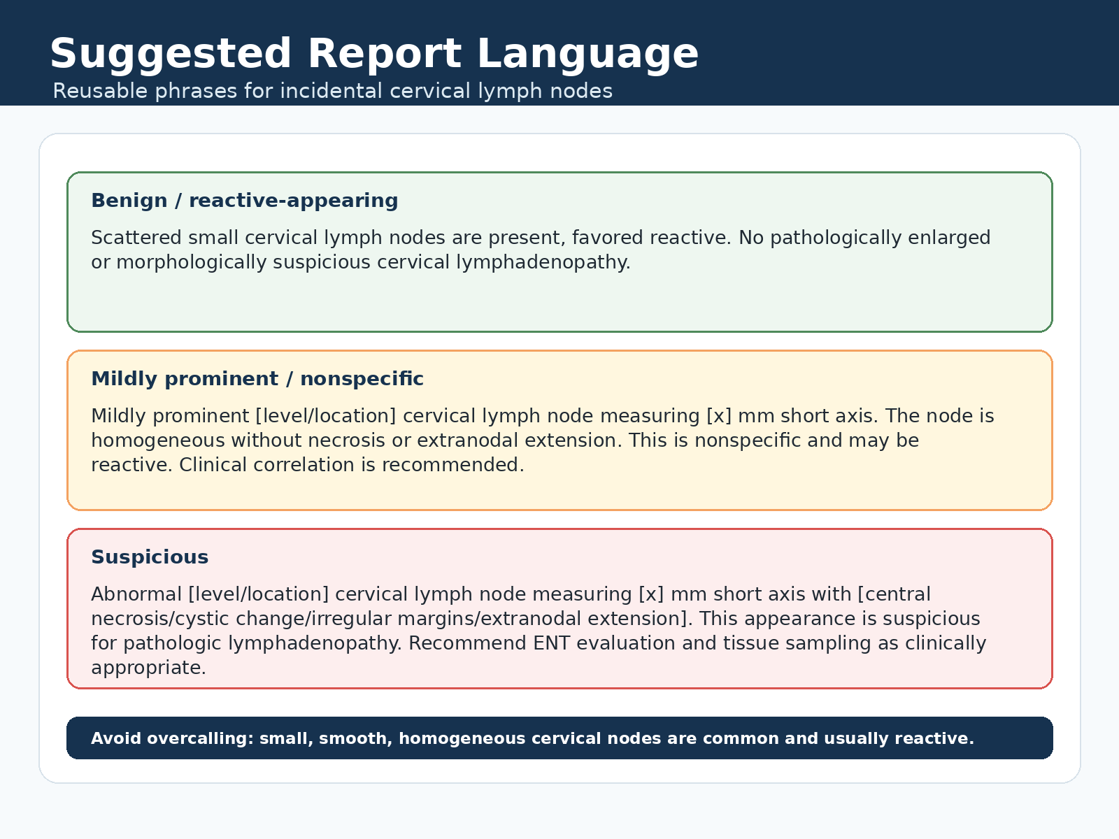
- Benign/reactive: Scattered small cervical lymph nodes are present, favored reactive. No pathologically enlarged or morphologically suspicious cervical lymphadenopathy.
- Mildly prominent/nonspecific: Mildly prominent [level/location] cervical lymph node measuring [x] mm short axis, homogeneous without necrosis or extranodal extension. Clinical correlation is recommended.
- Suspicious: Abnormal [level/location] cervical lymph node measuring [x] mm short axis with [necrosis/cystic change/irregular margins/extranodal extension], suspicious for pathologic lymphadenopathy.
Next steps
- Likely reactive nodes usually need no imaging follow-up.
- Indeterminate nodes: clinical correlation and ENT exam if persistent, enlarging, or unexplained.
- Suspicious nodes: ENT referral and tissue sampling, typically ultrasound-guided FNA/core biopsy, as clinically appropriate.
- Known or suspected cancer: dedicated staging work-up or oncology pathway.