Mounier-Kuhn syndrome
Key Images
Look For First
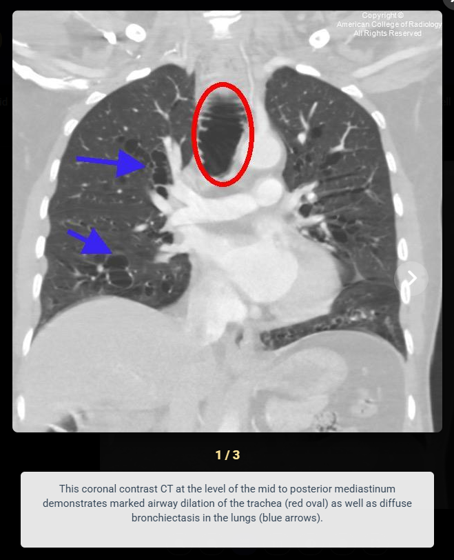
- Marked dilatation of the trachea with transverse diameter >25 mm in men (>21 mm in women)
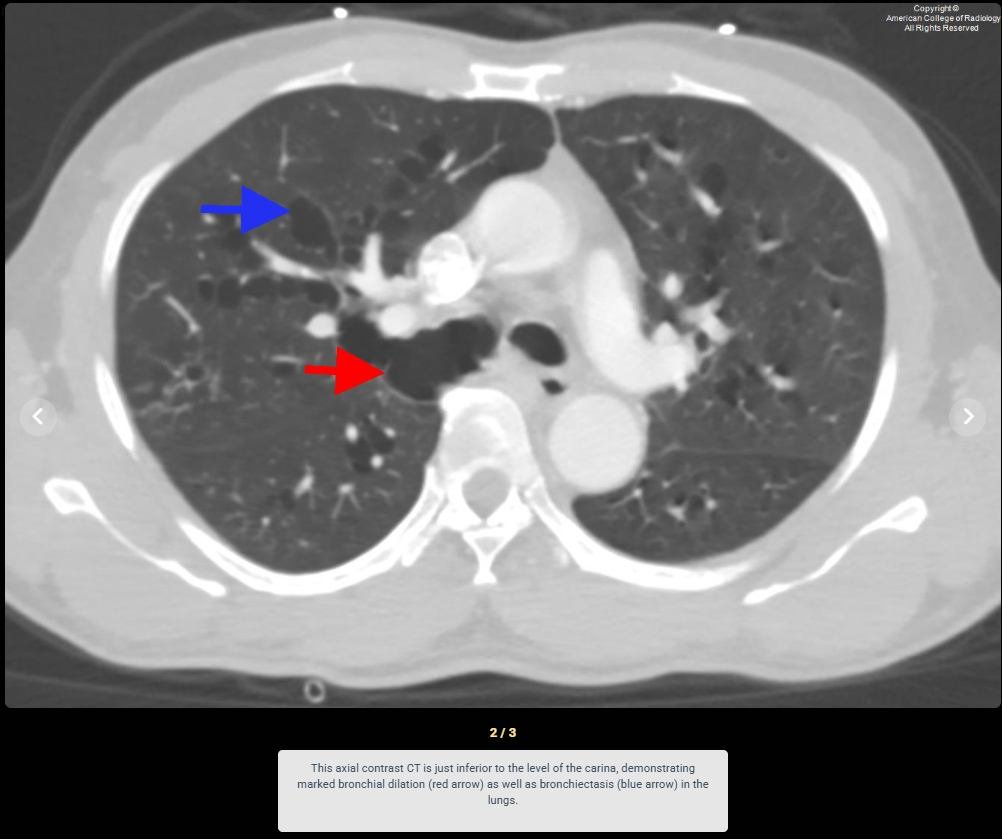
- Dilatation of proximal main bronchi with diameter >18-21 mm in men (>17.4-19.8 mm in women)
- Diffuse bronchiectasis throughout the lungs
- Characteristic diverticula between tracheal and bronchial cartilage rings
Key Image Findings
- Transverse and sagittal tracheal diameter greater than 25 and 27 mm respectively in men, or greater than 21 and 23 mm in women, indicating severe tracheomegaly.
- Left and right main bronchus diameters exceeding 18 and 21 mm in men, or 17.4 and 19.8 mm in women, demonstrating proximal bronchomegaly.
- Formation of diverticula between the rings of cartilage in the trachea and bronchi resulting from atrophy of elastic fibers and muscularis.
- Diffuse bronchiectasis affecting multiple lung segments with bronchial wall thickening.
- Increased wall compliance and loss of structural support causing potential collapse of the tracheobronchial tree.
- Chest X-ray and CT readily demonstrate the characteristic findings of tracheobronchomegaly and associated bronchiectasis.
Differential Diagnosis
- Bronchiectasis: distinguished by the presence of severe tracheobronchomegaly disproportionate to the degree of bronchiectasis in Mounier-Kuhn syndrome.
- Williams-Campbell syndrome: presents with bronchiectasis but lacks the marked tracheobronchomegaly that is pathognomonic for Mounier-Kuhn syndrome.
- Chronic obstructive pulmonary disease: causes airway changes but does not produce the severe structural dilatation and diverticula characteristic of tracheobronchomegaly.
- Tracheomalacia: presents with airway collapse but typically without the permanent structural dilatation and diverticula seen in Mounier-Kuhn syndrome.
- Bronchitis: presents with airway inflammation but lacks the structural abnormalities of marked tracheal and bronchial dilatation.
- Cystic fibrosis: may be associated with Mounier-Kuhn syndrome but is distinguished by different genetic and clinical features.
Discussion
Mounier-Kuhn syndrome results from atrophy of longitudinal elastic fibers and surrounding muscularis within the tracheobronchial tree, leading to increased wall compliance and characteristic diverticula formation.
The etiopathogenesis is multifactorial, combining genetic factors (suspected autosomal recessive inheritance) with acquired causes including smoking, environmental pollutants, mechanical ventilation, and complications of infections.
Associations with Ehlers-Danlos syndrome, Marfan syndrome, cystic fibrosis, and primary ciliary dyskinesia suggest underlying connective tissue or ciliary dysfunction may predispose to disease development.
Diagnosis relies primarily on imaging recognition using specific radiological criteria rather than clinical presentation, as symptoms are nonspecific and overlap with common respiratory diseases.
Treatment is severity-dependent: asymptomatic patients benefit from smoking cessation and vaccination, while symptomatic patients may require mucolytic therapy, airway clearance techniques, or in severe cases, stent placement or laser tracheoplasty.
Chronic recurrent lower respiratory tract infections result from increased wall compliance, collapse of the airway, and impaired mucociliary clearance in the dilated tracheobronchial tree.
Reporting Pearls
Clearly document the absolute tracheal and main bronchial diameters in millimeters, comparing against sex- and age-specific diagnostic criteria, and describe the distribution and degree of associated bronchiectasis and diverticula to establish the diagnosis of Mounier-Kuhn syndrome with confidence.
Pitfalls
- Failing to compare measured tracheal and bronchial diameters to sex-specific diagnostic criteria, which differ between men and women and may lead to missed or incorrect diagnosis.
- Attributing the bronchiectasis to primary bronchiectasis rather than recognizing it as a secondary finding in Mounier-Kuhn syndrome.
- Overlooking the characteristic diverticula between cartilage rings, which are a distinguishing feature that helps differentiate this condition from other causes of airway dilatation.
- Confusing Mounier-Kuhn syndrome with tracheomalacia or bronchitis without recognizing the permanent structural changes and marked dilatation that define tracheobronchomegaly.