Brainstem Anatomy
Key levels, cranial nerves, nuclei, surface landmarks, and major tracts. Select the diagram to enlarge it, then use the level guide below to review each labeled structure.
Labeled Brainstem Diagram
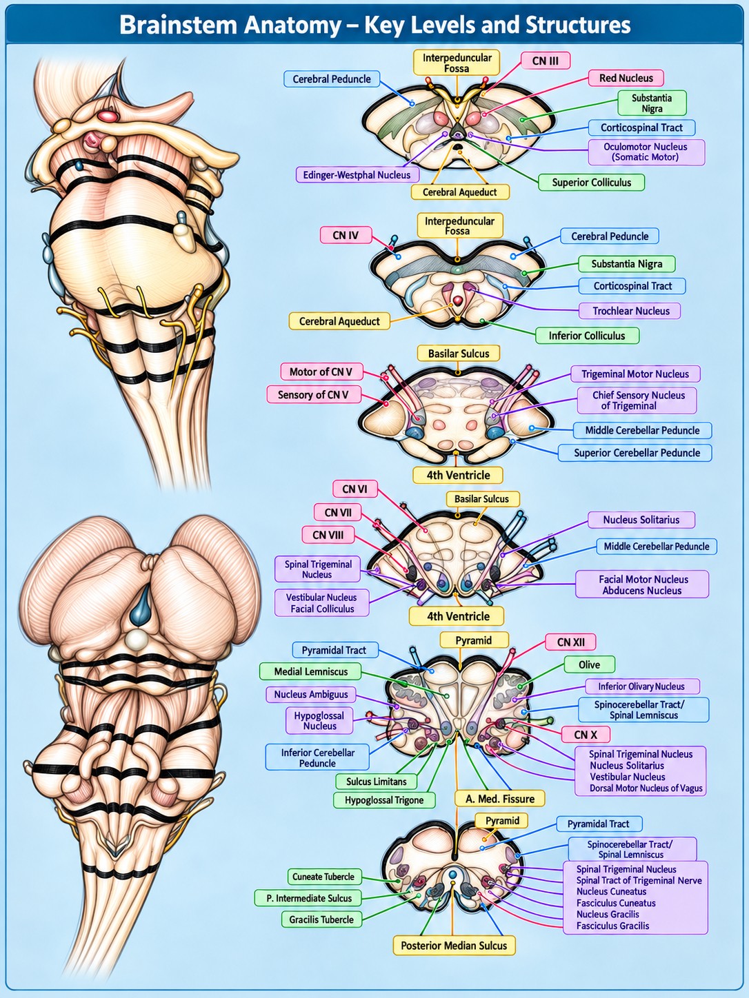
Select the image to enlarge · Press your browser’s back button or select Close to return
Midbrain
Superior- and inferior-colliculus levels
- Cerebral peduncle
- Ventral midbrain bundle carrying descending corticospinal, corticobulbar, and corticopontine fibers.
- Interpeduncular fossa
- Ventral midline depression between the cerebral peduncles where CN III emerges.
- CN III (oculomotor nerve)
- Motor supply to most extraocular muscles and levator palpebrae; carries parasympathetic fibers for pupil constriction and accommodation.
- Red nucleus
- Motor-related tegmental nucleus involved in coordination and cerebellar pathways.
- Substantia nigra
- Pigmented midbrain nucleus involved in basal ganglia movement circuits.
- Corticospinal tract
- Major descending voluntary motor pathway from cortex through brainstem to spinal cord.
- Oculomotor nucleus (somatic motor)
- CN III motor nucleus for most extraocular muscles and levator palpebrae superioris.
- Edinger–Westphal nucleus
- Parasympathetic nucleus associated with CN III for pupillary constriction and accommodation.
- Cerebral aqueduct
- Narrow CSF channel through the midbrain connecting the third and fourth ventricles.
- Superior colliculus
- Dorsal midbrain structure involved in visual reflexes and orienting eye and head movements.
- CN IV (trochlear nerve)
- Motor nerve to the superior oblique muscle; the only cranial nerve that exits dorsally.
- Trochlear nucleus
- CN IV motor nucleus; its fibers decussate before dorsal exit.
- Inferior colliculus
- Dorsal midbrain auditory relay projecting toward the thalamus.
Pons
Trigeminal and facial-colliculus levels
- Motor root of CN V
- Trigeminal motor fibers supplying the muscles of mastication and other first-arch derivatives.
- Sensory root of CN V
- Trigeminal fibers carrying facial touch, pain, temperature, and proprioception.
- Basilar sulcus
- Ventral midline groove of the pons accommodating the basilar artery.
- Trigeminal motor nucleus
- CN V motor nucleus supplying the muscles of mastication.
- Chief sensory nucleus of trigeminal
- Processes discriminative touch and pressure from the face.
- Middle cerebellar peduncle
- Large pontocerebellar connection carrying fibers important for motor planning and coordination.
- Superior cerebellar peduncle
- Major cerebellar outflow pathway toward the midbrain and thalamus.
- Fourth ventricle
- CSF-filled space between the brainstem and cerebellum, continuous superiorly with the aqueduct.
- CN VI (abducens nerve)
- Motor nerve to the lateral rectus muscle, which abducts the eye.
- CN VII (facial nerve)
- Motor supply to facial-expression muscles; also carries taste and parasympathetic fibers.
- CN VIII (vestibulocochlear nerve)
- Conveys hearing and balance information from the inner ear.
- Spinal trigeminal nucleus
- Receives facial pain, temperature, and crude-touch information; extends into the medulla and upper cervical cord.
- Vestibular nucleus
- Processes head-motion and balance input for posture and eye movements.
- Facial colliculus
- Floor-of-fourth-ventricle prominence formed by the abducens nucleus and looping facial nerve fibers.
- Nucleus solitarius
- Visceral sensory nucleus receiving taste and other visceral afferents.
- Facial motor nucleus
- CN VII motor nucleus for muscles of facial expression.
- Abducens nucleus
- CN VI motor nucleus for lateral rectus and coordinated horizontal gaze.
Medulla
Open medulla and olivary level
- Pyramidal tract
- Descending corticospinal fibers within the medullary pyramids.
- Medial lemniscus
- Ascending vibration, fine-touch, and conscious-proprioception pathway from the contralateral body.
- Nucleus ambiguus
- Branchial motor nucleus supplying the pharynx, larynx, and soft palate via CN IX, X, and cranial XI.
- Hypoglossal nucleus
- CN XII motor nucleus supplying intrinsic and extrinsic tongue muscles.
- Inferior cerebellar peduncle
- Medulla–cerebellum connection carrying proprioceptive, vestibular, and olivocerebellar inputs.
- Sulcus limitans
- Floor-of-fourth-ventricle groove marking the developmental motor–sensory boundary.
- Hypoglossal trigone
- Surface prominence in the fourth-ventricle floor overlying the hypoglossal nucleus.
- Anterior median fissure
- Ventral midline groove separating the two medullary pyramids; labeled “A. Med. Fissure” in the figure.
- Pyramid
- Ventral medullary prominence formed by corticospinal fibers.
- CN XII (hypoglossal nerve)
- Pure motor nerve to the tongue.
- Olive
- Ventrolateral medullary surface bulge over the inferior olivary nucleus.
- Inferior olivary nucleus
- Motor-learning relay projecting climbing fibers to the cerebellum.
- Spinocerebellar tract / spinal lemniscus
- The figure combines two pathways: unconscious proprioception to the cerebellum, and anterolateral pain, temperature, and crude-touch fibers toward the thalamus.
- CN X (vagus nerve)
- Mixed motor, sensory, and parasympathetic nerve to the pharynx, larynx, thorax, and much of the abdomen.
- Dorsal motor nucleus of vagus
- CN X parasympathetic nucleus sending preganglionic fibers to thoracic and abdominal viscera.
- Fourth ventricle (“4th V”)
- CSF space at the open-medulla level; the diagram uses both “4th Ventricle” and “4th V.”
Caudal Medulla & Dorsal Columns
Closed medulla level
- Spinal tract of trigeminal nerve
- Descending trigeminal sensory fibers carrying mainly facial pain and temperature to the spinal trigeminal nucleus.
- Spinal trigeminal nucleus
- Processes facial pain, temperature, and crude touch; also labeled at the pontine level.
- Nucleus cuneatus
- Dorsal column nucleus receiving fine touch, vibration, and proprioception from the upper body.
- Fasciculus cuneatus
- Ascending dorsal column tract from the upper trunk and limbs.
- Nucleus gracilis
- Dorsal column nucleus receiving fine touch, vibration, and proprioception from the lower body.
- Fasciculus gracilis
- Ascending dorsal column tract from the lower trunk and legs.
- Cuneate tubercle
- Dorsal surface prominence over the nucleus cuneatus.
- Posterior intermediate sulcus
- Dorsal groove between medial fasciculus gracilis and lateral fasciculus cuneatus; labeled “P. Intermediate Sulcus.”
- Gracilis tubercle
- Dorsal surface prominence over the nucleus gracilis.
- Posterior median sulcus
- Midline dorsal groove separating the posterior columns.
Cranial Nerves Labeled
CN III, CN IV, the motor and sensory roots of CN V, CN VI, CN VII, CN VIII, CN X, and CN XII appear in the diagram. CN IX and XI are mentioned in the nucleus ambiguus description but are not separately labeled in the figure.
Sources & Notes
Diagram and structure descriptions were supplied for this page. Anatomy descriptions were cross-checked against NCBI Bookshelf: Neuroanatomy, Brainstem and RadioGraphics: Midbrain, Pons, and Medulla. The illustration is a study aid and should be interpreted with standard anatomical references.